Paper Menu >>
Journal Menu >>
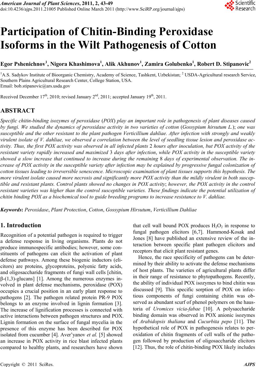 American Journal of Plant Sciences, 2011, 2, 43-49 doi:10.4236/ajps.2011.21005 Published Online March 2011 (http://www.SciRP.org/journal/ajps) Copyright © 2011 SciRes. AJPS 43 Participation of Chitin-Binding Peroxidase Isoforms in the Wilt Pathogenesis of Cotton Egor Pshenichnov1, Nigora Khashimova1, Alik Akhunov1, Zamira Golubenko1, Robert D. Stipanovic2 1A.S. Sadykov Institute of Bioorganic Chemistry, Academy of Science, Tashkent, Uzbekistan; 2 USDA-Agricultural research Service, Southern Plains Agricultural Research Center, College Station, USA. Email: bob.stipanovic@ars.usda.gov Received December 17th, 2010; revised January 2nd, 2011; accepted January 19th, 2011. ABSTRACT Specific chitin-binding isozymes of peroxidase (POX) play an important role in pathogenesis of plant diseases caused by fungi. We studied the dynamics of peroxidase activity in two varieties of cotton (Gossypium hirsutum L.); one was susceptible and the other resistant to the plant pathogen Verticillium dahliae. After infection with strongly and weakly virulent isolate of V. dahliae, we observed a correlation between the level of seedling tissue lesion and peroxidase ac- tivity. Thus, the first POX activity was observed in all infected plants 2 hour s after inoculation, but POX activity of the resistant variety rapidly increased and maximized 3 days after infection, while POX activity in the susceptible variety showed a slow increase that continued to increase during the remaining 8 days of experimental observation. The in- crease of POX activity in the susceptib le variety after infection may be explain ed by progressive fungal colon ization of cotton tissues leading to irreversible senescence. Microscopic examination of plant tissues supports this hypothesis. The more virulent isolate caused more necrosis and significantly more POX activity than the mildly virulent in both suscep- tible and resistant plants. Control plants showed no changes in POX activity; however, the POX activity in the control resistant varieties was higher than the control susceptible varieties. These findings indicate the potential utilization of chitin binding POX as a biochemical tool to guide breeding programs to increase resistance to V. dahliae. Keywords: Peroxidase, Plant Protection, Cotton, Gossypium Hirsutum, Verticillium Dahliae 1. Introduction Recognition of a potential pathogen is required to trigger a defense response in living organisms. Plants do not produce immunospecific antibodies; however, some con- stituents of pathogens can elicit the activation of plant defense pathways. Among these biogenic inductors (eli- citors) are proteins, glycoproteins, polyenic fatty acids, and oligosaccharide fragments of fungi wall cells [chitin, β-(1,3)-glucans] [1]. Among the numerous enzymes in- volved in plant defense mechanisms, peroxidase (POX) occupies a crucial position in an early plant response to pathogens [2]. The pathogen related protein PR-9 POX belongs to an enzyme involved in lignin formation [3]. The increase of lignification processes is connected with active interactions between pathogen structures and POX. Lignin formation on the surface of fungal mycelia in the presence of this enzyme has been described for POX isolated from cucumber [4]. Aver’yanov et al. [5] showed an increase in POX activity in rice blast infected plants compared to healthy plants, and researchers have shown that cell wall bound POX produces H2O2 in response to fungal pathogen elicitors [6,7]. Hammond-Kosak and Jones [8] have published an extensive review of the in- teraction between specific plant pathogen elicitors and receptors that elicit plant resistant genes. Hence, the race specificity of pathogens can be deter- mined by their ability to activate the defense mechanisms of host plants. The varieties of agricultural plants differ in their range of resistance to phytopathogens. Recently, the ability of individual POX isozymes to bind chitin was discussed [9]. This specific sorption of POX on infec- tious components of fungi containing chitin was ob- served as abundant scurf of phenol polymers on the haus- toria of Uromices vicia-fabae [10]. A polysaccharide binding domain was observed in POX anionic isozymes of Arabidopsis thaliana and Cucurbita pepo [11]. The hypothetical role of POX in pathogenesis relates to per- oxidation of chitin fragments of cell walls of the patho- gen followed by production of oligosaccharide elicitors [12]. Thus, the role of chitin-binding POX likely includes  Participation of Chitin-Binding Peroxidase Isoforms in the Wilt Pathogenesis of Cotton Copyright © 2011 SciRes. AJPS 44 initiation of plant defense mechanisms after pathogen attack. Herein we report our investigation of the dynamic activation of POX in seedlings from two cotton varieties (Gossypium hirsutum L.) varieties triggered by the action of two different strains of the fungal wilt pathogen Verti- cillium dahliae and discuss its POX activation. 2. Materials and Methods 2.1. Biological Material Cottonseed (Gossypium hirsutum L.) of AN-Bayaut-2 and C-4727 varieties were grown in an experimental field at the Institute of Cotton Breeding and Seed Pro- duction (Tashkent, Uzbekistan). AN-Bayaut-2 is consid- ered to be highly resistant to V. dahliae and C-4727 is considered to be susceptible [13]. Conidia of V. dahliae Kleb. (weakly virulent T-4 and highly virulent AN-3 isolates) were originally isolated from the diseased cotton and are from the collection housed at the Institute of Ge- netics, Academy Sciences of Republic of Uzbekistan, Tashkent, Uzbekistan. The pathogen was grown on Czapek’s agar. 2.2. General Chitin from crab shells (Fluka Chemical Company) was used for chromatography. Isoelectric focusing of proteins was carried out on an LKB Multiphor-2117 apparatus. Sigma Chemical Company’s IEF mix 3.5-10.6 was used as the isoelectric focusing marker. Isoelectrofocusing of POX was carried out on a horizontal plate containing 7% polyacrylamide gel, 0.016% N, N-methylene-bis-acryla- mide, 10% glycerol, 1.5% ampholines pH 3.5-10 (LKB, Sweden) and 0.033% ammonium persulfate in 8 M urea. The anode was 0.5% HCI, and the cathode was 0.5% NaOH. The POX activity was ascertained on the gel us- ing a 0.1% solution of benzidine dihydrochloride. POX isozymes were quantitated by reaction with a 0.01% ben- zidine dihydrochloride solution (Reachim Company, Russia) and 0.005% Н202 in a 0.1% sodium acetate buffer (рН 4.7), and the absorption maxima at 620 nm was measured after one minute of reaction time. The POX activity of each isozyme was determined by meas- uring the color intensity using a LKB Densitometer. The total protein concentration was determined by the method of Lowry [15]. Assays were performed using an optical microscope [Neofot-2 (Carl Zeiss, Germany)] at 90 x magnification. All experiments were repeated at least three times. Data were subjected to analysis of variance (ANOVA), and differences between treatments assessed by Student’s two-sample t-test at P < 0.05. 2.3. Infection of Cotton Seedlings Cottonseed were cleansed with H2SO4, rapidly washed with running water and kept in water overnight. Seed- lings were wrapped in paper towels for 7 days at 27˚C. Infection of seedlings was initiated by placing the towels in flasks containing a conidial suspension (107 conidia per mL) of V. dahliae . 2.4. Lesion Assay Cottonseeds were sterilized by treatment with a solution containing 36 % H2O2 and 96 % ethanol (1:1). V. dahliae (AN-3 and 4-T strains) conidia were incubated in sepa- rate tubes (2.0x20 cm) with Czapek’s agar (1 × 10⁶ per tube; 2.0-2.5 cm columns of media) for 10 days. Then a warm agar solution was added to the tubes and a single sterilized seed was placed in this layer in each of six tubes for each treatment. The lesion on seedlings was evaluated by visual observation and plant damage was assayed by measuring the height of the seedlings and the size of the necrotic zones on the surfaces of damaged seedlings. 2.5. Crude Enzyme Preparation The enzyme preparation was a modification of that pre- viously described [14]. That is, raw plant material was ground in liquid nitrogen and stirred with 0.1M sodium phosphate buffer (pH 6.6), and 0.5M NaCl (5 mL per 1 g of plant tissue) with a magnetic mixer 1 h at 4˚C. The precipitate was removed by centrifugation (6000 g, 20 min). The supernatant solution was used for further in- vestigations. 2.6. Adsorption of POX Isozymes on Chitin and V. dahliae Conidia All procedures were carried out at 4˚C. The supernatant obtained as described above was treated with ammonium sulfate to 30% saturation. The supernatant solution was collected and brought to 70% (NH4)2SO4 saturation. The residue was collected by centrifugation (6000 g, 20 min), resuspended in a minimal volume of twice-distilled water and desalted in 0.01M sodium phosphate buffer (pH 6.6) using “Vivaspin 20” tubes (Sartorius, Germany) at 6000 g. The chitin adsorption assay was performed on a chro- matographic column (2 cm × 6 cm) packed with chitin previously equilibrated with 0.1M sodium phosphate buffer (pH 6.6). The crude protein preparation was ap- plied to the top of the column and the column was washed with 0.1 M phosphate buffer with a flow of 20 mL/h to remove the unbound protein. Fractions were collected and protein fractions were detected by moni- toring the UV absorption at 280 nm using a flow cell on an Uvicord system (LKB, Sweden). When all of the un- bound protein had been eluted, the bound protein was eluted with 1 M NaCl in 0.01 M phosphate buffer (pH 6.6). The V. dahliae conidia adsorption assay was carried  Participation of Chitin-Binding Peroxidase Isoforms in the Wilt Pathogenesis of Cotton Copyright © 2011 SciRes. AJPS 45 out in “volume”. Conidia (500 mg) were previously treated with 1N NaOH (5 min) and spun down (6000 g, 20 min.) and then washed with 0.1M sodium phosphate buffer (pH 6.6). After a second spin (6000 g, 20 min.) the desalinated total protein preparation was suspended with conidia in a minimal volume of the same buffer (30 min), and conidia were eluted with some portions of buffer. The column was eluted with 1M NaCl in 0.01M sodium phosphate buffer (pH 6.6). Fractions were monitored at 280 nm as indicated above. 3. Results and Discussion 3.1. Peroxidase Activity in Seedlings Segments of the two varieties of 7-day-old cotton seed- lings were harvested each hour after infection with V. dahliae conidia (AN-3 strain) up to 6 hour (Figure 1(a)) and then daily for 8 days (Figure 1(b)). POX activity in control samples was not statically different at any period compared to time zero. In the case of the AN-Bayaut-2 cotton variety, we observed an increase in POX activity at 2 hours after infection in all segments (Figure 1(a)). At 3 hours the POX activity had slightly decreased and further significant increase was observed after 3 days (Figure 1(b)) with a gradual lost of activity up to 8 days. The POX activity of the C-4727 was not significantly different than the control after 2 hour and 8 days in any of the time segments; there was a slight increase in POX activity beginning 3 days after infection in C-4727 (Fig- ure 1(a), (b)) and continuing until 8 days. This is proba- bly due to the additional tissue that is infected with time. In the case of AN-Bayaut-2, the pathogen infection is quickly contained and additional tissues are not infected; thus, the level of POX decreases. 3.2. Lesion of Seedlings The effect of the V. dahliae isolates on the two cotton cultivars are shown in Table 1. The C-4727 cotton seed- lings were 1.8 cm shorter than the control seedlings when they were treated V. dahliae isolate 4-T, while those for AN-Bayaut-2 were only 0.5 cm shorter than the control when treated with the same isolate. The V. dahlia e AN-3 isolate had a greater effect on these cotton varieties. That is, C-4727 was 2.6 cm shorter than the control and AN-Bayaut-2 was 2.2 cm shorter. Surface symptoms of infection appeared for C-4727 cotton seedlings after 4-5 days as small dark necrotic lesions on stems, cotyledon leaves, and roots. These le- sions were about 1 cm in length on stem and cotyledon tissues with desiccation of tissues. Fifteen to eighteen days after infection of the C-4727 cultivar with the highly virulent V. dahliae isolate AN-3 the seedlings were dead. Thus, the C-4727 cultivar was highly suscep- (a) (b) Figure 1. Dynamics of POX activity in 7-days seedlings of two cotton varieties susceptible (C-4727) and resistant (AN- Bayaut-2) after infection (a – in hours, and b – in days) with V. dahliae (AN-3 isolate) conidia. Table 1. Growth inhibition of cotton seedlings after infec- tion with different strains of V. dahliae (AN-3, highly viru- lent; 4-T weakly virulent). Height of seedlings after 8 days (cm) V. dahliae isolate C-4727 AN-Bayaut-2 Control 14.5 ± 0.6 13.5 ± 1.2 4-T 12.7 ± 0.8 13.0 ± 1.6 AN-3 11.9 ± 0.5 11.3 ± 1.3 tible to the AN-3 isolate. In comparison, when the AN-Bayaut-2 cotton seedlings were infected with the AN-3 isolate, some growth inhibition was observed as well as the appearance of necrotic zones in hypocotyl tissue (about 2 mm length) 15 to 18 days after infection. The isolate 4-T did not cause any surface symptoms ex- cept a small inhibition in growth for AN-Bayaut, and only a 1.8 cm inhibition in growth in the C-4727. 3.3. Optical Microscopic Assay of Cotton Seedling Tissues after Infection with V. dahliae Pathogen penetration sites on root rhizoderma of AN-  Participation of Chitin-Binding Peroxidase Isoforms in the Wilt Pathogenesis of Cotton Copyright © 2011 SciRes. AJPS 46 Bayaut-2 cotton seedlings appeared under the micro- scope as zones of necrosis around live cells. Figure 2(a) shows the cytoplasm of root cortex cells of plants inocu- lated with isolate 4-T. These cells were filled with opti- cally dense material. In the case of AN-3 isolate, the mycelia extended to the root cortex parenchyma (Figure 2(b)). After infection of C-4727 cotton seedlings with both 4-T and AN-3 the mycelia of V. dahliae isolates had more thoroughly permeated the root parenchyma. Hypo- cotyl microscopic sections of AN-Bayaut-2 and C-4727 cotton seedlings had clear differences in pathogen pene- tration. Hyphae penetrated both the inter- and intracellu- lar C-4727 cotton seedlings. The pathogen freely pene- trated the vascular tissues of susceptible C-4727 and can be easily visualized. In the fourth day after infection of C-4727 seedlings we observed colonization of intercel- lular spaces with AN-3 fungal hyphae (Figure 3(a)). In comparison, vascular cells of AN-Bayaut-2 cotton seed- lings were thickened and separated from this pathogen (Figure 3(b)), and pathogen penetration was not observed. Tissues of C-4727 vascular system also exhibited cell plasmolysis, i.e. plasmolemma discharge from vascular cell wall (Figure 4(a)). A histochemical analysis of a non fixed preparation of AN-Bayaut-2 vascular cells shows that POX activity is localized, in external and in- ternal surfaces of cell wall and in the zones of necrosis (Figures 3(b) and 4(b)). (a) (b) Figure 2. V. dahliae (4-T, a and AN-3, b isolates) penetration of the root rhizoderma of AN-Bayaut-2 cotton seed- lings. Arrows indicate nodules in cell wall and necrosis. (a) (b) Figure 3. Verticillium dahliae (AN-3 isolate) penetration sites of hypocotyls of C-4727 (a) and cotton seedlings (AN-Bayaut-2) (b). Arrows indicate hyphae between cells of C-4727 (a) and cell wall and necrotic zone of AN-Bayaut-2 (b).  Participation of Chitin-Binding Peroxidase Isoforms in the Wilt Pathogenesis of Cotton Copyright © 2011 SciRes. AJPS 47 (a) (b) Figure 4. Tissues of C-4727 vascular system after infection with AN-3 isolate of Verticillium dahliae (a). Histo- chemical analysis of POX localization in AN-Bayaut-2 vascular cells infected with AN-3 isolate of V. dahliae (b). 3.4. Isozymes with POX Activity in 7-Day-Old Cotton Seedlings Anionic isozymes of POX accumulate during cell wall lignifications. Zheng [16] and Passardi [17] showed that resistant plants rapidly accumulated lignin after infection with fungal pathogens. The pathogen penetrates through intercellular space by hyphal growth. Chitin biosynthesis is localized in the apical zone of hyphae and its frag- ments may penetrate to the intercellular space inducing activity of extracellular anionic isozymes of POX. Thus, these POXs may bind with and localize the fungal pathogens. We designed an experiment which modeled the interaction between the fungal pathogen and cotton POX enzymes using V. dahliae conidia as a chroma- tographic matrix. Isoelectrofocusing shows that 7-days after cotton seedlings of AN-Bayaut-2 had two isozymes that bind to both chitin and V. dahlia e conidia (Figure 5, lane 3) while C-4727 had only one of these types of isozyme (Figure 5, lane 6). This data suggest that chitin binding isozymes of POX are signaling molecules in plant defense mechanisms which identify the oligosac- charide containing phytopathogens. 4. Conclusions Expression of resistance genes to phytopathogens is best observed during the infection process when plant cells are exposed to the pathogen. The riposte reaction pre- supposes induction of such resistance factors. In our study we examined the dynamics of POX activity in seedlings of two different cotton varieties infected with weak (T-4) and highly virulent (AN-3) isolates of V. dahliae. When infected with the highly virulent isolate AN-3, we observed higher POX activity in the infected AN-Bayaut-2 variety as compared to the susceptible C-4727. An increase of POX activity of cotton seedlings infected with V. dahliae had a biphasic characteristic during 6 to 8 days. The first enzyme activation of both varieties was noted 2 h after infection. This activation may be described as Figure 5. Isozyme spectrum of POX from 7-day-old cotton seedlings (guaiacol/H2O2 stained): a. AN-Bayaut-2 cotton seedlings: 1 – total POX fraction; 2 – POX fraction not ad- sorbed by chitin or V. dahliae conidia; 3 – chitin-specific isozymes; b. C-4727 cotton seedlings: 4 – total POX fraction; 5 – POX fraction not adsorbed on chitin and V. dahliae co- nidia; 6 – chitin specific isozyme; M – pI markers (Coo- massie BB G-250 stained).  Participation of Chitin-Binding Peroxidase Isoforms in the Wilt Pathogenesis of Cotton Copyright © 2011 SciRes. AJPS 48 super sensitivity to the pathogen. The second activation of POX was observed during the next few days, and ex- hibited significant differences in activity. That is, POX activity of AN-Bayaut-2 rapidly increased and maxi- mized 3 days after infection. This was followed by a gradual decreased in activity. POX activity in the C-4727 variety after infection showed a slow increase that con- tinued to increase during the remaining days. The control samples showed no changes in POX activity during the experiment period, however, the POX activity was higher in the resistant AN-Bayaut-2 plant compared to the sus- ceptible C-4727. The increase of POX activity in the C-4727 variety after infection with V. dahliae may be explained by progressive fungal colonization of cotton tissues leading to irreversible senescence. Microscopic examination supports this hypothesis. Thus, our data correlates well with the level of resis- tance of cotton varieties to fungi pathogens. In addition, the amount of tissue exhibiting necrosis also correlated with the dynamics of POX activity in different parts of the seedlings. The difference of these parameters was dependent on the isolate of V. dahliae which was used for infection. That is, the AN-3 isolate, which is the more virulent, caused more necrosis and significantly more POX activity than the 4-T strain. The hypersensitivity plant reaction to pathogen attack causes POX gene expression and a biphasic accumula- tion of mRNA transcripts which encode biosynthesis of anionic POX [18]. The first phase occurs 2 to 9 hours after infection with the pathogen. The second phase of POX transcript accumulation differs for susceptible and resistant genotype. This phase in susceptible plants oc- curs slower and in resistant plants occurs in 24 to 48 hours after the first phase. Thus, susceptible plants have a slower response allowing greater cell damage. Pathogen penetration and penetration inside the root tissues is ob- served for both susceptible and resistant cotton varieties, but in the resistant variety the surface symptoms do not appear. However, the depth of pathogen penetration de- pends on the resistance of cotton variety. Observation of surface symptoms of wilt infection depends on over- growth of fungal hyphae in plant cells. POX localization around cell wall shows active lignifications which blocks fungal penetration. In this case, chitin, the component of cell walls of V. dahliae, may induce lignifications. Free radicals produced by the action of POX are highly reac- tive and form covalent bounds with proteins and carbo- hydrates of fungi cell walls. Thus, POX may be consid- ered as a defense factor in wilt pathogenesis [11]. These findings offer the potential utilization of chitin binding POX as a biochemical tool to guide breeding programs to increase resistance to V. dahliae. 5. Acknowledgements This work was supported by a grant from the Department of Science and Technologies Development, Republic of Uzbekistan. REFERENCES [1] N. Benhamou, “Elicitor-Induced Plant Defense Path- ways,” Trends in Plant Science , Vol. 1, No. 7, July 1996, pp. 233-240. [2] C. C. Lin and C. H. Kao, “Abscisic Acid Induced Changes in Cell Wall Peroxidase Activity and Hydrogen Peroxide Level in Roots of Rice Seedlings,” Plant Sci- ence, Vol. 160, No. 2, 2001, pp. 323-329. doi:10.1016/S0168-9452(00)00396-4 [3] L. C. Van Loon, “Induced Resistance in Plants and Role of Pathogenesis-Related Proteins,” European Journal of Plant Pathology, Vol. 103, 1997, pp. 735-765. doi:10.1023/A:1008638109140 [4] R. Hammerschmidt and J. Kuc, “Lignification as a Mechanism for Induced Systemic Resistance of Cucum- ber,” Physiological and Molecular Plant Pathology, Vol. 20, 1982, pp. 61-71. doi:10.1016/0048-4059(82)90024-8 [5] A. A. Aver’yanov, V. P. Lapikova, O. N. Nikolaev and A. I. Stepanov, “Active Oxygen-Associated Control of Rice Blast Disease by Riboflavin and Roseoflavin,” Biochem- istry (Moscow), Vol. 65, No. 11, November 2000, pp. 1292-1298. [6] G. P. Bolwell, K. A. Blee, V. S. Butt, P. R. Davies, S. L. Gardner, C. Gerrich, F. Minilaeva, E. G. Rowntree and P. Woitaszek, “Recent Advances in Understanding the Ori- gin of the Apoplastic Oxidative Burst in Plant Cells,” Free Radical Research, Vol. 31, December 1999, pp. 137-145. doi:10.1080/10715769900301431 [7] C. S. Bestwick, I. R. Brown, M. H. R. Bennett and J. M. Mansfield, “Localization of Hydrogen Peroxidase Accu- mulation during the Hypersensitive Reaction of Lettuce Cells to Pseudomonas syringae pv. phaseolicola,” The Plant Cell, Vol. 9, No. 2, February 1997, pp. 209-221. [8] K. E. Hammond-Kosak and J. D. Jones, “Resistant Gene- Dependent Plant Defense Responses,” The Plant Cell, Vol. 8, No. 10, October 1996, pp. 1773-1791. [9] I. V. Maksimov, E. A. Cherepanova, I. E. Akhmetova and R. M. Khairullin, “Contribution of Chitin and Its Oli- gomers to the Induced Resistance of Plants against Phy- topathogens,” Agricultural Chemistry, Vol. 8, 2004, pp. 77-89. [10] C. Schweikert, A. Liszkay and P. Schopher, “Polysaccha- ride Degradation by Fenton Reaction- or Peroxidase- Gene-Rated Hydroxyl Radicals in Isolated Plant Cell Walls,” Phytochemistry, Vol. 61, No. 1, September 2002, pp. 31-35. doi:10.1016/S0031-9422(02)00183-8 [11] C. Dunand, M. Tognolli, M. Meyer, P. Simon and C. Penel, “Identification and Characterization of Ca2+ —  Participation of Chitin-Binding Peroxidase Isoforms in the Wilt Pathogenesis of Cotton Copyright © 2011 SciRes. AJPS 49 Pectate Binding Peroxidase in Arabidopsis thailiana,” Journal of Plant Physiology, Vol. 159, No. 11, November 2002, pp. 1165-1171. doi:10.1078/0176-1617-00768 [12] P. Bonatti, P. Medeghini, G. Lorenzini, R. Formasiero, R. Boroni, C. Nali and E. Sgarbi, “Cytochemical Detection of Cell Wall Bound Peroxidase in Rust Infected Broad Bean Leaves,” Journal of Phytopathology, Vol. 140, No. 4, April 1994, pp. 319-325. doi:10.1007/BF02980704 [13] Sh. I. Ibragimov, “Catalogue of Cotton Varieties. Volume 2,” Tashkent, Uzbekistan, 1993. [14] Z. Golubenko, A. Akhunov, N. Khashimova, Yu. Beres- neva, E. Mustakimova, F. Ibragimov, N. Abdurashidova and R. Stipanovic, “Induction of Peroxidase as a Disease Resistance Response in Resistant (Hibiscus trionum) and Susceptible (Althea armeniaca) in the Family Malvaceae,” Phytoparasitica, Vol. 35, No. 4, August 2007, pp. 401-413. doi:10.1007/BF02980704 [15] O. N. Lowry, N. Rosebrouhg and R. I. Randall, “Protein Measurement with the Folin Phenol Reagent,” Biolgical Chemistry, Vol. 193, November 1951, pp. 265-275. [16] H. Z. Zheng, C. L. Cui, Yu. Zhang, D. Wang, Yu. Jing and K. Y. Kim, “Active Changes of Lignification-Related Enzymes in Pepper Response to Glomus Introduces and/or Phytophthoracapsici,” Journal of Zhejiang Uni- versity Science, Vol. 6B, No. 8, July 2005, pp. 778-786. doi:10.1631/jzus.2005.B0778 [17] F. Passardi, C. Penel and C. Dunand, “Performing the Paradoxical: How Plant Peroxidase Modify the Cell Wall,” Trends in Plant Science, Vol. 9, No. 11, Novem- ber 2004, pp. 534-540. [18] P. Kawalleck, E. Schmelzer, K. Hahlbrock and I. E. Somssich, “Two Pathogen-Responsive Genes in Parsley Encode a Tyrosine-Rich Hydroxyproline Glycoprotein (HRGP) and Anionic Peroxidase,” Molecular and Gen- eral Genetics, Vol. 247, No. 4, 1995, pp. 444-452. doi:10.1007/BF00293146 |