Open Journal of Inorganic Non-Metallic Materials, 2014, 4, 7-11
Published Online January 2014 (http://www.scirp.org/journal/ojinm)
http://dx.doi.org/10.4236/ojinm.2014.41002
OPEN ACCESS OJINM
The Surface Fractal Structure of Fish Scales
Godzhaev Eldar, Abasov Abbasali, Aliyeva Sharafkxanim, Charuxcev David
Department of Physics, Azerbaijan Technical University, Baku, Azerbaijan
Email: geldar-04@mail.ru
Received August 23, 2013; revised September 30, 2013; accepted October 7, 2013
Copyright © 2014 Godz haev Eldar et al. This is an open access article distributed under the Creative Commons Attribution License,
which permits unrestricted use, distribution, and reproduction in any medium, provided the original work is properly cited. In accor-
dance of the Creative Commons Attribution License all Copyrights © 2014 are reserved for SCIRP and the owner of the intellectual
property Godzhaev Eldar et al. All Copyright © 2014 are guarded by law and by SCIRP as a guardian.
ABSTRACT
By atomic force , microscopy studied the morphology of the surface of the scale of the Caspian Kutum. Analysis
of the structure of channels of sensory systems showed that the nanostructures are not only fractal, but also
self-similar. Fractal trajectories originate from the primary nucleus and extend to the sprawling low-density
structure radically outwards scales like Lichtenberg figures. However, in contrast to the figures, Lichtenberg
fractal band scales of fish each year are surrounded by concentric threads. The most likely mechanism for the
formation of structural self-organization on the surface of the scales can be a model Witten-Sander.
KEYWORDS
Fractals; Scales; Fragments; Aggregation; Channels
1. Introduction
Similarity of the surface structures, some biological ob-
jects with fractals, is a v ery interesting fact in itself . Lan-
guage fractal geometrycan be extremely useful to the
description of certain biological objects of living beings.
For example, the shape of the lateral line of the fish is
very convenient to describe a fractal language according
to [1-3]. This approach can be very rewarding. It is
known that the bodies of the lateral line of the fish carry
a distinct mechanoreceptor function. Actually all known
physical and chemical and biotic configurations arising
in a reservoir, are accepted by tools of wel l -developed
sensory systems of fish. The essential role in behavior of
fishes is played with sense organsthe lateral line, or
seismotouch system. It unites all sensitive receptor cells
of shift which can be met in various sites of a body and
the head [3,4]. The fishes catch earthquake before t he most
sensitive devices [4]. Live organisms including bodies of
fishes look expedient(adapted for living conditions)
simply because they die of inadaptation [2,3]. Studying
morphology scales of fishes by a method of the atomic
power microscopy (APM) would bring a certain clarity
in distribution of touch channels.
The aim of the work was to identify the nature of the
morphological features of the surface structures of the
fish scales (for example, Kutum) by optical and atomic
force microscopy (AFM).
2. Experiment and Discussion
The technique of research surface scales with AFM im-
ages in 2D and 3D is given in work [5]. Kutums lateral
line represents a certain channel, which passes along all
ridge, it begins from the head and comes to the end only
at a tail. All surface consists of the weight of sensitive
sensors which are connected with external receptors,
located on scales and the nervous terminations of the fish.
Sensitive cages of receptors of bodies of the studied ob-
jects of the lateral line form the isolated clusters in skin,
but more often they are found through some intervals in
fillets or channels in scales. The channels are completely
closed depressions in the skin, or are in the form of open
fractal grooves. Scale channels are filled with slime that
promoted by porous and fractal character of morphology
of this surface. Sensitive cells of the lateral line are col-
lected in kidney-shaped groups and hidden to channels
on scales of fishes who are washed by water. Removed
on an optical microscope (OP) the morphology of a sur-
face is given in Figures 1 and 2. The fragments of year
rings on a surface scales from under the first fin, touch
channels, pictures with a optic microscope are given in
Figures 3 and 4. OP-images of scales under the top fin
G. ELDAR ET AL.
OPEN ACCESS OJINM
8
connected with annual rings are given in Figure 5 . AFM
fragments of nanodistributions round year rings of fish
Kutum were considered in Figure s 6-9. During AFM in-
vestigations mucus from the surface of scales was wa-
shed off with water. Some characteristics of morphology
are clearly visible: growth of branching on scale begins
with a certain place not being in the scales center, but is
closer to its edge watched in Figures 8 and 9. Growth
dynamics of the surface areas can be considered as a spe-
cial case of aggregation model that is suitable for descri-
bing the growth surface for biological systems.
Figure 1. Morphology of a surface of scales of the Caspian
Kutum. Vertical lines proceeding from the center of scales
noted nanogrooves (channels) in which liquids of special
structure are shipped. In the depth of these channels pass
the nervous terminations on which various signals are tran-
smitted.
Figure 2. Fragments of the optical images of the scales of
Kutum. The upper and lower fragments marked fractal
outputs of the individual bands on the surface (the boun-
dary marked by horizontal bands, and the vertical arrows
marked zigzag s hap ed strips, in out put ) .
Figure 3. The photo made by an optical microscope of a
surface of scales of Kutum. By the bottom arrow (No. 5) it is
specified (an ellipsoidal form) the main channel in the scales
center. From this channel in the radial direction strips (2 - 4,
7-creep away 8). From below in skin of fish they are con-
nected with the nervous terminations transmitting certain
signals to the think-tank. Thus, the main fragments of the
sensory system are special channels on the scales of fish.
Figure 4. Morphology of the central part of a lateral surface
of scales of Kutum. The bottom big arrow defines location
of the lateral line. In Figure 1 they are noticeable as dashed
lines). Between two horizontal shooters fragments of the
rarefied structures with cross channels above are allocated.
The same cross channels (it not annual Rings) are noted by
horizontal shooters (on the right drawing). In the left from
below the arrow noted thinner small radial structures of the
line. They carry out a role of touch systems located on a
scales surface.
Lines stretching from grain to edges of scales can be
described by model of Witten-Sander [6-8]. Active sur-
face in the course of growth the lines on it similar light-
nings at the description across Witten-Sandra has abso-
lutely other structure, than in Edens model [7]. This dis-
tinction r eflects an essential role of diffusion and is shown
that clusters in model of Witten-Sander externally look
G. ELDAR ET AL.
OPEN ACCESS OJINM
9
Figure 5. Morphology of a surface of OP of scales, with
Kutum’s allocated annual rings.
Figure 6. AFM fragment—images in 3Dthe scale of a
surface of scales of Kutum: through channels (they are not-
ed by longitudinal lines) carry out communication from the
head to a tail. It also is a fragment of the coherent lateral
line on a scales surface. Separate nanoislands are noted by
vertical lines. The black line noted the aggregated nano-
structures—bioclusters. (Scan size: 7 × 7 mkm).
Figure 7. A fragment of the AFM image in 3-D-scale of the
surface scales of the Caspian Kutum. In the center of nano-
pore allocated round the edges of the vertical arrows indi-
cate the nanoprotrusions.
Figure 8. Longitudinal section of AFM—the image in 2D
the scale of a surface of scales of Kutum: pore sizes ranging
≈ 80-nm, the maximum height of structures within ≈ 100
nm.
Figure 9. AFM image of the surface of the 2D—scale of the
scales of Kutum: profilogram bottom surface of the channel
shows its nano dimension: height of the bottom lines of
scales ranging from 10 nm to 30 nm.
as fractals (see Figures 1-9). Ensembles of particles ag-
gregating form larger objects with strongly branched
rarefied structu re, threadlike strea ms are thus formed, they
are well visible in Figure 4. These thread s as it is visible
from AFM-images are the aggregated fractal clusters of
the various sizes (see Figures 6-10).
On a surface of fish in information transfer nanodi-
mensional channels of scales (them it is possible carry to
touch sensors) have to play a leading role. In the pre-
sented fragments of OPimages in Figures 4 and 5
G. ELDAR ET AL.
OPEN ACCESS OJINM
10
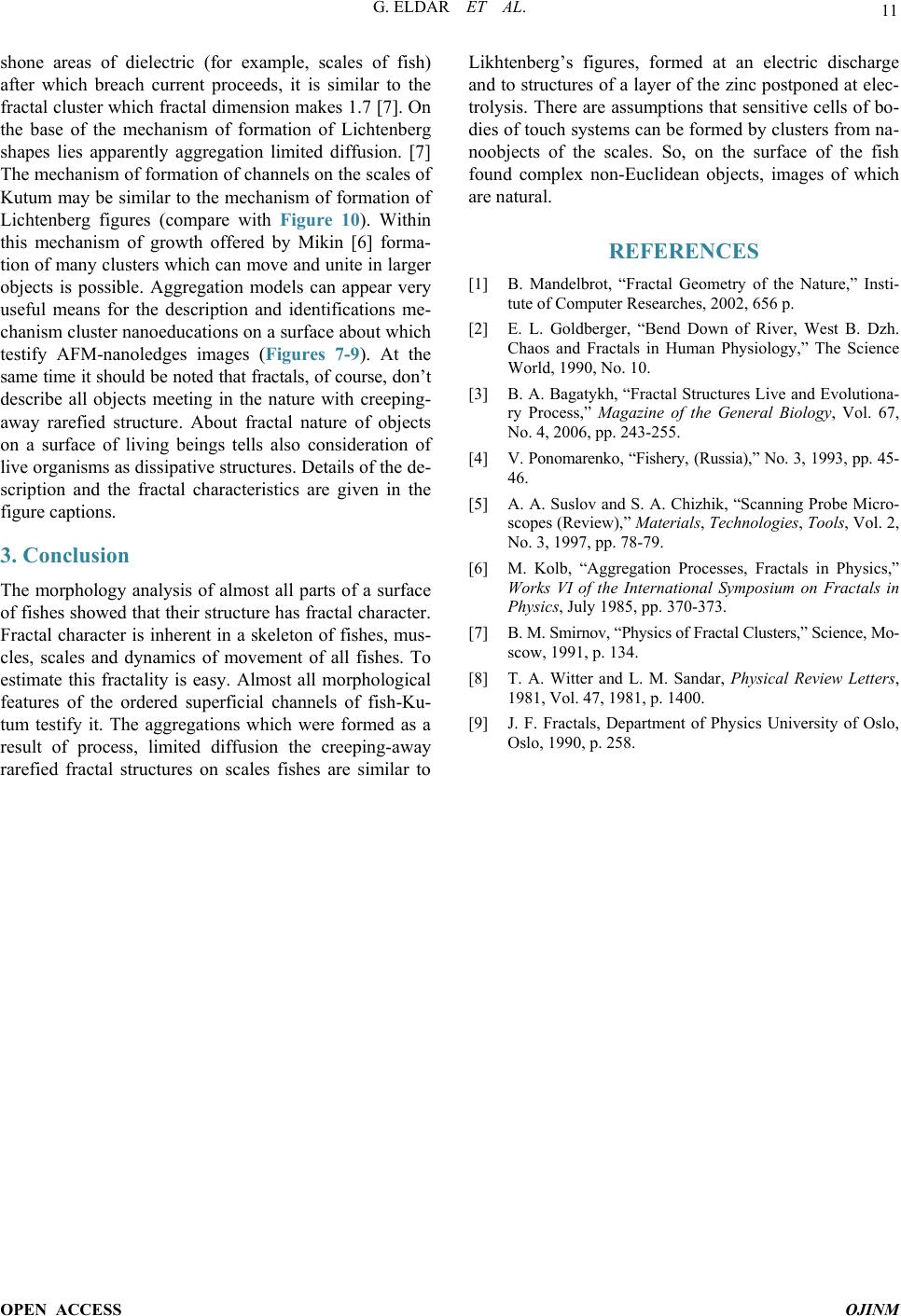
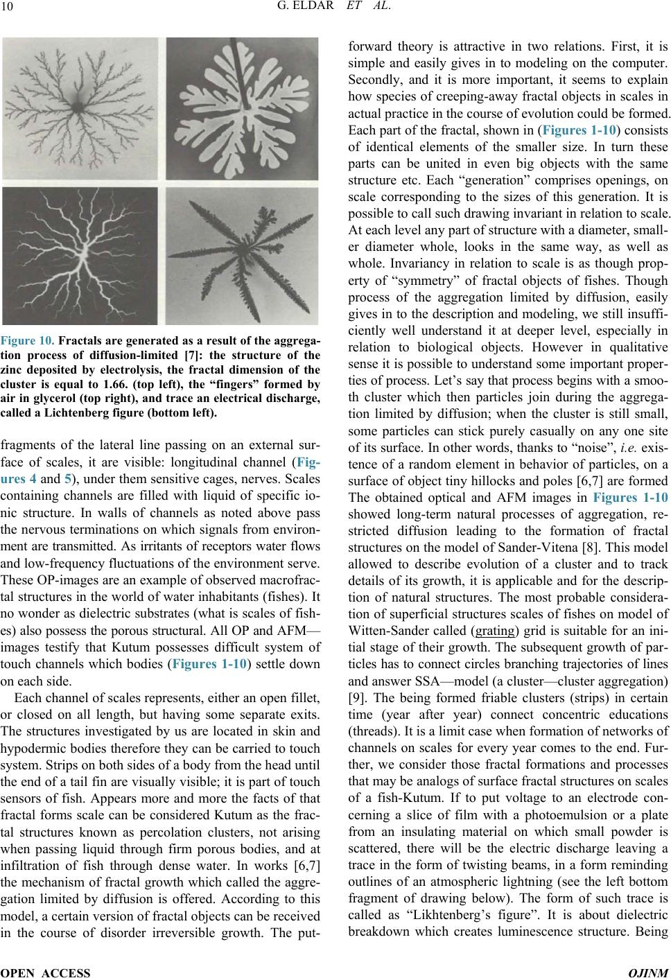
Figure 10. Fractals are generated as a result of the aggrega-
tion process of diffusion-limited [7]: the structure of the
zinc deposited by electrolysis, the fractal dimension of the
cluster is equal to 1.66. (top left), the “fingers” formed by
air in glycerol (top right), and trace an electrical discharge,
called a Lichtenberg figure (bottom left).
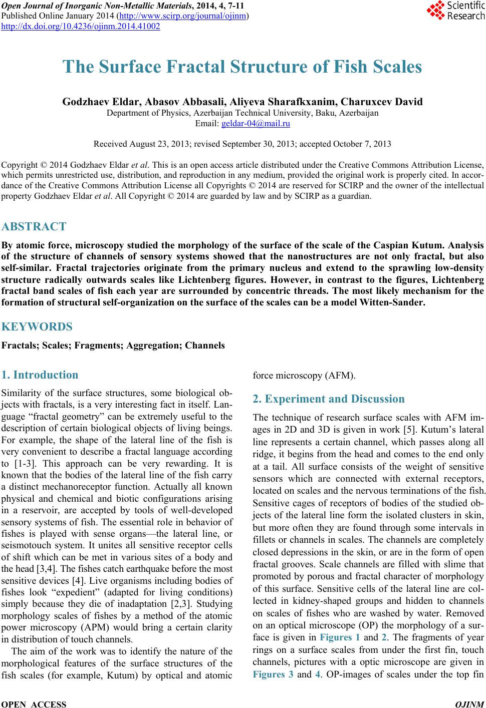
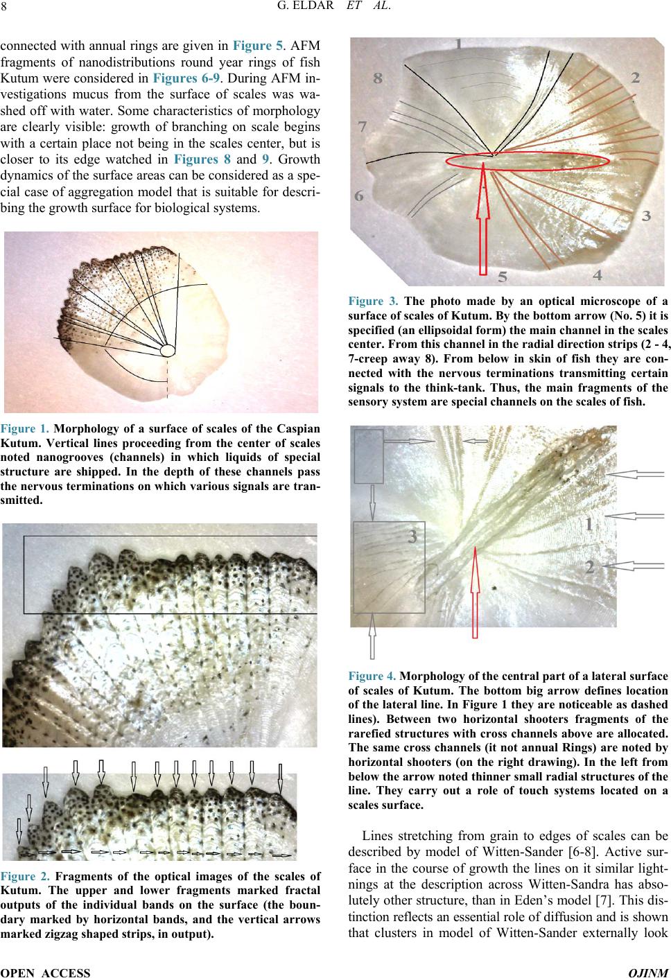
fragments of the lateral line passing on an external sur-
face of scales, it are visible: longitudinal channel (Fig-
ures 4 and 5), under them sensitive cages, nerves. Scales
containing channels are filled with liquid of specific io-
nic structure. In walls of channels as noted above pass
the nervous terminations on which signals from environ-
ment are transmitted. As irritants of receptors water flows
and low-frequency fluctuations of the environment serve.
These OP-images are an example of observed macrofrac-
tal structures in the world of water inhabitan ts (fishes). It
no wonder as dielectric substrates (what is scales of fish-
es) also possess the porous structural. All OP and AFM
images testify that Kutum possesses difficult system of
touch channels which bodies (Figures 1-10) settle down
on each side.
Each channel of scales represents, either an open fillet,
or closed on all length, but having some separate exits.
The structures investigated by us are located in skin and
hypodermic bodies therefore they can be carried to touch
system. Strips on both sides of a body from the head until
the end of a tail fin are visually visib le; it is part of touch
sensors of fish. Appears more and more the facts of that
fractal forms scale can be considered Kutum as the frac-
tal structures known as percolation clusters, not arising
when passing liquid through firm porous bodies, and at
infiltration of fish through dense water. In works [6,7]
the mechanism of fractal growth which called the aggre-
gation limited by diffusion is offered. According to this
model, a certain version of fractal objects can be received
in the course of disorder irreversible growth. The put-
forward theory is attractive in two relations. First, it is
simple and easily gives in to modeling on the computer.
Secondly, and it is more important, it seems to explain
how species of creeping-away fractal objects in scales in
actual practice in the course of evolution could be formed.
Each part of the fractal, shown in (Figures 1-10) consists
of identical elements of the smaller size. In turn these
parts can be united in even big objects with the same
structure etc. Each generationcomprises openings, on
scale corresponding to the sizes of this generation. It is
possible to call such drawing invariant in relation to scale.
At each level any part of structure with a diameter, small-
er diameter whole, looks in the same way, as well as
whole. Invariancy in relation to scale is as though prop-
erty of “symmetry” of fractal objects of fishes. Though
process of the aggregation limited by diffusion, easily
gives in to the description and modeling, we still insuffi-
ciently well understand it at deeper level, especially in
relation to biological objects. However in qualitative
sense it is possible to understand some important proper-
ties of process. Lets say that process begins with a smoo-
th cluster which then particles join during the aggrega-
tion limited by diffusion; when the cluster is still small,
some particles can stick purely casually on any one site
of its surface. In other words, thanks to noise”, i.e. exis-
tence of a random element in behavior of particles, on a
surface of object tiny hillocks and poles [6,7] are formed
The obtained optical and AFM images in Figures 1-10
showed long-term natural processes of aggregation, re-
stricted diffusion leading to the formation of fractal
structures on the model of Sander-Vitena [8]. This model
allowed to describe evolution of a cluster and to track
details of its growth, it is applicable and for the d escr ip-
tion of natural structures. The most probable considera-
tion of superficial structures scales of fishes on model of
Witten-Sander called (grating) grid is suitable for an ini-
tial stage of their growth. The subsequent growth of par-
ticles has to connect circles branching trajectories of lines
and answer SSA—model (a clu ster—cluster aggregation)
[9]. The being formed friable clusters (strips) in certain
time (year after year) connect concentric educations
(threads). It is a limit case when formation of networks of
channels on scales for every year comes to the end. Fur-
ther, we consider those fractal formations and processes
that may be analogs of surface fractal structures on scales
of a fish-Kutum. If to put voltage to an electrode con-
cerning a slice of film with a photoemulsion or a plate
from an insulating material on which small powder is
scattered, there will be the electric discharge leaving a
trace in the form of twisting beams, in a form reminding
outlines of an atmospheric lightning (see the left bottom
fragment of drawing below). The form of such trace is
called as “Likhtenberg’s figure. It is about dielectric
breakdown which creates luminescence structure. Being
G. ELDAR ET AL.
OPEN ACCESS OJINM
11
shone areas of dielectric (for example, scales of fish)
after which breach current proceeds, it is similar to the
fractal cluster which fractal dimension makes 1.7 [7]. On
the base of the mechanism of formation of Lichtenberg
shapes lies apparently aggregation limited diffusion. [7]
The mechanism of formation of channels on the scales of
Kutum may be similar to the mechanism of formation of
Lichtenberg figures (compare with Figure 10). Within
this mechanism of growth offered by Mikin [6] forma-
tion of many clusters which can move and unite in larger
objects is possible. Aggregation models can appear very
useful means for the description and identifications me-
chanism cluster nanoeducations on a surface about which
testify AFM-nanoledges images (Figures 7-9). At the
same time it should be noted that fractals, of course, don’t
describe all objects meeting in the nature with creeping-
away rarefied structure. About fractal nature of objects
on a surface of living beings tells also consideration of
live organisms as dissipativ e structur es. Details of the de-
scription and the fractal characteristics are given in the
figure captions.
3. Conclusion
The morphology analysis of almost all parts of a surface
of fishes showed that their structure has fractal character.
Fractal character is inherent in a skeleton of fishes, mus-
cles, scales and dynamics of movement of all fishes. To
estimate this fractality is easy. Almost all morphological
features of the ordered superficial channels of fish-Ku-
tum testify it. The aggregations which were formed as a
result of process, limited diffusion the creeping-away
rarefied fractal structures on scales fishes are similar to
Likhtenbergs figures, formed at an electric discharge
and to structures of a layer of the zinc postponed at elec-
trolysis. There are assumptions that sensitiv e cells of bo-
dies of touch systems can be formed by clusters from na-
noobjects of the scales. So, on the surface of the fish
found complex non-Euclidean objects, images of which
are natural.
REFERENCES
[1] B. Mandelbrot, “Fractal Geometry of the Nature,” Insti-
tute of Computer Researches, 2002, 656 p.
[2] E. L. Goldberger, “Bend Down of River, West B. Dzh.
Chaos and Fractals in Human Physiology,” The Science
World, 1990, No. 10.
[3] B. A. Bagatykh, “Fractal Structures Live and Evolutiona-
ry Process,” Magazine of the General Biology, Vol. 67,
No. 4, 2006, pp. 243-255.
[4] V. Ponomarenko, Fishery, (Russia ),” No. 3, 1993, pp. 45-
46.
[5] A. A. Suslov and S. A. Chizhik, “Scanning Probe Micro-
scopes (Review),” Materials, Technologies, Tools, Vol. 2,
No. 3, 1997, pp. 78-79.
[6] M. Kolb, “Aggregation Processes, Fractals in Physics,”
Works VI of the International Symposium on Fractals in
Physics, July 1985, pp. 370-373.
[7] B. M. Smirnov, Physics of Fractal Cluste rs,” Science, Mo-
scow, 1991, p. 134.
[8] T. A. Witter and L. M. Sandar, Physical Review Let t e rs,
1981, Vol. 47, 1981, p. 1400.
[9] J. F. Fractals, Department of Physics University of Oslo,
Oslo, 1990, p. 258.