Paper Menu >>
Journal Menu >>
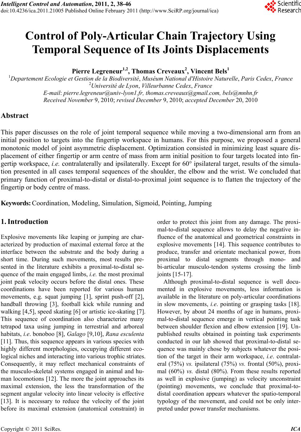 Intelligent Control and Automation, 2011, 2, 38-46 doi:10.4236/ica.2011.21005 Published Online February 2011 (http://www.SciRP.org/journal/ica) Copyright © 2011 SciRes. ICA Control of Poly-Articular Chain Trajectory Using Temporal Sequence of Its Joints Displacements Pierre Legreneur1,2, Thomas Creveaux2, Vincent Bels1 1Departement Ecologie et Gestion de la Biodiversité, Muséum National d'Histoire Naturelle, Paris Cedex, France 2Université de Lyon, Villeurbanne Cedex, France E-mail: pierre.legreneur@univ-lyon1.fr, thomas.creveaux@gmail.com, bels@mnhn.fr Received November 9, 2010; revised December 9, 2010; accepted December 20, 2010 Abstract This paper discusses on the role of joint temporal sequence while moving a two-dimensional arm from an initial position to targets into the fingertip workspace in humans. For this purpose, we proposed a general monotonic model of joint asymmetric displacement. Optimization consisted in minimizing least square dis- placement of either fingertip or arm centre of mass from arm initial position to four targets located into fin- gertip workspace, i.e. contralaterally and ipsilaterally. Except for 60° ipsilateral target, results of the simula- tion presented in all cases temporal sequences of the shoulder, the elbow and the wrist. We concluded that primary function of proximal-to-distal or distal-to-proximal joint sequence is to flatten the trajectory of the fingertip or body centre of mass. Keywords: Coordination, Modeling, Simulation, Sigmoid, Pointing, Jumping 1. Introduction Explosive movements like leaping or jumping are char- acterized by production of maximal external force at the interface between the substrate and the body during a short time. During such movements, most results pre- sented in the literature exhibits a proximal-to-distal se- quence of the main engaged limbs, i.e. the most proximal joint peak velocity occurs before the distal ones. These coordinations have been reported for various human movements, e.g. squat jumping [1], sprint push-off [2], handball throwing [3], football kick while running and walking [4,5], speed skating [6] o r artistic ice-skating [7]. This sequence of coordination also characterize many tetrapod taxa using jumping in terrestrial and arboreal habitats, i.e. bonoboo [8], Galago [9,10], Rana esculenta [11]. Thus, this sequence appears in various species with highly different morphologies, occupying different eco- logical niches and interacting into various tro phic striates. Consequently, it may reflect mechanical constraints of the musculo-skeletal systems engaged in animal and hu- man locomotions [12]. The more the joint approaches its maximal extension, the less the transformation of the segment angular velocity into linear velocity is effective [13]. It is necessary to reduce the velocity of the joint before its maximal extension (anatomical constraint) in order to protect this joint from any damage. The proxi- mal-to-distal sequence allows to delay the negative in- fluence of the anatomical and geometrical constraints in explosive movements [14]. This sequence contributes to produce, transfer and orientate mechanical power, from proximal to distal segments through mono- and bi-articular musculo-tendon systems crossing the limb joints [15-17]. Although proximal-to-distal sequence is well docu- mented in explosive movements, less information is available in the literature on poly-articular coordinations in slow movements, i.e. pointing or grasping tasks [18]. However, by about 24 months of age in humans, proxi- mal-to-distal sequence emerge in vertical pointing task between shoulder flexion and elbow extension [19]. Un- published results obtained in pointing task experiments conducted in our lab showed that proximal-to-distal se- quence was mainly chose by subjects whatever the posi- tion of the target in their arm workspace, i.e. contralat- eral (75%) vs. ipsilateral (75%) vs. frontal (50%), proxi- mal (60%) vs. distal (80%). From these results reported as well in explosive (jumping) as velocity unconstraint (pointing) movements, we conclude that proximal-to- distal coordination appears whatever the spatio-temporal typology of the movement, and could not be only inter- preted under power transfer mechanisms.  P. LEGRENEUR ET AL. Copyright © 2011 SciRes. ICA 39 As shown in Figure 1, a common characteristic of all previously described movements is the linear or quasi- linear shape of the body centre of mass (CoM) of the subjects or the distal extremity of the moving poly-arti- cular chain, e.g. the fingertip in pointing task. Fingertip trajectory concavity depends also on the spatial position of the target to reach and increase from ipsilateral to contralateral sides [20]. In human jumping and animal leaping, we showed that, whereas the magnitude of the velocity vector increased all along the CoM trajectory during push-off, its orientation was reached at 20% of total push-off time in Microcebus murinus [21] and at 40% in humans [22] and then remained constant. In other words, after an orien tation phase of the body CoM, these species tend to maintain their trajectory linear. Trajecto- ries of fingertip or CoM result from spatio-temporal or- ganizations of limb segments, i.e. shoulder, elbow and wrist in pointing task and hip, knee, ankle and meta- tarso-phalangeal joint in jumping. If we suppose that these joints move through rotations in a strictly mono- tonic manner, i.e. exclusive flexion or extension, it is obvious that simultaneous displacements of joints com- posing a poly-articular chain will induce angular dis- placement of its distal extremity or CoM. Thus, we can predict that linear trajectory of distal end or CoM of a poly-articular chain results necessary from temporal joints sequence. The main purpose of the present theo- retical study was then to test this pred iction. 2. General Model of Joint Displacement Throughout this paper, we state that the joint angle dis- placement is a monotonic function of time and that no regulation of the tr ajectory by the central ner vous system occurs during the whole joint displacement. Conse- quently, joint displacement was modelled through a sig- moid profile (Figure 2(a)) and a velocity bell-shaped shape (Figure 2(b)) [23,24]. These shapes account for synergistic actuators activations at a joint, i.e. agonist and antagonist musculo-tendon systems. At the start of the movement, the kinematics of the joint is gi ven by Figure 1. Stick diagrams of pointing or jumping in human and animals. Arrows indicate the sense of the movements. (a) Arm pointing task in human to a ipsilateral and distal target. Red circles represent the fingertip; (b) Push-off phase in human squat jumping. Red circles represent the body centre of mass; (c) and (d) Push-off phase in maximal high leaping in Micro- cebus murinus and Anolis carolinensis. Red circles represent their body mass centres assimilated to their iliac crest.  P. LEGRENEUR ET AL. Copyright © 2011 SciRes. ICA 40 time ( c ) dis p lacemen t velocit y acceleratio n α β κ α α (a) time (b) time Figure 2. Normalized time histories of general joint kinematics. (a) Normalized angular displacement: angle is achieved at instant ; (b) Normalized angular velocity: peak velocity is achieved at instant ; (c) Normalized angular accelera- tion allowed characterizing a biphasic joint displacement: instant corresponds to zero acceleration. 0 0 ii ii i t ttt t (1) Similarly, at the end of the joint rotation, 0 0 ff f f f t ttt t (2) Thus, the general formulation of joint displacement is given by τ with τ ii ff ifi ifi tt tt tttt t ; if ttt (3) where is a function defined on [0, 1] such as 0110 . This function is at least of class C², i.e. is at least twice continuous and differentiable. More- over, has to account for the asymmetric profile of joint angular velocity reported experimentally [25]. It should be noted that this asymmetry should be modified by several constraints. Thus, it increases with spatial accuracy demands [26]. Moreover, the faster is the movement, the longer is the acceleration phase [27]. In other words, the asymmetry of the velocity peak should be inverted if the velocity exceeds a specific threshold. Many equations are available in the literature to model symmetric sigmoid shapes. Fewer equations were de- veloped for asymmetric shapes, i.e. plant growth [28] or handwriting [29]. However, they are of class C1 or do not satisfy equation (3). So, we developed an original sig- moid model of class C as: ,, 0 1 ,, 0 a a hydy hydy (4) ,, , ,, , ,,1 0,0, 0,,1 0,0, 11 0,,1 a a a fygya y fya y hyfya y fyg yay (5)  P. LEGRENEUR ET AL. Copyright © 2011 SciRes. ICA 41 where represents the normalized time at which nor- malized angular peak velocity is attained. This in- stant corresponds to no rmalized joint position . These are given by 0i f i tt tt (6) 0i f i (7) f i fi K tt (8) where K repr esents absolute an gular peak velocity. Thu s, these parameters are linked through the following rela- tions: 0 (9) , f and ,a g are given by ,1 1 , , 00 0,1, 10 n yy n f yfye f (10) And , 1 0 10,0, 1 0,,1 t at a a gyea y ay (11) Solving Equation (9) allows posing 1 1 (12) From Equations (9) and (11), we can deduce , f : 11 e (13) Typical time courses of both joint displacement and angular velocity are given in Figure 3 for various values. That illustrates the flexibility of the model for simulating asymmetric shaped curves on both left (0.5 ) and right (0.5 ) sides. 3. Poly-Articular Chain Model Considering a poly-articular chain composed of a set of p points p A such as 0 A is the origin of the reference frame 0,, A ij R, angular positions of the articulations are given by Figure 3. Absolute time histories of general joint displacement for various parameterizations. (a) Angular displacement and velocity profiles with instant of velocity peak 0.2,0.4,0.6,0.8 . i t, f t, i θ, f, a and were fixed at 0, 1, 0, 90, 1 and respectively; (b) Angular displacement and velocity profiles with beginning of the movement 0.1,0.2,0.3,0.4 i t. f t, i θ, f, , a and were fixed at 1, 0, 90, 0.5, 1 and 0.5 respectively.  P. LEGRENEUR ET AL. Copyright © 2011 SciRes. ICA 42 101 21 1 , AA 2,,, , i ipAAAA (14) with the constraint 1,,, , iii ip (15) Segment lengths i l are defined as 1 1,,, iii iplAA (16) Thus, the coordinates of j A in the reference frame R are deduced from 11 0 1 1 cos sin ji j ii ik jji j ii k i xl AA yl (17) To model a human upper limb, we considered one subject of 1.80 m height. Lengths and positions of CoM of the segments constituting the upper limb were deter- mined from anthropometric tables [30]. Lengths are pre- sented as percent of total body height (0.108, 0.146 and 0.186 for the hand, forearm and upper arm respectively) as well as distance between proximal end of segments and their CoM (0.506, 0.430 and 0.436 for the hand, fo- rearm and upper arm respectively) and their masses (0.006, 0.016 and 0.028 for the hand, forearm and upper arm respectively). One degree of freedom was consid- ered per arm joint, i.e. shoulder abduction/adduction, elbow flexion/extension and wrist abduction/adduction. The angles corresponding to minima and maxima of joints displacement are respectively –60°/120° for the shoulder (S ), 0°/130° for the elbow ( E ) and –10°/25° for the wrist (W ) (Figure 4(a)). 0° corresponds to joint extension. These degrees of freedom of the arm allow defining the workspace of the fingertip in a two-dimen- sional space (Figure 4 (b)) [31]. For all targets, initial arm position was set so that the fingertip was located in front of the shoulder at a dis- tance of 30% of the arm length. The target distance was set at 80% of arm length for all targets. The spatial an- gles of the four distal targets were set at 20º and 60º ipsi- laterally, and 120º and 160º contralaterally (Figure 4(b)). At the beginning of the movement, angular positions of the shoulder, the elbow and the wrist were determined using classical inverse geometrical procedure (0 S , 130 E , 25 S ). 4. Upper Limb Joints Coordinations in Simulated Pointing Task For each joint of the arm, three control parameters were considered, i.e. , , a and f . Optimization consisted in minimizing least square displacement of either finger- tip or arm CoM. Each position was calculated over 1 s movement at a frequency of 100 Hz. Consequently, per- formance criterion J is given by : Figure 4. (a) Planar schematic representation of the arm and its reference frame. S θ, E and W represent the shoulder, elbow and wrist joint positions. Maximal extension corresponds to 0°; (b) Initial and final positions of the arm into the work- space of the fingertip (area traced in gray). Four targets are considered (T1 to T4). T1 and T2 are located ipsilaterally. T3 and T4 are located contralaterally.  P. LEGRENEUR ET AL. Copyright © 2011 SciRes. ICA 43 101 22 11 2iiii i Jxxyy (18) The fingertip trajectory was constrained by the mini- mal distance between its position at the end of the point- ing task and the target. Moreover, the fingertip final po- sition was constrained to be at most 5 mm far from the target. Optimization procedure was performed under Matlab® 7.3.0 software (Mathworks Inc., Natick, MA, USA). Results of the si mulation are p resen ted in Table 1, and kinematics of the arm is showed in Figure 5 for 20° ip- silateral target when either fingertip or arm CoM trajec- tories are optimized. The use of continuous and mono- tonic functions for joints' displacements induced that the fingertip is unable to follow a straight line (Figure 5a). Indeed, the hand path presented a curvature, as usually observed in the literature [20]. The purpose of the opti- mization procedure was to flatten fingertip or arm CoM trajectories. Considering instant of peak velocity , for both optimizations (fingertip vs arm CoM) and all targets, joints moved simultaneously only for 60° ipsilateral tar- get and arm CoM optimization. In all others cases, tem- poral sequences are predicted. Regarding to shoulder and elbow joints, these sequences are either proximal-to- distal into the ipsilateral space and distal-to-proximal into the contralateral one. Concerning the wrist, the se- quence was inconsistent, i.e. the wrist moved before the elbow or after. 5. Conclusions The trajectory shape of a point of a poly-articular chain, or its CoM depends on the temporal organisation of its constitutive joints displacements. Moreover, we demon- strated that minimization of path length, or linearization of this trajectory imposes a temporal sequence of its joints displacements if these are continuous and mono- tonic. This sequence is either proximal-to-distal or dis- tal-to-proximal in function of the location of the ending position into the work space. In dynamic locomotio n, like jumping, coordinations are always proximal-to-distal. That supports the idea that the primary function of proximal-to-distal sequence in dynamic movement is to flatten the animal CoM. The secondary function of this sequence will be to transfer muscle power produce by mono-articular muscles to the ground through bi-arti- cular muscles [1] as well as orientate reaction force [16]. Moreover, like model where the magnitude of the jerk was minimized [24], this theoretical analysis, based only on the kinematics of the movement, without any optimi- zation of the musculo-tendon systems activation, is able to successfully reproduce hand motion. Table 1. Control parameters values of arm joint angle kinematics for each ending position of the fingertip. For each position, values are given for digit and arm centre of mass (CoM) trajectory optimizations. 20° 60° 120° 160° Digit CoM Digit CoM Digit CoM Digit CoM θf -25 -22 15 14 74 74 114 114 1.50 0.99 1.32 1.05 1.31 0.60 0.85 0.63 1.50 1.25 0.80 0.96 1.32 1.39 1.41 1.46 a –1.11 –1.02 –1.00 –1.00 –0.20 –1.02 –0.65 –1.06 Shoulder 0.40 0.45 0.56 0.51 0.43 0.42 0.42 0.41 θf 74 78 75 81 81 79 76 78 1.50 1.05 0.72 0.95 1.39 1.47 1.33 1.50 0.64 0.76 0.67 1.04 1.26 0.68 1.15 0.63 a –0.96 –0.98 –1.02 –1.00 –2.31 –0.99 –1.16 –1.01 Elbow 0.60 0.57 0.44 0.49 0.47 0.60 0.52 0.61 θf 7 –10 5 –10 –9 –5 1 –2 1.50 1.00 0.96 1.00 1.50 1.03 1.08 1.09 0.38 0.99 0.95 1.00 1.12 0.95 0.96 0.88 a –0.88 –1.00 –1.00 –1.00 –1.35 –1.00 –1.04 –0.99 Wrist 0.73 0.50 0.51 0.50 0.47 0.51 0.51 0.53  P. LEGRENEUR ET AL. Copyright © 2011 SciRes. ICA 44 Figure 5. Predicted kinematics of pointing task for 20° ipsilateral target. (a) and (b) correspond to fingertip and arm centre of mass (CoM) trajectory optimizations respectively. Stick diagrams of the arm motion are compared with fingertip and CoM trajectories in case of simultaneous displacements of the joints (thin lines). Below are represented time histories of shoulder, elbow and wrist angular displacements and velocities. 6. Acknowledgements This project was conducted under the program ANR06- BLAN-132-02 and the ATM program of the Museum National d’Histoire Naturelle (Paris, France) entitled “Formes possibles, formes réalisées”. We would like to thank Jérôme Bastien for helping us to model joint dis- placement. 7. References [1] L. Gregoire, H. E. Veeger, P. A. Huijing and G. J. van I. Schenau, “Role of Mono- and Biarticular Muscles in Ex- plosive Movements,” International Journal of Sports Me-  P. LEGRENEUR ET AL. Copyright © 2011 SciRes. ICA 45 dicine, Vol. 5, No. 6, 1984, pp. 301-305. doi:10.1055/s- 2008-1025921 [2] R. Jacobs and G. J. van I. Schenau, “Intermuscular Coor- dination in a Sprint Push-off,” Journal of Biomechanics, Vol. 25, No. 9, 1992, pp. 953-965. doi:10.1016/0021-92 90(92)90031-U [3] H. Jöris, A. Muyen, G. J. van I. Schenau and H. Kemper, “Force, Velocity and Energy Flow during the Overarm Throw in Female Handball Players,” Journal of Biome- chanics, Vol. 18, No. 6, 1985, pp. 409-414. doi:10.1016/ 0021-9290(85)90275-1 [4] C. Putnam, “A Segment Interaction Analysis of Proximal- to-Distal Sequential Segment Motion Patterns,” Medicine and Science in Sports and Exercise, Vol. 23, No. 1, 1991, pp. 130-144. doi:10.1249/00005768-199101000-00019 [5] J. Van Der Kamp, “A Field Simulation Study of the Ef- fectiveness of Penalty Kick Strategies in Soccer: Late Alterations of Kick Direction Increase Errors and Reduce Accuracy,” Journal of Sports Sciences, Vol. 24, No. 5, 2006, pp. 467-477. doi:10.1080/02640410500190841 [6] G. J. van I. Schenau and K. Bakker, “A Biomechanical Model of Speed Skating,” Journal of Human Movement Studies, Vol. 6, 1980, pp. 1-18. [7] M. Haguenauer, P. Legreneur and K. M. Monteil, “Influ- ence of Figure Skating Skates on Vertical Jumping Per- formance,” Journal of Biomecha nics, Vol. 39, No. 4, 2006, pp. 699-707. doi:10.1016/j.jbiomech.2005.01.005 [8] M. N. Scholz, K. D’Aout, M. F. Bobbert and P. Aerts, “Vertical Jumping Performance of Bonobo (Pan Paniscus) Suggests Superior Muscle Properties,” Proceedings of Bi- ological Sciences, Vol. 273, No. 1598, 2006, pp. 2177- 2184. doi:10.1098/rspb.2006.3568 [9] P. Aerts, “Vertical Jumping in Galago Senegalensis: The Quest for an Obligate Mechanical Power Amplifer,” Phi- losophical Transactions of the Royal Society London B, Vol. 353, 1998, pp. 1607-1620. doi:10.1098/rstb.1998.03 13 [10] M. M. Gunther, “Biomechanische Voraussetzungen beim Absprung des Senegalgalagos,” Zeitschrift für Morpholo- gie und Anthropologie, Vol. 75, No. 3, 1985, pp.287-306. [11] S. Nauwelaerts and P. Aerts, “Propulsive Impulse as a Covarying Performance Measure in the Comparison of the Kinematics of Swimming and Jumping in Frogs,” Journal of Experimental Biology, Vol. 206, No. 23, 2003, pp. 4341-4351. [12] G. J. van I. Schenau, “From Rotation to Translation: Constraints on Multi-joint Movements and the Unique Action of Bi-articular Muscles,” Human Movement Sci- ence, Vol. 8, 1989, pp. 301-337. doi:10.1016/0167-9457 (89)90037-7 [13] R. M. Alexander, “Mechanics of Posture and Gait of Some Large Dinosaurs,” Zoological Journal of the Lin- nean Society, Vol. 83, 1985, pp. 1-25. doi:10.1111/j.109 6-3642.1985.tb00871.x [14] M. F. Bobbert and G. J. van I. Schenau, “Coordination in Vertical Jumping,” Journal of Biomechanics, Vol. 21, No. 3, 1988, pp. 249-262. doi:10.1016/0021-9290(88)90175-3 [15] G. J. van I. Schenau, M. F. Bobbert and R. H. Rozendal, “The Unique Action of Bi-articular Muscles in Complex Movements,” Journal of Anatomy, Vol. 155, 1987, pp. 1-5. [16] R. Jacobs and G. J. van I. Schenau, “Control of an Exter- nal Force in Leg Extensions in Humans,” Journal of Phy- siology, Vol. 457, 1992, pp. 611-626. [17] R. Jacobs, M. F. Bobbert and G. J. van I. Schenau, “Me- chanical Output from Individual Muscles during Explo- sive Leg Extensions: The Role of Biarticular Muscles,” Journal of Biomechanics, Vol. 29, No. 4, 1996, pp. 513- 523. doi:10.1016/0021-9290(95)00067-4 [18] F. Lacquaniti and J. F. Soechting, “Coordination of Arm and Wrist Motion during a Reaching Task,” Journal of Neuroscience, Vol. 2, No. 4, 1982, pp. 399-408. [19] J. Konczak, and J. Dichgans, “The Development toward Stereotypic Arm Kinematics during Reaching in the First 3 Years of Life,” Experimental Brain Research, Vol. 117, No. 2, 1997, pp. 346-354. doi:10.1007/s002210050228 [20] Y. W. Tseng and J. P. Scholz, “The Effect of Workspace on the Use of Motor Abundance,” Motor Control, Vol. 9, No. 1, 2005, pp. 75-100. [21] P. Legreneur, F. R. Thévenet, K. M. Monteil, S. J. Mon- tuelle, E. Pouydebat and V. Bels, “Hindlimb Interarticular Coordinations in Microcebus Murinus in Maximal Leap- ing,” Journal of Experimental Biology, Vol. 213, No. 8, 2010, pp. 1320-1327. doi:10.1242/jeb.041079 [22] M. Haguenauer, P. Legreneur, and K. M. Monteil, “Ver- tical Jumping Reorganization with Aging: A Kinematic Comparison between Young and Elderly Men,” Journal of Applied Biomechanics, Vol. 21, No. 3, 2005, pp. 236- 246. [23] J. F. Soechting and F. Lacquaniti, “Invariant Characteris- tics of a Pointing Movement in Man,” Journal of Neuro- science, Vol. 1, No. 7, 1981, pp. 710-720. [24] T. Flash and N. Hogan, “The Coordination of Arm Move- ments: An Experimentally Confirmed Mathematical Model,” Journal of Neuroscience, Vol. 5, No. 7, 1985, pp. 1688- 1703. [25] H. N. Zelaznik, R. A. Schmidt and S. Gielen, “Kinematic Properties of Rapid Aimed Hand Movements,” Journal of Motor Behavior, Vol. 18, No. 4, 1986, pp. 353-372. [26] T. E. Milner and M. M. Ijaz, “The Effect of Accuracy Constraints on Three-Dimensional Movement Kinemat- ics,” Neuroscience, Vol. 35, No. 2, 1990, pp. 365-374. doi:10.1016/0306-4522(90)90090-Q [27] R. Plamondon, “A Kinematic Theory of Rapid Human Movements,” Biological Cybernetics, Vol. 72, No. 4, 1995, pp. 309-320. doi:10.1007/BF00202786 [28] X. Yin, J. Goudriaan, E. A. Lantinga, J. Vos and H. J. Spiertz, “A Flexible Sigmoid Function of Determinate Growth,” Annals of Botany (London), Vol. 91, No. 3, 2003, pp. 361-371. doi:10.1093/aob/mcg029 [29] R. Plamondon and M. Djioua, “A Multi-level Represen- tation Paradigm for Handwriting Stroke Generation,” Human Movement Science, Vol. 25, No. 4-5, 2006, pp. 586-607. doi:10.1016/j.humov.2006.07.004  P. LEGRENEUR ET AL. Copyright © 2011 SciRes. ICA 46 [30] D. A. Winter, “Biomechanics and Motor Control of Hu- man Movement,” 4th Edition, John Wiley & Sons, Inc., Hoboken, 2009. doi:10.1002/9780470549148 [31] J. Bastien, P. Legreneur and K. Monteil, “A Geometrical Alternative to Jacobian Rank Deficiency Method for Pla- nar Workspace Characterisation,” Mechanism and Ma- chine Theory, Vol. 45, No. 2, 2010, pp. 335-348. doi:10. 1016/j.mechmachtheory.2009.09.007 |