Open Journal of Stomatology, 2013, 3, 510-514 OJST
http://dx.doi.org/10.4236/ojst.2013.39084 Published Online December 2013 (http://www.scirp.org/journal/ojst/)
Necessity of Magnetic Resonance Imaging (MRI) using an
appropriate sequence for diagnosis of trigeminal neuralgia
associated with intracranial tumor
Yoko Yamazaki1*, Tomoko Niimi1, Yuko Ando1, Daisuke Tomizawa1, Masahiko Shimada2
1Orofacial Pain Clinic, Tokyo Medical and Dental University Dental Hospital, Tokyo, Japan
2Orofacial Pain Management, Oral Restitution, Oral Health Sciences, Graduate School of Medical and Dental Sciences, Tokyo
Medical and Dental University, Tokyo, Japan
Email:*yamazaki-yo.ofpm@tdm.ac.jp
Received 18 November 2013; revised 19 December 2013; accepted 28 December 2013
Copyright © 2013 Yoko Yamazaki et al. This is an open access article distributed under the Creative Commons Attribution License,
which permits unrestricted use, distribution, and reproduction in any medium, provided the original work is properly cited.
ABSTRACT
Aims: Trigeminal neuralgia is generally caused by
neurovascular compression. In rare cases intracranial
tumors may also lead to this condition. The present
study was conducted to identify clinical symptoms
and testing methods that are useful for early detection
of trigeminal neuralgia associated with intracranial
tumor. Methods: Five patients with trigeminal neu-
ralgia suspected to be due to intracranial tumor, who
visited our department for the first time during the
period between February 2007 and March 2009, were
examined. We analyzed the medical records and MRI
findings of these patients. The clinical symptoms of
subjects were compared to those presented at the In-
ternational Classification of Headache Disorders. Re-
sults: There were no feature symptoms to trigeminal
neuralgia caused by intracranial tumors compared
with trigeminal neuralgia in general. None of the pa-
tients complained of spontaneous headache and nau-
sea, which are clinical symptoms characteristic of
brain tumor. Head MRI at our hospital was the most
accurate method to detect intracranial tumors. Fi-
nally four of five patients received brain surgery to
remove tumors. Conclusion: Small tumors and roots
of the trigeminal nerve may not create accurate im-
ages by regular head MRI. Therefore, MRI using the
imaging sequence which enables accurate visualiza-
tion of roots of the trigeminal nerve is essential to
confirm the presence of tumors in patients with sus-
pected trigeminal neuralgia.
Keywords: Trigeminal Neuralgia; Intracranial Tumor;
Benign Tumor; MRI; Neurosurgery
1. INTRODUCTION
Trigeminal neuralgia is an excruciating type of pain that
occurs in the face and oral cavity. It greatly interferes
with daily activities of patients, including eating and face
washing. Neurovascular compression is a widely known
cause of trigeminal neuralgia [1]. In rare cases intra-
cranial tumors may also lead to this condition. For the
treatment of trigeminal neuralgia associated with intra-
cranial tumors, the tumors are often removed by neuro-
surgical procedures in order to eliminate pain [2]. How-
ever, the surgery must be performed early because com-
plete removal of the tumor may become impossible or
the surgery may damage other nerves as the size of the
tumor increases.
In trigeminal neuralgia pain arises in the areas of the
jaw, oral cavity and face; as a result some patients first
visit a dentist for treatment of the condition. If clinical
features or testing methods to identify trigeminal neural-
gia caused by tumors were known, dentists could easily
refer patients to neurosurgeons and this could contribute
to early detection of tumors.
The present study was conducted to identify clinical
symptoms and testing methods that are useful for early
detection of intracranial tumors. We retrospectively ex-
amined the clinical symptoms and imaging tests of pa-
tients with trigeminal neuralgia treated at the Orofacial
Pain Clinic, Tokyo Medical and Dental University Hos-
pital of Dentistry, whose neuralgia was speculated to be
caused by intracranial tumors.
2. METHODS
Five patients with trigeminal neuralgia suspected to be
due to intracranial tumor, who visited our department for
the first time during the period between February 2007
*Corresponding author.
OPEN ACCESS
Y. Yamazaki et al. / Open Journal of Stomatology 3 (2013) 510-514 511
and March 2009, were examined. The medical records
and MR images of each patient were retrospectively
analyzed. Data obtained from the medical record in-
cluded sex, age, duration of disease before the first visit,
past and current medical history, characteristics of pain
and the diagnosis and treatment provided at our depart-
ment and the Department of Neurosurgery. The clinical
symptoms of subjects were compared to those of classi-
cal trigeminal neuralgia presented at the International
Classification of Headache Disorders.
3. RESULTS
3.1. Background of Patients
All the patients were females. There was no particular
tendency in their characteristics, such as age, duration of
disease before the first visit to our department and past
history (Table 1).
3.2. Clinical Symptoms
The clinical symptoms of trigeminal neuralgia reported
by the subjects of this study were compared to those of
classical trigeminal neuralgia presented at the Interna-
tional Classification of Headache Disorders (Table 2).
There was no feature unique to trigeminal neuralgia
caused by intracranial tumors in the factors that induce
pain as well as character and duration of pain. This con-
dition did not greatly differ from trigeminal neuralgia in
general. None of the patients complained of spontaneous
headache or nausea, which are clinical symptoms char-
acteristic of brain tumor. Only one patient reported diz-
ziness.
All the patients also visited the Department of Neuro-
surgery, Tokyo Medical and Dental University Hospital
of Medicine and were diagnosed with intracranial tumor.
The diagnoses made at our department and at the De-
partment of Neurosurgery are shown in Table 3. None of
the subjects had clinical symptoms of brain stem tumor,
such as hemiplegia and hearing loss.
3.3. Effect of Drugs
All five patients had received the prescription for car-
bamazepine, which was effective. However, in 4 cases
the dose could not be increased due to adverse reactions,
making it difficult to eliminate pain with this drug
(Table 4).
Table 1. Background of patients.
Age Sex Duration of disease Past history
Case 1 24 female About 3 weeks
Case 2 67 female About 3 years
Transient ischemic attack
Drug eruption
(carbamazepine)
Case 3 40 female About 2 years
Case 4 71 female About 7 months
Temporomandibular joint disorder
Osteoporosis
Glaucoma
Rheumatism
Case 5 63 female About 3 months
Uterine myoma
Ischemic colitis
Hypertension
: nothing particular.
Table 2. The comparixon between clinical symptoms of trigeminal neuralgia caused by intracranial tumors and classical trigeminal
neuralgia.
Trigeminal neuralgia caused by intracranial tumors Classical trigeminal neuralgia
Pain inducing factor
Eating (5 cases)
Talking (4 cases)
Washing the face (2 cases)
Brushing the teeth (2 cases)
Trivial stimuli including washing, shaving, smoking, talking
and/or brushing the teeth (trigger factors)
Character of pain
Unilateral location
The first, second and/or third division of the trigeminal
nerve
Intense, electric shock-like pain, shooting, stabbing
Unilateral disorder
The distribution of one or more divisions of the trigeminal
nerve
Brief electric shock-like pains
Intense, sharp, superficial or stabbing
Duration of pain Flashing (4 cases)
2 - 3 minutes (1 cases) Fraction of a second to 2 minutes
Spontaneous pain Aching pain (2 cases) Dull background pain may persist in some long-standing cases
Copyright © 2013 SciRes. OPEN ACCESS
Y. Yamazaki et al. / Open Journal of Stomatology 3 (2013) 510-514
512
Table 3. Diagnosis at first visit, preoperative and postoperative diagnosis.
Diagnosis (first visit) Diagnosis (neurosurgery) Operation Diagnosis (postoperative)
Case 1 Left trigeminal neuralgia (V1) Schwannoma
Case 2 Left trigeminal neuralgia (V2) Epidermoid Epidermoid
Case 3 Left trigeminal neuralgia (V3)
Left persistent idiopathic facial pain
Petrous bone
meningioma Meningioma
Case 4 Left trigeminal neuralgia (V3)
Left glossopharyngeal neuralgia Cerebellopontine angle tumor Meningioma
Case 5 Right trigeminal neuralgia (V3)
Glossodynia Tumor nearby facial and trigeminal nerve Apophysis
In Case 1, operation was not performed. Thus, postoperative diagnosis was unknown.
Table 4. Medication and effect of drugs.
Drugs Effect
Case 1 Carbamazepine (600 mg)
No change of pain intensity
Reduction of pain frequency
Drug eruption
Case 2 Zonisamide (100 mg)
Carbamazepine (100 - 200 mg)
Zonisamide failed to control of pain
Carbamazepine made pain reduction
Drug eruption (Carbamazepine)
Case 3 Carbamazepine (800 mg)
Zonisamide (400 mg)
Zonisamide failed to control of pain
Carbamazepine made pain reduction
Leukopenia (Carbamazepine)
Case 4 Goreisan (TJ-17) (7.5 g)
Carbamazepine (100 - 200 mg)
Goreisan failed to control of pain
Carbamazepine made pain reduction
Case 5
Carbamazepine (400 mg)
Goreisan (TJ-17) (7.5 g)
Zonisamide (200 mg)
Gabapentin (1200 mg)
Carbamazepine made pain reduction
Hepatic insufficiency (Carbamazepine)
Zonisamide failed to control of pain
Gabapentin made pain reduction
Goreisan (TJ-17) is Kampo: traditional Japanese herbal medicine.
In Case 1 administration of carbamazepine was dis-
continued due to drug eruption, but the relief from pain
was obtained immediately after discontinuation and fur-
ther drug therapy became unnecessary. In Cases 2, 3 and
5 the dose of carbamazepine could not be increased be-
yond the reported levels because of adverse reactions.
Zonisamide was used instead of carbamazepine, but pain
could not be adequately alleviated. Gabapentin was fi-
nally effective in Case 5, still this drug was impossible to
eliminate the patient’s pain. One patient (Case 4) refused
to take carbamazepine prescribed at our department and
therefore pain control by Goreisan (TJ-17) was started.
Subsequently, when the patient was referred to the De-
partment of Neurosurgery, carbamazepine was pre-
scribed and the patient agreed to take it.
3.4. Past Treatments and Imaging Tests
Some patients underwent treatment at other healthcare
facilities before visiting our department. These patients
had received dental treatment, such as occlusal adjust-
ment, root canal treatment of teeth in the area where pain
occurred and extraction of the teeth was performed.
Moreover medical treatment such as infraorbital nerve
block as well as head CT or MRI were execued. Patients
were prescribed antimicrobial agents, antiinflammatory
agents and carbamazepine. Although some patients un-
derwent imaging tests, the results did not reveal any tu-
mors. Two patients were diagnosed with trigeminal neu-
ralgia, but they were treated with medication or nerve
block without performing head MRI. In all patients in-
tracranial tumors were detected for the first time when
MRI was performed at our hospital.
3.5. Neurosurgeries
As a result of examination at the Department of Neuro-
surgery, brain surgery to remove tumors was performed
in all cases except Case 1. The diagnoses after the sur-
geries are shown in Table 3. In Case 5 craniotomy re-
vealed that the object suspected to be a tumor was an
apophysis, which was impossible to remove. After the
surgeries pain disappeared in Cases 2, 3 and 4. Case 1
was followed up without surgery. In Case 5, pain was
alleviated but did not disappear because neurovascular
compression was slightly resolved; however, the apophy-
Copyright © 2013 SciRes. OPEN ACCESS
Y. Yamazaki et al. / Open Journal of Stomatology 3 (2013) 510-514 513
sis could not be removed.
4. DISCUSSION
Trigeminal neuralgia is one of the most painful afflic-
tions of humanity [3], which substantially lowers the
patient’s quality of life. It is roughly divided into classi-
cal and symptomatic trigeminal neuralgia [4]. Trigeminal
neuralgia caused by tumors as examined in this study is a
rare type of symptomatic trigeminal neuralgia and its
overall incidence is low [5]. Ono et al. reported that
among 243 patients with trigeminal neuralgia, pain was
attributed to pontine angle tumors in 25 patients (10.3%)
[6]. Other studies reported that about 10% of trigeminal
neuralgia cases were caused by brain tumors [2,7]. At
our department about 2.9% of trigeminal neuralgia cases
were caused by brain tumors during the study period.
All five patients examined in this study visited the
Department of Neurosurgery and four of them later un-
derwent craniotomy. After the tumors were removed, the
pain disappeared in three patients, indicating that pain
control in trigeminal neuralgia caused by intracranial
tumors requires tumor resection. On the other hand, in
Case 5 with a bony prominence compressing nerves, pain
was alleviated but not eliminated after the surgery. The
compression of the trigeminal nerve remained, because
the bony prominence could not be resection.
Tumors that cause trigeminal neuralgia include sch-
wannoma, meningioma and epidermoid cyst, which are
mostly benign [8]. Because benign tumors grow slowly
and are unlikely to cause intracranial hypertension, clini-
cal symptoms such as nausea and dizziness do not de-
velop so often [9]. However, when tumors grow and oc-
cupy a large volume in the vicinity of brain stem, they
can not only affect the trigeminal nerve but also influ-
ence activities of daily life. Moreover, when tumors in-
crease in size, complete resection would be difficult be-
cause of the possibility of damaging various nerves dur-
ing the surgery. Therefore, it could be essential that early
detection of intracranial tumors is performed and a con-
tinuous follow-up is necessary.
There were no characteristic clinical symptoms in this
study. Other investigators reported that there was no
clinical feature characteristic of trigeminal neuralgia
caused by tumors [9]. Some reports suggest that vascular
compression or tumoral compression produces the same
pathologic findings.
Therefore, the present study indicates that it is difficult
to speculate about the presence or absence of tumors
based only on clinical symptoms.
The present study demonstrated that head MRI was
the most accurate method to detect tumors. In the algo-
rithm for diagnosis of trigeminal neuralgia proposed by
Kleef et al., MRI is given high priority in the diagnostic
procedure [10]. There is also a report suggesting that
MRI was useful in diagnosis of symptomatic trigeminal
neuralgia [11,12].
In our department head MRI is performed for almost
all patients with suspected trigeminal neuralgia. The
MRI device used in Tokyo Medical and Dental Univer-
sity Hospital (Magnetom Vision; Siemens, Erlangen,
Germany) creates images with a magnetic force of 1.5
Tesla. Because early-stage tumors are small and roots of
the trigeminal nerve occupy a very limited area, regular
head MRI may not create accurate images. Therefore,
MRI using an imaging sequence that allows for accurate
visualization of roots of the trigeminal nerve would be
necessary. We use fast imaging with steady-state preci-
sion (FISP) and constructive interference in steady state
(CISS) as well as T1 and T2-weighted images. These
imaging sequences use the following paramaters; FISP:
39/6.5 (repetition time msec/echo time msec), 20˚ flip
angle, CISS: 12.25/5.9, 70˚ flip angle on CISS. In par-
ticular CISS enables visualization of not only arteries but
also veins [13]. In FISP and CISS we enlarge the image
of the area of pons and visualize it with a slice width of 1
mm. By doing so, we can see the details of the area of
roots of the trigeminal nerve (Figure 1). Therefore, small
tumors that cannot be confirmed by regular MRI could
be detected using this method, so head MRI would be the
most accurate method to detect intracranial tumors at our
hospital whether or not patients are examined with regu-
lar MRI at other hospitals.
Figure 1. This image is MRI using an imaging sequence of
CISS in Case 2. The arrow is an epidermoid tumor in the left
cerebellopontine angle. On CISS imaging, the blood vessel and
the nerve were shown as structures with a low signal intensity,
and the cerebrospinal fluid was shown as a structure with a
high signal intensity. The structures were clearly visualized.
Copyright © 2013 SciRes. OPEN ACCESS
Y. Yamazaki et al. / Open Journal of Stomatology 3 (2013) 510-514
Copyright © 2013 SciRes.
514
[7] Nomura, T., Ikezaki, K., Matsushima, T. and Fukui, M.
(1994) Trigeminal neuralgia: Differentiation between in-
tracranial mass lesions and ordinary vascular compres-
sion as causative lesions. Neurosurgical Review, 17, 51-
57. http://dx.doi.org/10.1007/BF00309988
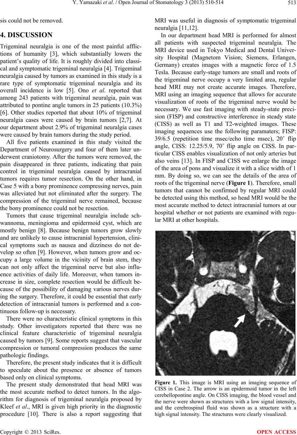
Thus, MRI using an appropriate imaging sequence
seems to be indispensable for the detection of tumors.
5. ACKNOWLEDGEMENTS
[8] Love, S. and Coakham, H.B. (2001) Trigeminal neuralgia:
Pathology and pathogenesis. Brain, 124, 2347-2360.
http://dx.doi.org/10.1093/brain/124.12.2347
The authors thank the Department of oral and maxillofacial radiology,
Tokyo Medical and Dental Hospital of dentistry for presenting details
of MRI.
[9] Cirak, B., Kiymaz, N. and Arslanoglu. A. (2004) Trige-
minal neuralgia caused by intracranial epidermoid tumor:
Report of a case and review of the different therapeutic
modalities. Pain Physician, 7, 129-132.
REFERENCES
[1] Kabatas, S., Karasu, A., Civelek, E., Sabanci, A.P., Hep-
gul, K.T. and Teng, Y.D. (2009) Microvascular decom-
pression as a surgical management for trigeminal neural-
gia: Long-term follow-up and review of the literature.
Neurosurgical Review, 32, 87-93.
http://dx.doi.org/10.1007/s10143-008-0171-3
[10] van Kleef, M., van Genderen, W.E., Narouze, S., Nur-
mikko, T.J., van Zundert, J., Geurts, J.W. and Mekhail, N.
(2009) 1. Trigeminal neuralgia. Pain Practice: The Offi-
cial Journal of World Institute of Pain, 9, 252-259.
[11] Goh, B.T., Poon, C.Y. and Peck, R.H. (2001) The impor-
tance of routine magnetic resonance imaging in trigemi-
nal neuralgia diagnosis. Oral Surgery, Oral Medicine,
Oral Pathology, Oral Radiology, and Endodontics, 92,
424-429. http://dx.doi.org/10.1067/moe.2001.115130
[2] Barker 2nd, F.G., Jannetta, P.J., Babu, R.P., Pomonis, S.,
Bissonette, D.J. and Jho, H.D. (1996) Long-term outcome
after operation for trigeminal neuralgia in patients with
posterior fossa tumors. Journal of Neurosurgery, 84, 818-
825. http://dx.doi.org/10.3171/jns.1996.84.5.0818 [12] Gronseth, G., Cruccu, G., Alksne, J., Argoff, C., Brainin,
M., Burchiel, K., Nurmikko, T. and Zakrzewska, J.M.
(2008) Practice parameter: the diagnostic evaluation and
treatment of trigeminal neuralgia (an evidence-based re-
view): Report of the Quality Standards Subcommittee of
the American Academy of Neurology and the European
Federation of Neurological Societies. Neurology , 71, 1183-
1190.
http://dx.doi.org/10.1212/01.wnl.0000326598.83183.04
[3] Okeson, J.P. (2005) Neuropathic pains. In: Harmon, L.,
Ed., Bells orofacial pains: The clinical management of
orofacial pain, Sixth Edition, Quintessence, Tokyo, 449-
517.
[4] Headache Classification Subcommittee of the Interna-
tional Headache Society (2004) The international classi-
fication of headache disorders: 2nd edition. Cephalalgia,
24, 9-160. [13] Yoshino, N., Akimoto, H., Yamada, I., Nagaoka, T.,
Tetsumura, A., Kurabayashi, T., Honda, E., Nakamura, S.
and Sasaki, T. (2003) Trigeminal neuralgia: Evaluation of
neuralgic manifestation and site of neurovascular com-
pression with 3D CISS MR imaging and MR angiogra-
phy. Radiology, 228, 539-545.
http://dx.doi.org/10.1148/radiol.2282020439
[5] Cirak, B., Kiymaz, N. and Arslanoglu, A. (2004) Trige-
minal neuralgia caused by intracranial epidermoid tumor:
Report of a case and review of the different therapeutic
modalities. Pain Physician, 7, 129-132.
[6] Nakagawa, K., Aoyagi, M., Kawano, Y. and Ohno, K.
(2009) Clinical and operative findings in patients with
trigeminal neuralgia caused by brain tumors. No Shinkei
Geka, 37, 863-871.
OPEN ACCESS