Engineering, 2013, 5, 487-490
http://dx.doi.org/10.4236/eng.2013.510B100 Published Online October 2013 (http://www.scirp.org/journal/eng)
Copyright © 2013 SciRes. ENG
Cloning, Characterization and Bioinformatic Analysis of
the Gene Encoding the Larval Serum Protein 2 in Diapause
of the Onion Maggot, Delia Antiqua
Jingjing Xu, Bin Chen*, Zhengbo He, Youjin Hao
Institute of Entomology and Molecular Biology, College of Life Sciences, Chongqing Normal University,
Shapingba, Chongqing, China
Email: *c_bin@hotmail.com.
Received 2013
ABSTRACT
The full-length cDNA encoding Larval serum protein 2 (LSp-2) in the onion maggot, Delia antiqua, was cloned and
sequenced by rapid amplification of cDNA ends methods. The result showed that the cDNA was 2203 bp long and the
open reading frame (ORF) of 2106 bp encoded 701 amino acid with a calculated molecular weight of 80.5 kDa and an
isoelectric point of 5.87. The onion maggot LSp-2 shows highest homology (83%) to that of Calliphora vicina at amino
acid level. Its signal peptides, domains and structures were predicted and analyzed by using bioinformatic methods. The
amino acid sequence of LSP-2 suggests that it would be a typical hexamerin.
Keywords: Delia Antiqua; Diapause; Larval Serum Protein 2; Cloning ; Mo lecular Characterization; Bioinformatic
Analysis
1. Introduction
Larval serum protein 2 (LSP-2) belongs to a superfamily
of hexameric hemolymph proteins that have been found
in all insect species investigated [1]. In holometabolous
insects, hexamerins are thought to act mainly as storage
proteins that provide energy and amino acids during me-
tamorphosis [2,3]. Expression of LSP-2 is restricted to
the fat body cells, where it reabsorbs proteins and other
macromolecules that have accumulated in the haemo-
lymph during the larval feeding period [5,6]. Roughly
half of the cell population survives metamorphosis, indi-
cating a specific degree of differentiation during postem-
bryonic life. Diapause is a developmental strategy wide-
spread among insects and their arthropod relatives. The
onion maggot (Delia antiqua), a serious pest of onion
(Alliumcepa), passing over winters and/or summers as
diapausing pupae, is an excellent model for diapause
research [4]. LSP-2 as a kind of storage proteins is be-
lieved to provide with a means for the feeding insect to
build reserves of amino acids for use in protein synthesis
and energy metabolism during diapause stages.
2. Materials and Methods
2.1. Insects
The colony of D. antiqua was reared on an artificial diet
at 25˚C with a 16L:8D cycle and relativ e humidity 50% -
70%. Larvae were maintained at 25˚C with a 16L:8D
photocycle to induce SD and 16˚C with 12L:12D to in-
duce WD.
2.2. RNA Extraction and RT-PCR
Total RNAs were extracted with Trizol® reagent (Invi-
trogen Co., USA) from the pupae according to the man-
ufacturer’s instructions. The first -strand cDNA was syn-
thesized from 1 µg of total RNA in 20 µl reaction mix-
ture prepared with RevertAidTM First Strand cDNA Syn-
thesis Kit (Fermentas). One microliter of the reaction
mixture was a dded to 25 µl of PCR reaction system, PCR
amplification was performed using one specific primers
ku6contig11F (5’-CCACTGGCTTGACTCGCATG-3’)
and ku399R (5’-GCGGGCTCCAAAGTATTCA-3’) (Fig-
ure 1) based on known EST sequences in SSH cDNA
library conservative sequences with the following reac-
tion conditions: 1 min at 94˚C, 30 S at 55˚C, 30 S at 72˚C
with 35 cycles, then 10 min at 72˚C.
2.3. RACE Amplification
Specific primers were synthesized for 5’ and 3’RACE
based on the cDNA sequences obtained from internal
amplification. For 5’ and 3’RACE, the cDNA was syn-
thesized according to the manufacturer’s protocol
*Corresponding a uthor.
J. J. XU ET AL.
Copyright © 2013 SciRes. ENG
488
(SMARTer ™ R AC E cD N A Amplification Kit, Clontech).
5’ and 3’-RACE amplification was performed on 2.5 ml
of 3’-re ady-cDNA with UPM and GSPF. The reaction
mixture was subjected to 30 thermal cycles that consisted
of 94˚C, 1 min; 65˚C, 1 min; 72˚C, 1 mi n.
2.4. Cloning and Sequencing
The amplified DNAs were separated on a 1.5% agarose
gel and purified using DNA Gel Extraction Kit. DNA
fragments were cloned according to the manufacturer’s
protocol (TOPO TA Cloning Kit for Sequencing). Mul-
tiple sequencing reactions were run using both M13F and
M13R primers.
2.5. Phylogenetic Analysis
The program MEGA5.0 was used for the phylogenetic
tree reconstructions, and Neighbor-joining tree were in-
ferred with the program.
3. Results
3.1. Cloning of Onion Maggot LSP-2 cDNA
The tools provided by the ExPASy Molecular Biology
Server of the Swiss Institute of Bioinformatics (http://
www.expasy.org) were used for the analyses of DNA
and amino acid sequences. Onion maggot LSP-2 cDNA
contains a 2106 bp open reading frame (ORF) encoding a
701 amino acid precursor protein. The first 21 amino
acids are predicted for the sequence of a signal peptide
using SignalP Version 3.0 software (http://www.cbs.dtu.
dk/ser vic es/Sign alP). The 5’ untranslated region upstream
of the transcription start code (ATG) is about 23 nucleo-
tides. The ORF is terminated by a TAA stop code that is
followed by a 74bp 3’ untranslated region.
3.2. Bioinformatics Analysis
As determined by the use of protparam software (http://
web.expasy.org/protparam) the deduced amino acid se-
quence of 680 residues has a molecular mass of 80.5 kDa
and a pI of 5.87. Onion maggot LSP-2 was most similar
to LSP-2 from Drosophila melanogaster and Calliphora
vicina. Using the SMART software (http://smart.embl-
heidelberg.de) found domains (Figure 1). Putative gly-
cosylation and phosphorylation sites as well as the con-
served motifs ADKDFLXKQK (position 27; Gordadze et
al., 1999) and TMMRDPMFY (position 480; PROSITE,
Bairoch et al., 1997), found in several insect hexame-
rins, were identified in the LSP-2 amino acid sequence
[7].
The position of secondary-structure elements was de-
duced using UniProt (http://www.uniprot.org) (Figure
2).
Name Begin End E-value
Pfa m: Hemocyanin_N 31 156 6.80E-32
Pfa m: Hemocyanin_M 160 421 1.30E-53
Pfa m: Hemocyanin_C 427 694 1.30E-89
Figure 1. Predicted domain of LSP-2.
Figure 2. Protein secondary-structure of onion maggot
LSP-2.
J. J. XU ET AL.
Copyright © 2013 SciRes. ENG
489
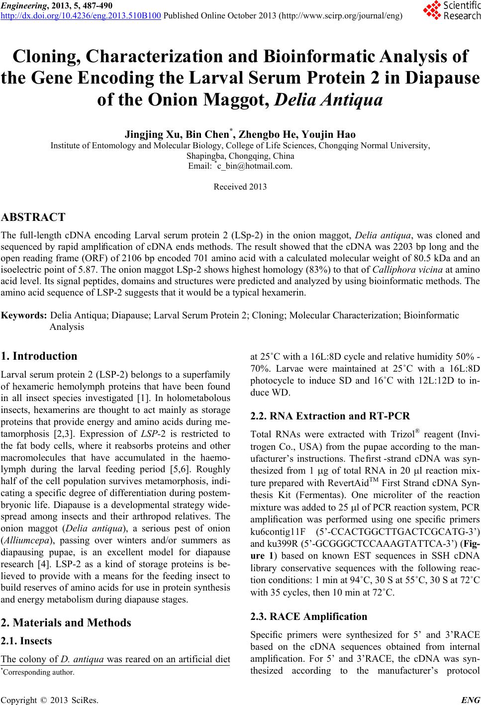
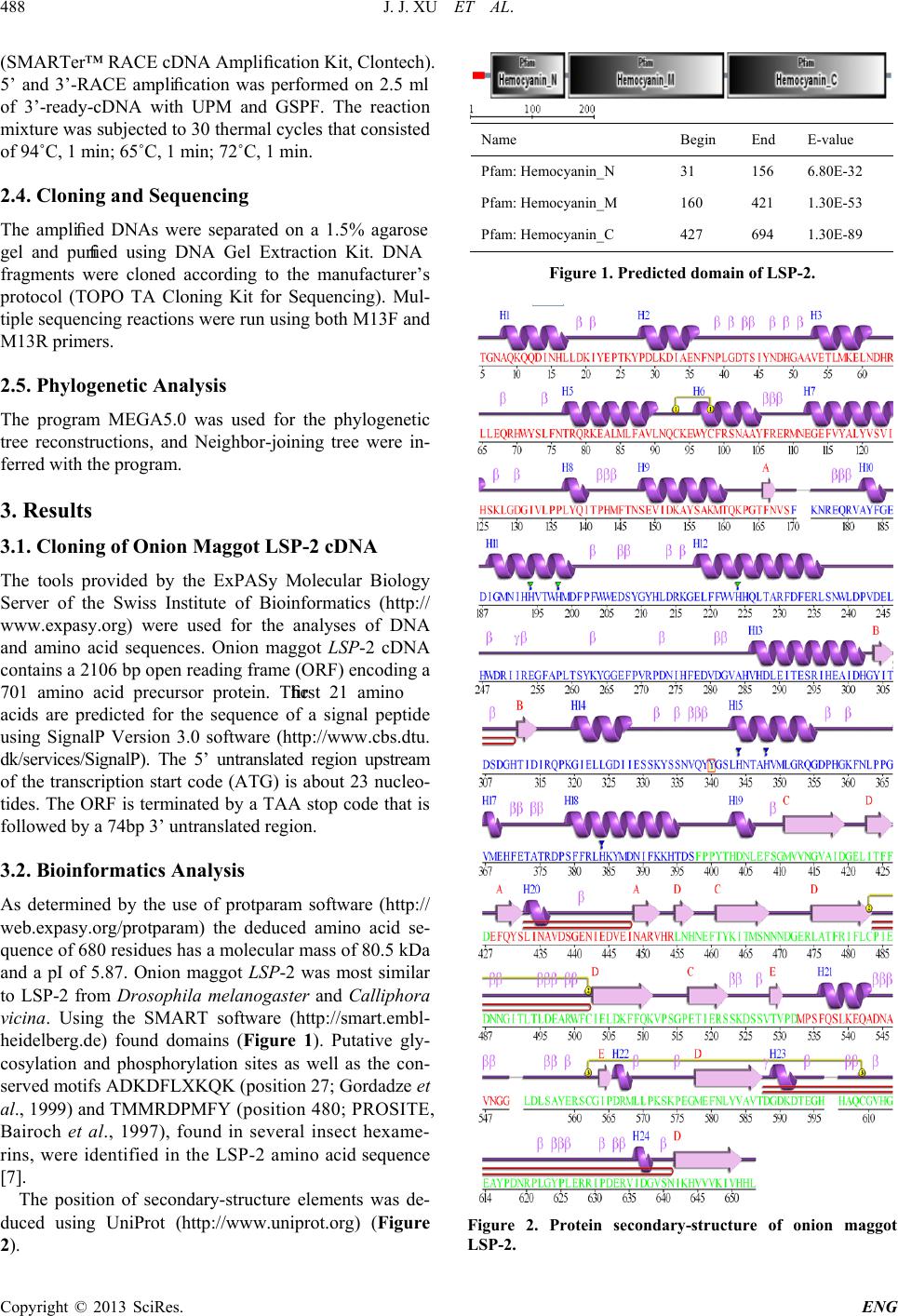
The position of tertiary-structure elements was de-
duced using UniProt (http://www.uniprot.org) (Figure
3).
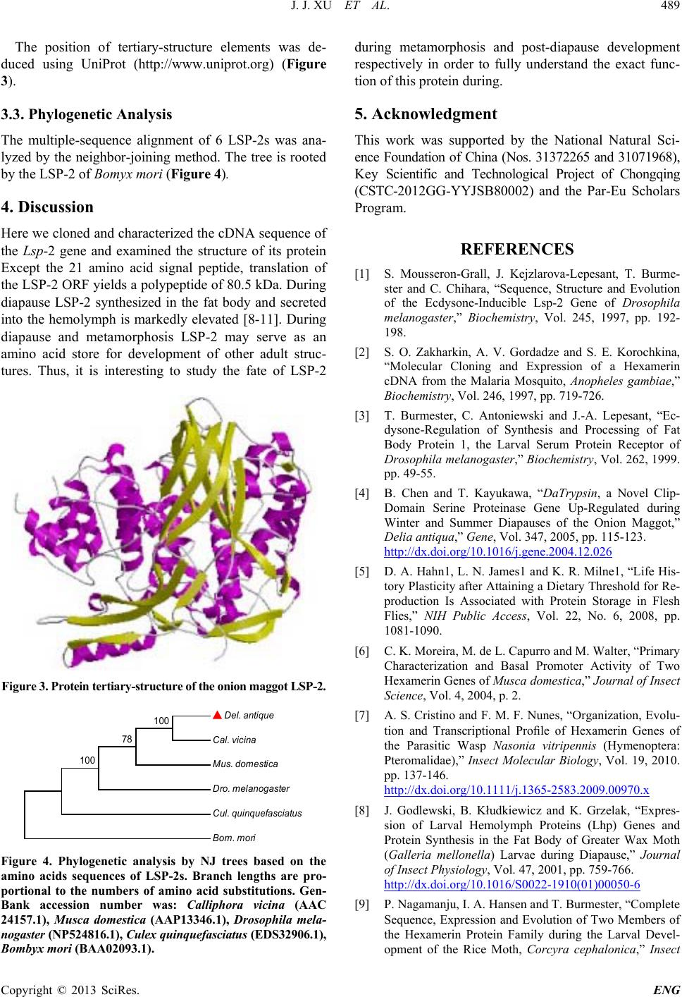
3.3. Phylogenetic Analysis
The multiple-sequence alignment of 6 LSP-2s was ana-
lyzed by the neighbor-joining method. The tree is rooted
by the LSP-2 of Bomyx mori (Figure 4).
4. Discussion
Here we cloned and characterized the cDNA sequence of
the Lsp-2 gene and examined the structure of its protein
Except the 21 amino acid signal peptide, translation of
the LSP-2 ORF yields a polypeptide of 80.5 kDa. During
diapause LSP-2 synthesized in the fat body and secreted
into the hemolymph is markedly elevated [8-11]. During
diapause and metamorphosis LSP-2 may serve as an
amino acid store for development of other adult struc-
tures. Thus, it is interesting to study the fate of LSP-2
Figure 3. Protein tertiary-structure of the on ion maggot LS P-2.
Figure 4. Phylogenetic analysis by NJ trees based on the
amino acids sequences of LSP-2s. Branch lengths are pro-
portional to the numbers of amino acid substitutions. Gen-
Bank accession number was: Calliphora vicina (AAC
24157.1), Musca domestica (AAP13346.1), Drosophila mela-
nogaster (NP524816.1), Culex qui nquefa sciatu s (EDS32906.1),
Bombyx mori (BAA02093.1).
during metamorphosis and post-diapause development
respectively in order to fully understand the exact func-
tion of thi s protei n during.
5. Acknowledgment
This work was supported by the National Natural Sci-
ence Foundation of China (Nos. 31372265 and 31071968),
Key Scientific and Technological Project of Chongqing
(CSTC-2012GG-YYJSB80002) and the Par -Eu Scholars
Program.
REFERENCES
[1] S. Mousseron-Grall, J. Kejzlarova-Lepesant, T. Burme-
ster and C. Chihara, “Sequence, Structure and Evolution
of the Ecdysone-Inducible Lsp-2 Gene of Drosophila
melanogaster,” Biochemistry, Vol. 245, 1997, pp. 192-
198.
[2] S. O. Zakharkin, A. V. Gordadze and S. E. Korochkina,
“Molecular Cloning and Expression of a Hexamerin
cDNA from the Malaria Mosquito, Anopheles gambiae,”
Biochemistry, Vol. 246, 1997, pp. 719-726.
[3] T. Burmester, C. Antoniewski and J.-A. Lepesant, “Ec-
dysone-Regulation of Synthesis and Processing of Fat
Body Protein 1, the Larval Serum Protein Receptor of
Drosophila melanogaster,” Biochemistry, Vol. 262, 1999.
pp. 49-55.
[4] B. Chen and T. Kayukawa, “DaTrypsin, a Novel Clip-
Domain Serine Proteinase Gene Up-Regulated during
Winter and Summer Diapauses of the Onion Maggot,
Delia antiqua,” Gene, Vol. 347, 2005, pp. 115-123.
http://dx.doi.org/10.1016/j.gene.2004.12.026
[5] D. A. Hahn1, L. N. James1 and K. R. Milne1, “Life His-
tory Plasticity after Attaining a Dietary Threshold for Re-
production Is Associated with Protein Storage in Flesh
Flies,” NIH Public Access, Vol. 22, No. 6, 2008, pp.
1081-1090.
[6] C. K. Moreira, M. de L. Capurro and M. Walter, “Prima ry
Characterization and Basal Promoter Activity of Two
Hexamerin Genes of Musca domestica,” Journal of Insec t
Science, Vol. 4, 2004, p. 2.
[7] A. S. Cristino and F. M. F. Nunes, “Organization, Evolu-
tion and Transcriptional Prole of Hexamerin Genes of
the Parasitic Wasp Nasonia vitripennis (Hymenoptera:
Pteromalidae),” Insect Molecular Biology, Vol. 19, 2010.
pp. 137-146.
http://dx.doi.org/10.1111/j.1365-2583.2009.00970.x
[8] J. Godlewski, B. Kłudkiewicz and K. Grzelak, “Expres-
sion of Larval Hemolymph Proteins (Lhp) Genes and
Protein Synthesis in the Fat Body of Greater Wax Moth
(Galleria mellonella) Larvae during Diapause,” Journal
of Insect Physiology, Vol. 47, 2001, pp. 759-766.
http://dx.doi.org/10.1016/S0022-1910(01)00050-6
[9] P. Nagamanju, I. A. Hansen and T. Burmester, “Complete
Sequence, Expression and Evolution of Two Members of
the Hexamerin Protein Family during the Larval Devel-
opment of the Rice Moth, Corcyra cephalonica,” Insect
Del. a n tiq ue
Cal. vi c i n a
Mus. d o mestic a
Dr o . melan og a s ter
Cul. q u inq u efasc ia tu s
Bom. mori
100
78
100
J. J. XU ET AL.
Copyright © 2013 SciRes. ENG
490
Biochemistry and Molecular Biology, Vol. 33, 2003, pp.
73-80. http://dx.doi.org/10.1016/S0965-1748(02)00178-9
[10] C. A. D. De Korta and A. B. Koopmanschapa, “Nucleo-
tide and Deduced Amino Acid Sequence of a cDNA
Clone Encoding Diapause Protein 1, an Arylphorin-Type
Storage Hexamer of the Colorado Potato Beetle,” Journal
of Insect Physiology, Vol. 40, 1994, pp. 527-535.
http://dx.doi.org/10.1016/0022-1910(94)90126-0
[11] J. Tungjitwitayakul, T. Singtripop, A. Nettagul, Y. Oda,
N. Tatun, T. Sekimoto and S. Sakurai, “Identification,
Characterization, and Developmental Regulation of Two
Storage Proteins in the Bamboo Borer Omphisa fusci-
dentalis,” Journal of Insect Physiology, Vol. 54, 2008, pp.
62-76.