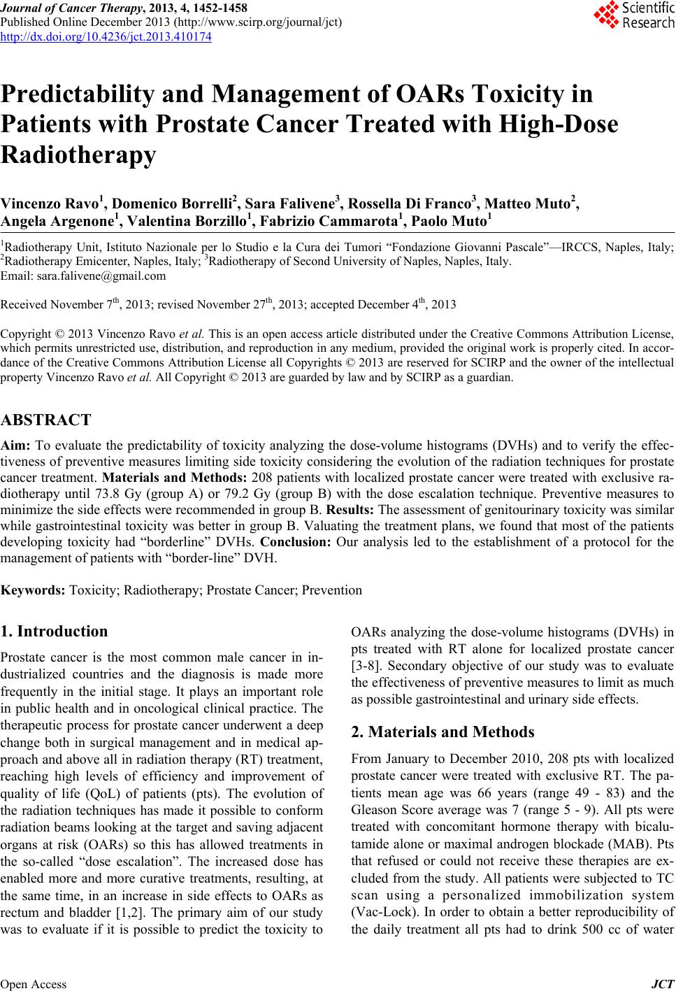
Predictability and Management of OARs Toxicity in Patients with
Prostate Cancer Treated with High-Dose Radiotherapy
1457
Prostate Cancer,” International Journal of Radiation On-
cology, Biology, Physics, Vol. 54, No. 5, 2002, pp. 1314-
1321.
http://dx.doi.org/10.1016/S0360-3016(02)03742-2
[4] R. Miralbell, D. Taussky, O. Rinaldi, A. Lomax, S. Ca-
nales, L. Escude, P. Nouet, O. Ozsoy and M. Rouzaud,
“Influence of Rectal Volume Changes during Radiother-
apy for Prostate Cancer: A Predictive Model for Mild-To-
Moderate Late Rectal Toxicity,” International Journal of
Radiation Oncology, Biology, Physics, Vol. 57, No. 5,
2003, pp. 1280-1284.
http://dx.doi.org/10.1016/S0360-3016(03)00749-1
[5] S. T. Peeters, W. D. Heemsbergen, W. L. van Putten, A.
Slot, H. Tabak, J. W. Mens, J. V. Lebesque and P. C.
Koper, “Acute and Late Complications after Radiother-
apy for Prostate Cancer: Results of a Multicenter Ran-
domized Trial Comparing 68 Gy to 78 Gy,” International
Journal of Radiation Oncology, Biology, Physics, Vol. 61,
No. 4, 2005, pp. 1019-1034.
http://dx.doi.org/10.1016/j.ijrobp.2004.07.715
[6] J. J. Nuyttens, S. Milito, P. F. Rust, A. T. Turrisi, “Dose-
Volume Relationship for Acute Side Effects during High
Dose Conformal Radiotherapy for Prostate Cancer,” Ra-
diotherapy & Oncology, Vol. 64, No. 2, 2002, pp. 209-
214. http://dx.doi.org/10.1016/S0167-8140(02)00185-8
[7] C. Fiorino, V. Vavassori, G. Sanguineti, C. Bianchi, G. M.
Cattaneo, A. Piazzolla, C. Cozzarini, “Rectum Contour-
ing Variability in Patients Treated for Prostate Cancer:
Impact on Rectum Dose-Volume Histograms and Normal
Tissue Complication Probability,” Radiotherapy & On-
cology, Vol. 63, No. 3, 2002, pp. 249-255.
http://dx.doi.org/10.1016/S0167-8140(01)00469-8
[8] J. J. Tuech, V. Chaudron, V. Thoma, J. C. Ollier, V. Tas-
setti, D. Duval and J. F. Rodier, “Prevention of Radiation
Enteritis by Intrapelvic Breast Prosthesis,” European
Journal of Surgical Oncology, Vol. 30, No. 8, 2004, pp.
900-904. http://dx.doi.org/10.1016/j.ejso.2004.06.012
[9] A. L. Zietman, M. L. Desilvio, J. D. Slater, C. J. Rossi Jr.,
D. W. Miller, J. A. Adams, W. U. Shipley, “Comparison
of Conventional-Dose vs High-Dose Conformal Radia-
tion Therapy in Clinically Localized Adenocarcinoma of
the Prostate: A Randomized Controlled Trial,” JAMA,
Vol. 294, No. 10, 2005, pp. 1233-1239.
http://dx.doi.org/10.1001/jama.294.10.1233
[10] D. P. Dearnaley, M. R. Sydes, J. D. Graham, E. G. Aird,
D. Bottomley, R. A. Cowan, R. A. Huddart, C. C. Jose, J.
H. Matthews, J. Millar, A. R. Moore, R. C. Morgan, J. M.
Russell, C. D. Scrase, R. J. Stephens, I. Syndikus, M. K.
Parmar and RT01 Collaborators “Escalated-Dose versus
Standard-Dose Conformal Radiotherapy in Prostate Can-
cer: First Results from the MRC RT01 Randomised Con-
trolled Trial,” The Lancet Oncology, Vol. 8, No. 6, 2007,
pp. 475-487.
http://dx.doi.org/10.1016/S1470-2045(07)70143-2
[11] I. Syndikus, R. C. Morgan, M. R. Sydes, J. D. Graham,
and D. P. Dearnaley, “Late Gastrointestinal Toxicity after
Doseescalated Conformal Radiotherapy for Early Prostate
Cancer: Results from the UK Medical Research Council
RT01 trial (ISRCTN47772397),” International Journal of
Radiation Oncology Biology Physics, Vol. 77, No. 3, 2010,
pp. 773-783,
http://dx.doi.org/10.1016/j.ijrobp.2009.05.052
[12] A. Al-Mamgani, W. L. van Putten, W. D. Heemsbergen,
van G. J. Leenders, A. Slot, M. F. Dielwart, L. Incrocci
and J. V. Lebesque, “Update of Dutch Multicenter Dose-
Escalation Trial of Radiotherapy for Localized Prostate
Cancer,” International Journal of Radiation Oncology,
Biology, Physics, Vol. 72, No. 4, 2008, pp. 980-988.
http://dx.doi.org/10.1016/j.ijrobp.2008.02.073
[13] A. Hille, N. Töws, H. Schmidberger and C. F. Hess, “A
Prospective Three-Dimensional Analysis about the Im-
pact of Differences in the Clinical Target Volume in
Prostate Cancer Irradiation on Normal-Tissue Exposure.
A Potential for Increasing the Benefit/Risk Ratio,”
Strahlentherapie und Onkologie, Vol. 181, No. 12, 2005,
pp. 789-795.
http://dx.doi.org/10.1007/s00066-005-1452-1
[14] R. Sauer, H. Becker, W. Hohenberger, C. Rödel, C. Wit-
tekind, R. Fietkau, P. Martus, J. Tschmelitsch, E. Hager,
C. F. Hess, J. H. Karstens, T. Liersch, H. Schmidberger
and R. Raab, “German Rectal Cancer Study Group-Pre-
operative versus Postoperative Chemoradiotherapy for
Rectal Cancer,” The New England Journ
Vol. 351, No. 17, 2004, pp. 1731-1740.
al of Medicine,
http://dx.doi.org/10.1056/NEJMoa040694
[15] M. S. Litwin, R. D. Hays, A. Fink, P. A. Ganz, B. Leake,
R. H. Brook, “The UCLA Prostate Cancer Index: Devel-
opment, Reliability, and Validity of a Health-Related
Quality of Life Measur
1998, pp. 1002-1012.
e,” Medical Care, Vol. 36, No. 7,
http://dx.doi.org/10.1097/00005650-199807000-00007
[16] J. T. Wei, R. L. Dunn, M. S. Litwin, H. M. Sandler and M.
G. Sanda, “Development and Validation of the Expanded
Prostate Cancer Index Composite (EPIC) for Comprehen-
sive Assessment of Health-Related Quality of Life in Men
with Prostat
899-905.
e Cancer,” Urology, Vol. 56, No. 6, 2000, pp.
http://dx.doi.org/10.1016/S0090-4295(00)00858-X
[17] S. T. Peeters, W. D. Heemsbergen, W. L. van Putten, A.
Slot, H. Tabak, J. W. Mens, J. V. Lebesque and P. C.
Koper, “Acute and Late Complications after Radiother-
apy for Prostate Cancer: Results of a Multicenter Ran-
domized Trial Comparing 68 Gy to 78 Gy,” International
Journal of Radiation Oncology
No. 4, 2005, pp. 1019-1034.
, Biology, Physics, Vol. 61,
http://dx.doi.org/10.1016/j.ijrobp.2004.07.715
[18] S. T. Peeters, M. S. Hoogeman, W. D. Heemsbergen, A.
A. Hart, P. C. Koper and J. V. Lebesque, “ Rectal Bleed-
ing, Fecal Incontinence, and High Stool Frequency after
Conformal Radiotherapy for Prostate Cancer: Normal
Tissue Complication Probability Modeling,” Interna-
tional Journal of Radiation Onc
Vol. 66, No. 1, 2006, pp. 11-19.
ology, Biology, Physics,
http://dx.doi.org/10.1016/j.ijrobp.2006.03.034
[19] M. J. Zelefsky, E. Aschkenasy, S. Kelsen and S. A. Lei-
bel, “Tolerance and Early Outcome Results of Post-
prostatectomy Three-Dimensional Conformal Radiother-
apy,” International Journal of Radiation Oncology, Biol-
Open Access JCT