Advances in Infectious Diseases, 2013, 3, 269-273
Published Online December 2013 (http://www.scirp.org/journal/aid)
http://dx.doi.org/10.4236/aid.2013.34041
Open Access AID
269
A Clinco-Bacteriological Study of Leprosy in Paediatric
Age Group*#
Leena Das1, Bijay Kumar Meher1, Nikunja Bihari Das2, Deepti Damayanty Pradhan1, S. Pradeep1,
Jayashree Mohanty3
1Department of Paediatrics, S.V.P. Post Graduate Institute of Paediatrics, SCBMCH, Cuttack, India; 2Pediatric Specialist, Commu-
nity Health Centre (CHC), Chikiti, Ganjam, India; 3Department of Dermatology, S.C.B. Medical College, Cuttack, India.
Email: bkmeher187@yahoo.co.in
Received October 23rd, 2013; revised November 23rd, 2013; accepted November 30th, 2013
Copyright © 2013 Leena Das et al. This is an open access article distributed under the Creative Commons Attribution License, which
permits unrestricted use, distribution, and reproduction in any medium, provided the original work is properly cited.
ABSTRACT
Clinico-bacteriological study was done in 51 leprosy patients below 14 years of age. Majority of the patients were males
in the age group of 11 - 14 years. Nearly 84% had not received any prior treatment because of lack of awareness and
financial constraints. Only 11.76% had a positive contact history. Skin lesions were present in all cases and 84.3% had
lesions mainly on the exposed areas of the body and their number was found to increase significantly with advancing
age (p < 0.005). These lesions were hypo-pigmented patches in 88% cases. 88% of cases had hypo-anesthesia and nerve
thickening was observed in 24% cases. The most common type of skin lesion was borderline tuberculoid (BT) in 53%
cases. Positivity of the skin smears increased significantly as the number of the skin lesions per patient increased (p <
0.001). Multibacillary cases were seen in 15.6% of cases and found only in the age group of 10 - 14 years of age. No
statistically significant association between BCG vaccination and prevention of leprosy was seen.
Keywords: Leprosy in Children; Tuberculoid Leprosy; BCG
1. Introduction
Leprosy is probably the oldest disease known to mankind.
It is a chronic infectious disease caused by Mycobacte-
rium leprae. It is one of the leading causes of physical
disabilities which contribute to intense social stigma, re-
sulting in discrimination of the patient and their families.
According to world statistics at the beginning of 2006,
the number of leprosy cases in the world was 22 million.
Today the highest burden of cases of world is concentrat-
ed in 6 countries and India remains at the top [1]. India
accounts for 55% of the new leprosy cases detected glo-
bally in 2010 [2]. According to the National Leprosy Eli-
mination Programme (NLEP) report of March 2012, there
were a total of about 0.13 million cases of leprosy in In-
dia, 9.7% of which were children. In Odisha, the preva-
lence rate (PR) of childhood leprosy is 1.06/10,000 popu-
lation [3]. The profile and magnitude of leprosy in pedi-
atric populations have an important bearing on the epide-
miology of the disease and reflect the level of control in a
community. A high child proportion signifies active and
recent transmission of the disease, making it a robust epi-
demiological indicator to assess the progress of leprosy
control programmes. It is expected to fall towards the eli-
mination phase of a leprosy control programme, suggest-
ing a decrease in transmission levels [4]. A multistage
cluster sampling survey in north Bangladesh demonstrat-
ed that the prevalence of previously undiagnosed leprosy
in children (5 - 14 years old) was 8.6 per 10,000 [5]. In
the pediatric age group, leprosy often presents as a single
hypopigmented patch, which may or may not be anesthe-
tic, and hence it is confused with other dermatological ail-
ments [6]. With this background, we carried out this pre-
sent work to study the clinico-bacteriological status of le-
prosy patients under 14 years of age.
Objective
Assessment of the Clinico-bacteriological status of
childhood leprosy patients at a tertiary level hospital.
*Conflict of interest: none.
#Funding: none. Smear positivity status in BCG vaccinated children.
A Clinco-Bacteriological Study of Leprosy in Paediatric Age Group
270
2. Materials and Methods
This study was carried out in the out-patient, in the de-
partment at Skin & V.D., S.C.B Medical College and Ho-
spital, Cuttack, Odisha. Fifty one patients under 14 year
of age were studied. In each case clinical history, physi-
cal examination like type of lesion, distribution, number,
colors of lesion and deformities were observed, periphe-
ral nerves cutaneous sensation were examined. Disability
assessment and grading were done as per WHO classifi-
cation [7]. Skin smear examination, Skin biopsy was done
in all cases.
Skin Smear Examination: In all patients with skin le-
sions, smear were taken from 3 sites i.e. one from ear
lobe and two from skin and stained by standard technique
as out lined by Jopling. The density of bacilli in the smear
was recorded using Ridley’s logarithmic scale [7].
Skin biopsy: In cases in whom the diagnosis was sus-
pected but was not definite on clinical grounds, biopsy
for histological confirmation were done and classifica-
tion was done by the standard technique [7]. Based on
these observations, patients were grouped under five heads
as suggested by Indian Association of Leprologist, Inde-
terminant (I), tuberculoid (TT), borderline (BB), Lepro-
matous (LL), and pure neuritic (P). The borderline group
was further subdivided into borderline tuberculoid (BT),
borderline-borderline (BB), borderline lepromatous (BL)
sub-groups [8].
3. Results
A total of 51 case 14 years were studied. About three
fourth of cases were males. 58.82% of cases fell in the
age group of 11 - 14 yrs. Majority of the children (56.8%)
has been brought to the hospital between 6 - 12 months
of onset of lesion. Eighty eight percent had an asymmet-
rical distribution of skin lesion (Figure 1) while rest had
symmetrical skin lesion. In our study single hypo pig-
mented skin lesion were observed in 60.78% (Table 1).
In 84% cases had initial skin lesion over the exposed part
of the body. Most of children (84%) had not received any
treatment. Only 12% of the children had positive contact
history. Skin sensation was unaffected in only 8% cases.
Examination of nerves revealed that 27.45% cases had
thickened nerves and the commonest nerve involved were
ulnar nerve followed by lateral popliteal nerve. As shown
in Table 1, there was an increase in number of skin le-
sion as the age advances (P < 0.005).
Based on skin biopsy ppatients were put under differ-
ent category. 2 cases were tuberculoid (TT) and rests were
borderline (BL) cases. Of the border line leprosy, 37 cas-
es were borderline tuberculoid (BT), 4 cases were BT-
type-1 reaction, 2 cases were borderline-borderline (BB),
2 cases were borderline lepromatous (BL), 4 cases were
BL-type-2 reaction. None of the cases were pure neuritic,
Table 1. Number of skin lesions in various age groups (N -
51).
Age (in yrs) 1 2 - 10 >10 Total
1 - 5 1.9 0 0 1.9
6 - 10 35.3 0 3.9 39.2
11 - 14 23.5 19.6 15.7 58.9
Total 60.78 19.6 19.6 100
Figure 1. Photograph of 12 yr old male child with large
multiple erythematous, infiltered edematous scaly plaques
lesions suggestive of Borderline Tuberculoid (BT) Lepra
type 1 reaction.
indeterminate or lepromatous leprosy (Table 2).
A total of 5 cases had Grade-II limb deformity in form
of claw hand and ulcer. No cases of foot drop were ob-
served. There was no case observed as nasal flattening or
ocular manifestation. There was no bony pathology or
limb amputation. The smear examination were negative
in majority cases (60.8%), however with increase in num-
ber of skin lesions per patient the chance of positivity of
smear increased significantly (<0.001), as shown in (Ta-
ble 3).
In two cases although bacteriological examination was
negative, as diagnosis of leprosy was strongly suspected
and skin biopsy was compatible with clinical diagnosis
were included in the study. Out of 20 smear positive
cases we had only 6 cases with BCG scar. (BCG scar is
taken as BCG vaccination.) But the association between
BCG vaccination and prevention of leprosy is not sig-
nificant in our study (p > 0.05) (Table 4).
4. Discussion
Leprosy is widely prevalent in India. As age advances the
number of lesions increased significantly (p < 0.005).
This could be explained by long incubation period of the
Open Access AID
A Clinco-Bacteriological Study of Leprosy in Paediatric Age Group 271
Table 2. Sex distribution according to type of pediatric lep-
rosy cases (N - 51).
Type of cases Male (%)
N = 37
Female (%)
N = 14
Total (%)
N = 51
TT 00 3.92 3.92
BT 52.94 19.6 72.54
BT (Type-1) 7.8 00 7.8
BB 00 3.92 3.92
BL 3.92 00 3.92
BL (Type-2) 7.8 00 7.8
Total 72.54 27.45 100
TT - Tuberculoid Leprosy, BT - Borderline Leprosy, BB - Borderline
Borderline, BL - Borderline leprosy.
Table 3. Number of skin smear positive lesion at the time of
presentation (N - 51).
Result of skin smear
Number of skin
lesions (+ve) (%) (ve) (%)
Total
1 11.76 49.00 60.76
2 - 10 11.76 7.80 19.56
>10 15.60 3.92 19.56
Total 39.20 60.78 100
Table 4. Skin smear status in bcg vaccinated children (N -
51).
BCG Scar Smear (+ve) (%) Smear (-ve) (%) Total (%)
Positive 11.76 29.40 41.17
Negative 27.45 31.30 58.82
Total 39.21 60.78 100
disease. 59% cases occurred in the age group of 11 - 14
yrs, as with other studies [9-11]. Males with the mean
age of presentation were 10.3 yrs. and females at 10.7 yrs.
Given a long incubation period it can be inferred that,
younger children are indeed at the greatest risk [9]. The
sex difference is greater in adults than in children [12].
we observed a male preponderance with male to female
ratio of 2.64:1 and males comprise 72.54% of total cases.
Similar observations were made by other studies [13].
Higher proportion among males was also observed by
Dayal R [9]. This may be due to their greater mobility and
increased opportunities for contact [14,15]. Moreover, de-
tection in girls may possibly be lower than boys due to
neglect of the female child.
In our study most common presentation was single
hypo pigmented macular lesion comparable with several
studies [9,10]. Selvasekhar A reported, 83% of the chil-
dren had a single patch, though Vara N [16] reported sin-
gle skin lesion in only 13.4% cases which is contrary to
our observation. Eighty five percent cases had lesions
over the exposed part of the body possibly due to direct
inoculation of organisms into the skin. Similar observa-
tion has been made by other authors [9,11]. However
Roy R et al. had more lesions over the covered areas
[17].
Majority of children (56.8%) presented from 6 month
to 1 yr after the onset of the disease and belongs to rural
society [18,19], signifies it needs more public awareness,
regarding the disease. A study conducted by A. Singal et
al. had duration of symptoms ranged from 2 months to 5
years. In 73% the duration was under one year [20].
About twelve percent had contact history positive inside
the family which is close to the observation made by Va-
ra N [16]. The lower incidence of contact might be due to
missing information. In our study borderline tuberculoid
was more common which is comparable with recent stu-
dies done by A. singal [20], VP shetty [21] (Table 1).
Commonest type of leprosy was borderline tuberculoid
similar to other studies [9,11]. This suggests that pauci-
bacillary skin lesions were the dominant type of lesion
and action for early diagnosis could be effective.
Type-1 reactions were observed in 4 cases and steroid
was given along with multidrug therapy. Type-II reac-
tions were observed in 4 cases. Lepromatous leprosy, pure
neuritic and intermediate cases were not seen. Peripheral
nerve involvement as thickened and/or tender was ob-
served in only 27.4% cases in our study, other authors
detected higher percentage (70.1%) of peripheral nerve
involvement [9,16,17]. We had Grade II deformities among
9.8% of our cases in the form of claw hands and ulcer. In
recent study by A. Singal found that 12.8% of deformi-
ties [18]. Vara N observed Grade-II deformities in 10%
of cases [16].
Smear Positivity and BCG Scar
In our study, smear examination was positive in 39.2% of
cases which is higher than 19.8% as found in the study
conducted by A. Singal. The positivity of the smear in-
creases significantly (p < 0.001) with increase in number
of skin lesions per patient. This is similar to observation
made by Dayal R [9]. This could be inferred that bacil-
lary load increases when the number of skin lesion in-
creases over the body. The proportions of smear positive
cases were less in number in scar positive patients as
compared to scar negative patients. In our study there
were no significant (p > 0.1) difference of leprosy in
BCG scar positive and negative individuals. However the
proportion of smear positive cases among the BCG scar
negative individuals was higher than that of BCG scar
positive individuals. There is now considerable evidence
that BCG vaccination can provide some protection against
Open Access AID
A Clinco-Bacteriological Study of Leprosy in Paediatric Age Group
272
leprosy [22]. This observation suggest to postulate that
though BCG is not protective totally, it may at least at-
tenuate the pathogenicity and modulates the immune-
status of the patients that foretell the high occurrence of
paucibacillary leprosy in BCG scar positive children. A
recent study showed increase in multibacillary cases
51.7% in A.singal [20] and 38% in VP shetty [21], but in
our study multi bacillary leprosy were only 15.6% of to-
tal cases. The study conducted by Dayal R also had less
multibacillary cases [9]. Multibacillary cases were found
in 10 - 14 yrs of age indicates multibacillary cases may
prevalent in latter part of the childhood.
5. Conclusion
An active surveillance is needed to identify the undetect-
ed hidden leprosy cases. Leprosy in children is essential-
ly a disease of school age children, suggesting school
survey as a tool for early detection and to prevent dis-
abilities. Borderline tuberculoid is the dominant variety
of presentation which responds to therapy. Early detec-
tion and effective treatment of leprosy patients in the pe-
diatric age group can prevent the development of more
severe devastating or crippling adult form of the disease.
6. Limitations
As this is a hospital based study, the results may not re-
flect the status childhood leprosy in a community. A rural
community based study would reflect the real scenario.
Sample size for the comparison of BCG and prevention
of leprosy should be large.
7. Contributions
LD & NBD drafted the initial manuscript and also engag-
ed in management of patients. BKM, DDP and PS re-
viewed the literature, LD & BKM was responsible for
designing the article, revision of manuscript and would
act as the guarantor for the paper. JM & NBD was in-
volved in doing skin biopsy.
8. Acknowledgements
The authors are grateful to the Superintendent, S.C.B. me-
dical college Cuttack for permitting them to publish the
study.
REFERENCES
[1] P. K. Leprosy, “Text Book of Preventive and Social Me-
dicine,” 19th Edition, M/S Banarasi Das bhanot, Jabalpur,
pp. 264-278.
[2] World Health Organization, “Global Burden of Leprosy
at the End of 2010,” The Weekly Epidemiological Record,
Vol. 86, 2011, pp. 389-400.
[3] WHO, “Weekly Epidemiological Record,” No. 28, 2000.
[4] M. B. Disability and Child Proportion, “Epidemiological
Significance and Interpretation. National Leprosy Eradi-
cation Program Guidelines.” http://nlep.nic.in/guide.html
[5] F. J. Moet, R. P. Schuring, D. Pahan, L. Oskam and J. H.
Richardus, “The Prevalence of Previously Undiagnosed
Leprosy in the General Population of Northwest Bang-
ladesh,” PLOS Neglected Tropical Diseases, Vol. 2, 2008,
p. e198. http://dx.doi.org/10.1371/journal.pntd.0000198
[6] S. Mahajan, K. Sardana, P. Bhushan, R. V. Koranne and
V. Mendiratta, “A Study of Leprosy in Children, from a
Tertiary Pediatric Hospital in India,” Leprosy Review, Vol.
77, 2006, pp. 160-162.
[7] W. H. Jopling, “Hand Book of Leprosy,” 3rd Edition, Wil-
liam-Heinemann, London, 1984.
[8] Dharmendra, “Leprosy,” Sammat and Company, Bombay,
1985.
[9] R. Dayal, A. K. Paliwal, R. Prasad, et al., “A Clinico-
Bacteriological Profile of Leprosy in Children,” Indian
Paediatric, Vol. 26, 1989, pp. 122-128.
[10] R. Dayal, P. K. Agarwal, K. Katra, et al., “Diagnostic Va-
lue of Gene Probes and Its Co-Relation with Clinical Pro-
file of Leprosy in Children,” Indian Pediatrics, Vol. 31,
1994, pp. 1521-1527.
[11] A. Selvasekhar, J. Geetha, K. Nisha, N. Manimozhi, K.
Jesudas and P. S. Rao, “Childhood Leprosy in an En-
demic Area,” Leprosy Review, Vol. 70, No. 1, 1999, pp.
21-27.
[12] R. Dayal, “Leprosy in Childhood,” Recent Advances in
Pediatrics, Vol. 1, 1991, p. 258.
[13] V. N. Sehgal and A. N. Chaudhary, “Leprosy in Children,
A Prospective Study,” 1993.
[14] C. Grover, S. Nanda, V. K. Garg and B. S. Reddy, “An
Epidemiologic Study of Childhood Leprosy from Delhi,”
Pediatric Dermatology, Vol. 22, 2005, pp. 489-490.
http://dx.doi.org/10.1111/j.1525-1470.2005.00124.x
[15] I. Kaur, S. Kaur, V. K. Sharma and B. Kumar, “Child-
hood Leprosy in Northern India,” Pediatric Dermatology,
Vol. 8, 1991, pp. 21-24.
http://dx.doi.org/10.1111/j.1525-1470.1991.tb00833.x
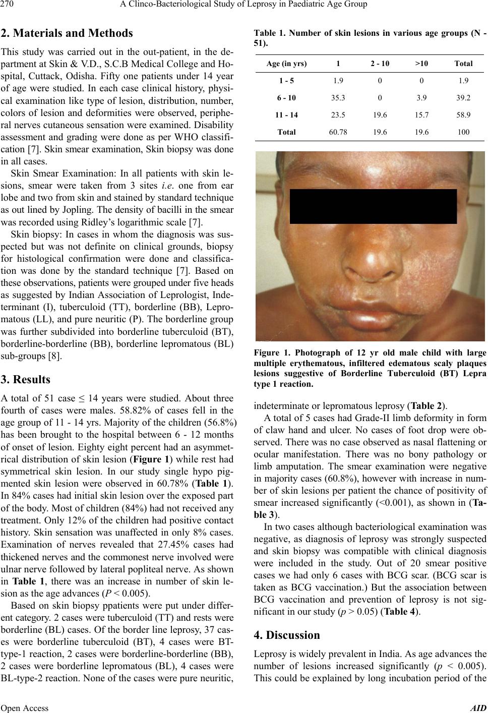
[16] N. Vara, “Profile of New Cases of Childhood Leprosy in
a Hospital Setting,” Indian Journal of Leprosy, Vol. 78,
No. 3, 2006, pp. 17-22.
[17] R. Roy and G. Kalla, “Pattern of Leprosy in Children in
Jodhpur,” Indian Journal of Leprosy, Vol. 69, No. 2,
1997, pp. 199-200.
[18] Van Braket Wh, et al., “Leprosy Review,” Vol. 63, 1992,
pp. 231-235.
[19] B. S. Bhavasar and N. R. Meheta, “An Epidemiological
of Leprosy through School Survey in Surat Dist. (South-
Gujurat),” Leprosy in India, Vol. 52, 1980, pp. 548-546.
[20] A. Singal, S. Sonthalia and D. Pandhi, “Childhood Le-
prosy in a Tertiary-Care Hospital in Delhi, India: A Re-
appraisal in the Post-Elimination Era,” Leprosy Review,
Vol. 82, 2011, pp. 259-269.
[21] V. P. Shetty, S. D. Ghate, A. V. Wakade, D. V. Thakur, U.
H. Thakar and E. D’souza, “Clinical, Bacteriological, and
Histopathological Characteristics of Newly Detected Chil-
Open Access AID
A Clinco-Bacteriological Study of Leprosy in Paediatric Age Group
Open Access AID
273
dren with Leprosy: A Population Based Study in a De-
fined Rural and Urban Area of Maharashtra, Western In-
dia,” Indian Journal of Dermatology, Venerology, Lepro-
logy, Vol. 79, No. 4, pp. 512-519.
[22] WHO, “Technical Report Series,” 1988, p. 768.