Journal of Computer and Communications, 2013, 1, 5-8
Published Online December 2013 (http://www.scirp.org/journal/jcc)
http://dx.doi.org/10.4236/jcc.2013.17002
Open Access JCC
5
Light Microbeams by Tapered Glass Capillaries for
Biological Irradiation
Wei-Guo Jin1, Kyohei Katoh1, Tatsuya Minowa1, Tokihiro Ikeda2
1Department of Physics, Faculty of Science, Toho University, 2-2-1 Miyama, Funabashi, Chiba 274-8510, Japan; 2RIKEN Nishina
Center, 2-1 Hirosawa, Wako, Saitama 351-0198, Japan.
Email: jin@ph.sci.toho-u.ac.jp
Received August 2013
ABSTRACT
Microbeams of visible ligh t were studied usin g a tapered g lass capillary. Trans mittance of laser ligh t through capillaries
with different inlet and outlet diameters was measured. About several % of the transmittance was obtained and larger
than 80% was achieved in combining with an optical lens. It was found that the obtained transmittance considerably
depended on the capillary shape, i.e., the taper angle. Densi ty enhancement of the extr acted beam was derived and show-
ed a strong foc usi ng abi lity for t he ta pered glass capill ary. Propagat ion of vi si ble li ght thr ough the capi ll ary wa s di scussed.
Keywords: Light Microbeam; Tapered Glass Capillary; Transmittance; Propagation of Light; Dens ity Enhan c ement
1. Introduction
In micro-surgery of single living cell, fluorescent mark-
ers conjunct to the nucleus or an organelle are needed.
Light microbeam provides a spot light so that the other
part within a microscope view is free from the bleaching
due to the undesired excitation light. If the microbeam is
strong enough, the beam itself can serve as a pin-point
knife. Although the conventional optical lens can focus
light into even several mm, it is hard to control the beam
size and the beam position on a living cell using the optical
lens.
Using tapered glass capillaries, many studies have re-
cently been reported on microbeam production of keV-
MeV accelerated ions [1-3]. Microbeams by the capilla-
ries have the advantages of low cost, easily positioning
on the target and preferable sizes of beams. However,
few studies about visible light with the glass capillary
have been reported and propagation of light through the
capillaries is not known. The capillary optics can be a
new method with above advantages to produce focused
microbeams of light in addition to optical lenses and fur-
ther is applicable to the UV and X-ray microbeam pro-
duction. The light propagation through the capillary is
also interesting in terms of light-matter interac tio n.
In this paper, we report light-microbeam production
using tapered glass capillaries for biological irradiation.
Transmittance is obtained for capillaries with different
inlet and outlet diameters, and results are discussed.
2. Experiment
Tapered glass capillaries were prepared using a puller
(PE-21, NARISHIGE Co. Ltd) by heating a straight glass
tube made of borosilicate and by pulling both ends with a
constant force. Glass tubes w ith the inlet diameters of 1.8
mm and 0.8 mm were used. Outlet diameter was deter-
mined by cutting the capillary with a Microforge (MF-
900, NARISHIGE Co. Ltd). Tens of images were taken
by a microscope for the whole capillary in order to obtain
the capillary shape and the taper angle.
Figure 1 shows the experimental setup for the trans-
mittance measurements. We used an Ar+ laser, a He-Ne
laser, and a diode la se r wi th a wavele ngth of 488 nm, 633
nm and 670 nm, respectively. The capillary was set on a
Figure 1. Experimental setup for transmittance measure-
ments.
Light Microbeams by Tapered Glass Capillaries for Biological Irradiation
Open Access JCC
6
5-axis (xyzqf) precise stage and fine adjustment with the
precision of mm could be made. In order to cut scattered
light, slits were located both upstream and downstream
of the capillary. The diffraction patter n of the transmitted
beam was used to align the capillary respec t to the initial
beam direction. Powers of transmitted beams through both
the tapered capillary and a straight glass tube were meas-
ured with a power meter.
In some measurements, an optical lens with a focal
length of 20 cm was set upstream of the capillary. The
lens together with the precise stage and the slits was set
on a straight rail and, therefore, the lens could be moved
along the beam direction in order to change the position
of the focal p oint.
3. Results and Discussion
We measured the powers of transmitted beams through
both the tapered capillary and a straight glass tube de-
fined as output power and input power, respectively.
Therefore, the transmittance T is defined as follows
output power
(%) 100.
input power
T= ×
(1)
3.1. Transmittance
Transmittance T was derived from the measurement for
capillaries with different outlet diameters. Results of T
for the 1.8 mm inlet diameter are shown in Figure 2 as a
function of the outlet diameter without the optical lens.
Transmittance T increases from about 3% to about 10%
when the outlet diameter rises from 17 mm to 100 mm
and, however, no obvious dependence on the wavelength
is found.
3.2. Combined with a Lens
The effect of an optical lens combined with capillaries
was investigated. Changing the position of the lens, i.e.,
Figure 2. Transmittance of laser beam through the capillary
of the 1.8 mm inlet diameter as a function of the outlet di-
ameter without the optical lens.
the position of the focal point, we measured T for differ-
ent capillaries. Dependence of T on the position of the
focal point at 488 nm is show in Figure 3 for the 1.8 mm
inlet diameter capillaries with three typical outlet diame-
ters of 28 mm, 37 mm and 67 mm. The origin of the x
axis is set at the position of the capillary outlet and the
position of the focal point moves to the inlet as x in-
creases.
It can be seen from Figure 3 that T strongly depends
on the position of the focal point particularly for small
outlet diameters and reaches maximum around x = 2 cm,
i.e., the region between the large taper angle and the
small one [4]. The capillaries with the outlet diameters of
28 mm, 37 mm and 67 mm show a same trend. Similar
behavior was also found for the wavelengths at 633 nm
and 670 nm.
Dependence of T on the outlet diameter with the opti-
cal lens was measured as shown in Figure 4, where the
Figure 3. Dependence of transmittance on the position of
the focal point for the 1.8 mm inlet diameter at 488 nm. The
origin of the x axis is set at the position of the capillary out-
let.
Figure 4. Transmittance of laser beam through the capillary
of the 1.8 mm inlet diameter as a function of the outlet di-
ameter with the optical lens.
Light Microbeams by Tapered Glass Capillaries for Biological Irradiation
Open Access JCC
7
position of the focal point is set at the region between th e
large taper angle and the small one, corresponding to the
maximum T. Rapid increase of T with the outlet diameter
can be found and T saturates to be larger than 80% in the
region beyond 40 mm.
3.3. Transmittance for Dif f erent Inlet Dia meters
Transmittance of light through the capillar y is considered
to be dependent on the capillary shape, i.e., the taper an-
gle of the capillary. In order to study this effect, we
measured transmittance of light through capillaries with
the 1.8 mm and 0.8 mm inlet diameters. Figure 5 shows
a comparison of transmittance between these two inlet
diameters at 633 nm. It can be seen from Figure 5 that
the transmittance for the 0.8 mm inlet diameter is consi-
derably larger than that for the 1.8 mm inlet diameter.
However, capillaries with the 0.8 mm inlet diameter have
a same trend as the 1.8 inlet diameter concerning the de-
pendence of the outlet diameter.
The capillary shape together with the taper angle was
analyzed using the capillary images taken . Figure 6 shows
a comparison of the inner radius of the capillary for the
1.8 mm and 0.8 mm inlet diameters in a direction to the
capillary outlet. The origin of the x axis is set to the posi-
tion before the taper starting. It is clearly found from
Figure 6 that the inner radius for the 1.8 mm inlet di-
ameter changes rapidly, corresponding a large taper an-
gle, compared with the 0.8 mm inlet diameter. This is the
reason of the small transmittance for the 1.8 mm inlet
diameter.
3.4. Density Enhancement
In order to check the focusing ability of the capillary, the
density enhancement was derived from densities at the
outlet and inlet and is shown in Figure 7 without the lens.
Figure 5. Comparison of transmittance through the capil-
lary between the 1.8 mm and 0.8 mm inlet diameters at 633
nm.
Figure 6. Comparison of the inner radius of the capillary
between the 1.8 mm and 0.8 mm inlet diameters.
Figure 7. Density enhancement of the capillary for the 1.8
mm inlet diameter as a function of the outlet diameter
without the optical lens.
The enhancement is about 30 at 100 mm, and reaches
about 400 at 17 mm, increasing when the outlet diameter
decreases. This shows that the capillary has strong fo-
cusing ability; the smaller outlet diameter the stronger
focusing.
The obtained experimental results can be qualitatively
interpreted. We introduce a model which divides a real
capillary shape into two regions: region 1 with large ta-
per angle of about 7˚ and region 2˚, near the outlet, with
a small taper angle of about 0.2˚. It is known that reflec-
tance of light by a glass surface strongly depends on the
incident angle; several % for the incident angle of small-
er than 60˚ while almost 100% for the incident angle
close to 90˚ [5]. The region 1 has, therefore, almost no
contribution to the transmission and reflection at the re-
gion 2 results in several % of the transmittance as expe-
rimentally obtained. In the case of the combination with
the optical lens, however, light is focused by the lens
directly into the region 2 and this yields the large trans-
mittance.
Light Microbeams by Tapered Glass Capillaries for Biological Irradiation
Open Access JCC
8
4. Summary
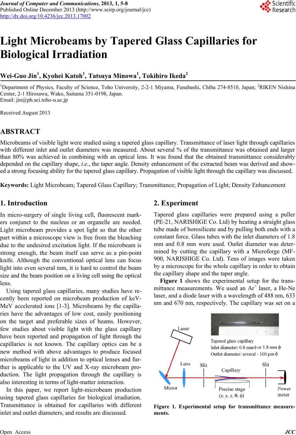
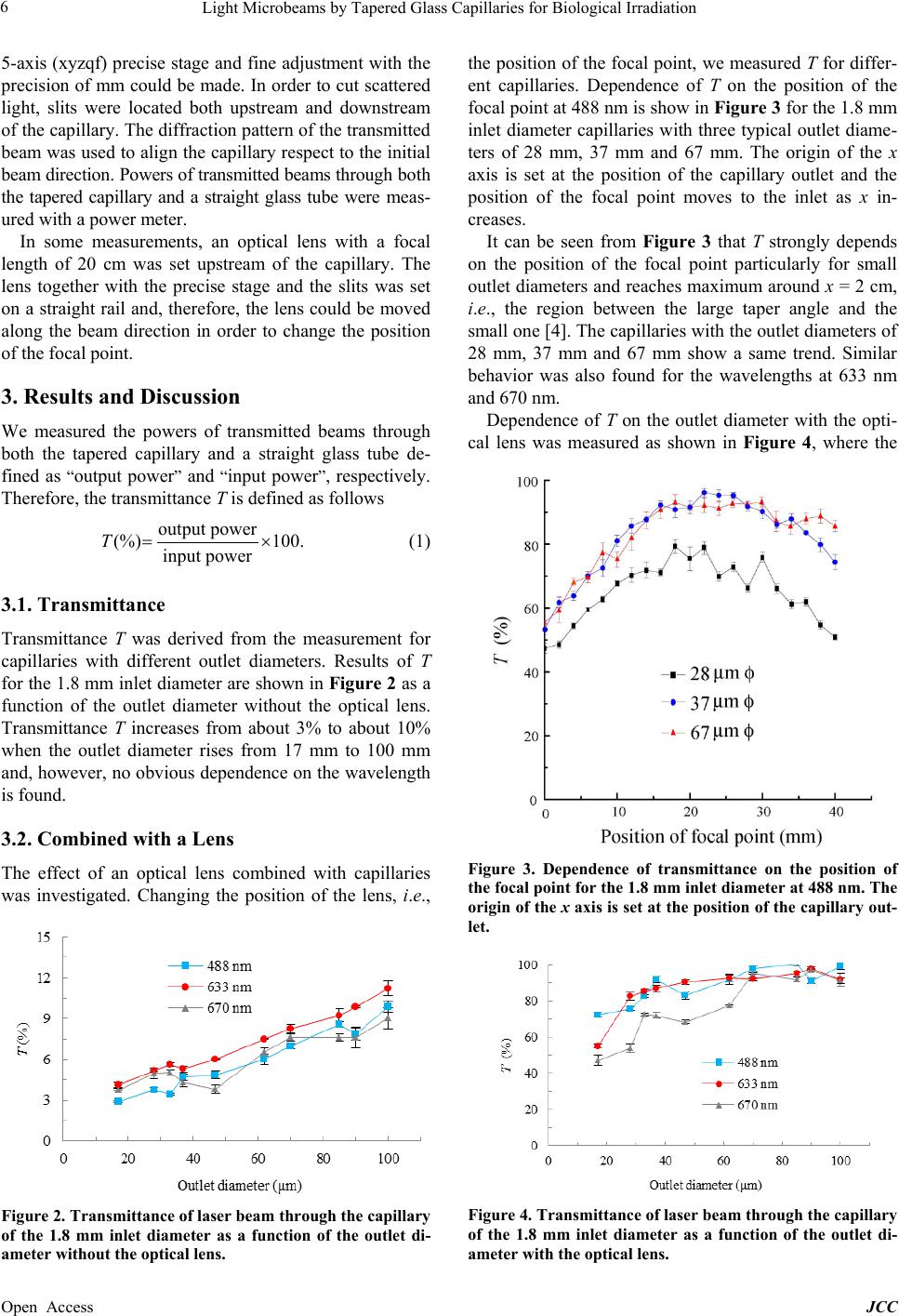
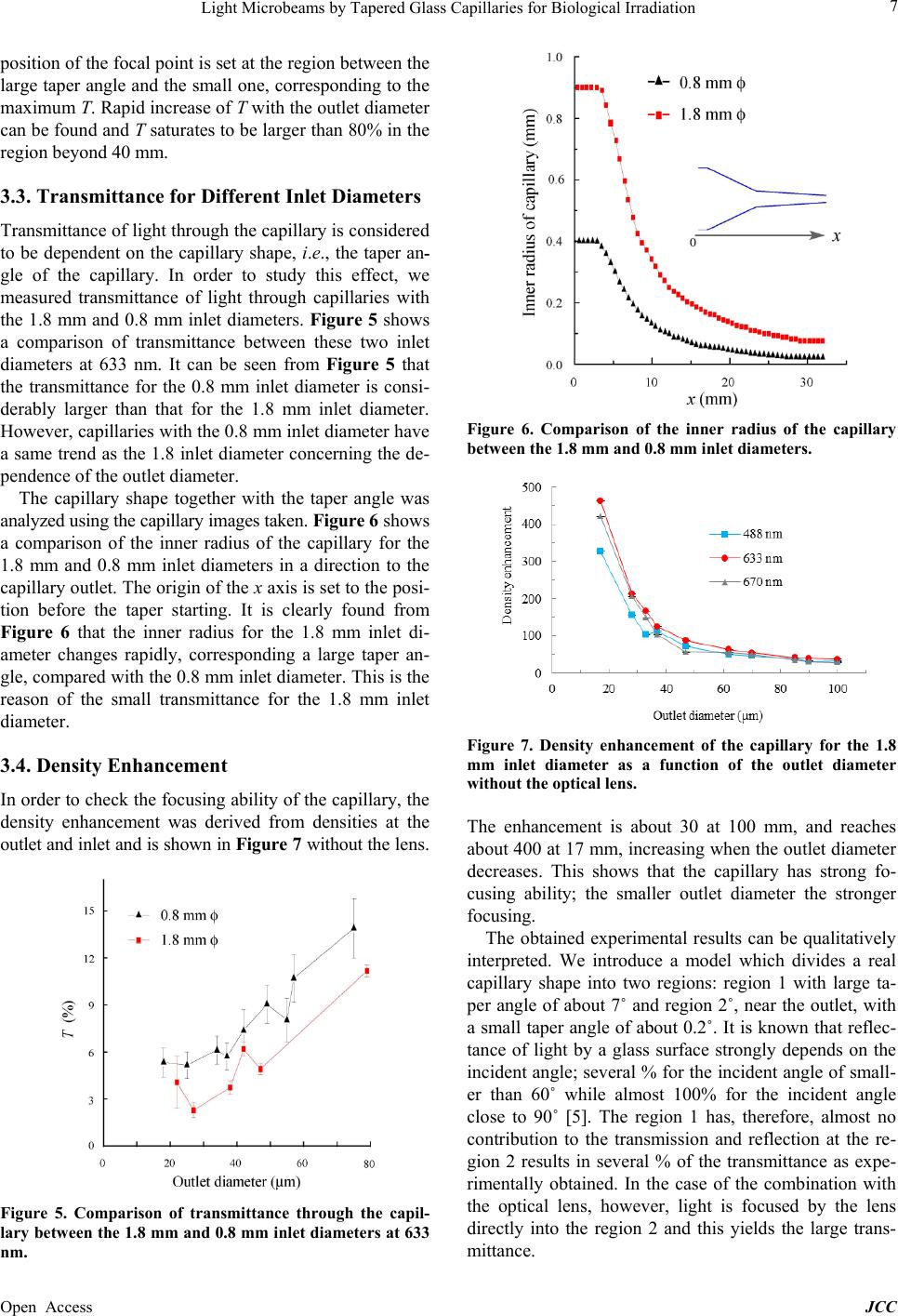
Light microbeams were produced using tapered glass
capillaries for biological irradiation. Propagation of visi-
ble light through the capillary was studied. Transmittance
was measured for the capillaries with different inlet and
outlet diameters. The transmittance was found to be about
several % without the optical lens and larger than 80%
with the optical lens, depending on the outlet diameter.
Measurements with and without the lens provide infor-
mation to find the op timum lens-free shapes of the capil-
laries. The transmittance for the 0.8 mm inlet diameter
was measured to be considerably larger than that for the
1.8 mm inlet diameter, and was attributed to the differ-
ence of the capillary shape which was analyzed using the
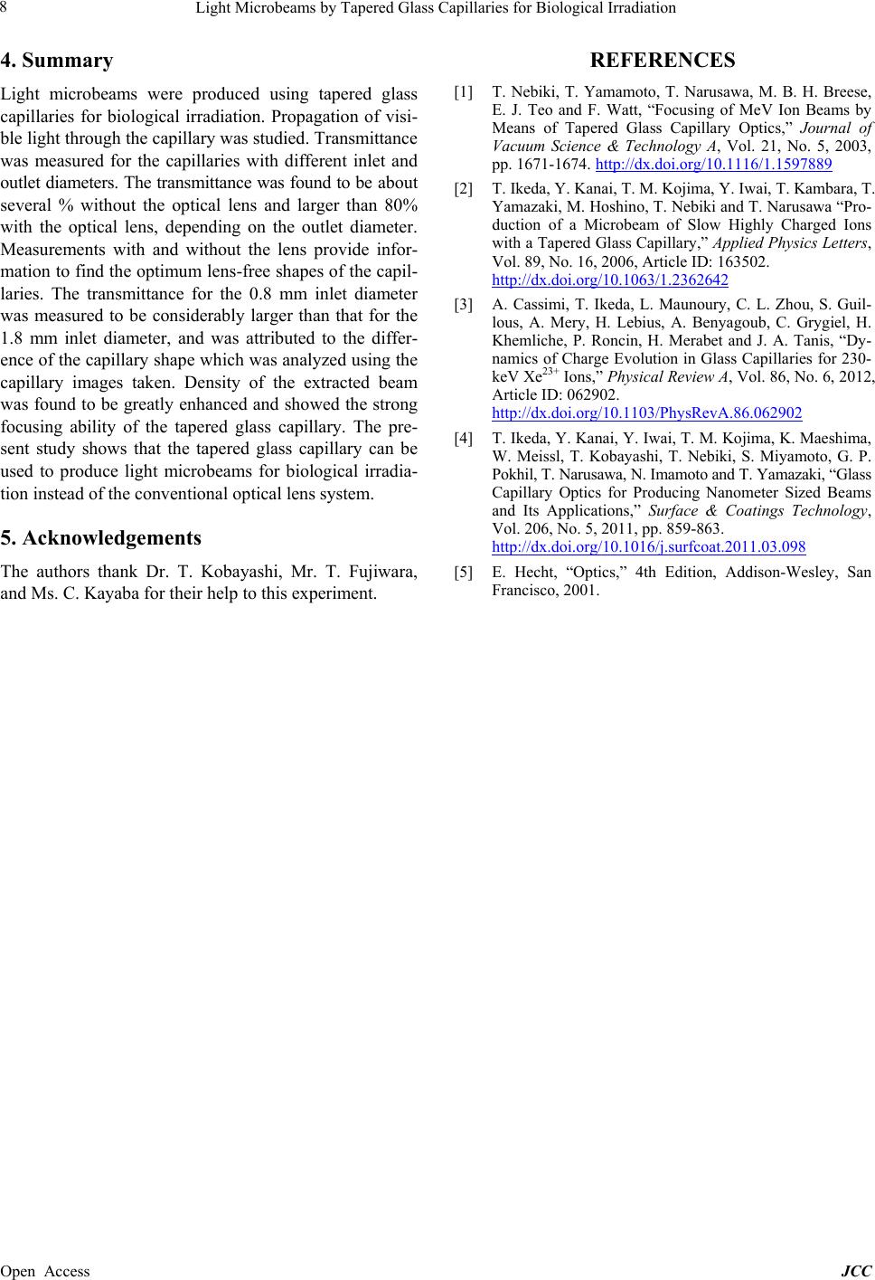
capillary images taken. Density of the extracted beam
was found to be greatly enhanced and showed the strong
focusing ability of the tapered glass capillary. The pre-
sent study shows that the tapered glass capillary can be
used to produce light microbeams for biological irradia-
tion instead of the conventional optical lens system.
5. Acknowledgements
The authors thank Dr. T. Kobayashi, Mr. T. Fujiwara,
and Ms. C. Kayaba for their help to this experiment.
REFERENCES
[1] T. Nebiki, T. Yamamoto, T. Narusawa, M. B. H. Breese,
E. J. Teo and F. Watt, “Focusing of MeV Ion Beams by
Means of Tapered Glass Capillary Optics,” Journal of
Vacuum Science & Technology A, Vol. 21, No. 5, 2003,
pp. 1671-1674. http://dx.doi.org/10.1116/1.1597889
[2] T. Ikeda, Y. Kanai, T. M. Kojima, Y. Iwai, T. Kambara, T.
Yamazaki, M. Hoshino, T. Nebiki and T. Narusawa “Pro-
duction of a Microbeam of Slow Highly Charged Ions
with a Tapered Glass Capillary,” Applied Physics Letters,
Vol. 89, No. 16, 2006, Article ID: 163502.
http://dx.doi.org/10.1063/1.2362642
[3] A. Cassimi, T. Ikeda, L. Maunoury, C. L. Zhou, S. Guil-
lous, A. Mery, H. Lebius, A. Benyagoub, C. Grygiel, H.
Khemliche, P. Roncin, H. Merabet and J. A. Tanis, “Dy-
namics of Charge Evolution in Glass Capillaries for 230-
keV Xe23+ Ions,” Physical Review A, Vol. 86, No. 6, 2012,
Article ID: 062902.
http://dx.doi.org/10.1103/PhysRevA.86.062902
[4] T. Ikeda, Y. Kanai, Y. Iwai, T. M. Kojima, K. Maeshima,
W. Meissl, T. Kobayashi, T. Nebiki, S. Miyamoto, G. P.
Pokhil, T. Narusawa, N. Imamoto and T. Yamazaki, “Glass
Capillary Optics for Producing Nanometer Sized Beams
and Its Applications,” Surface & Coatings Technology,
Vol. 206, No. 5, 2011, pp. 859-863.
http://dx.doi.org/10.1016/j.surfcoat.2011.03.098
[5] E. Hecht, “Optics,” 4th Edition, Addison-Wesley, San
Francisco, 2001.