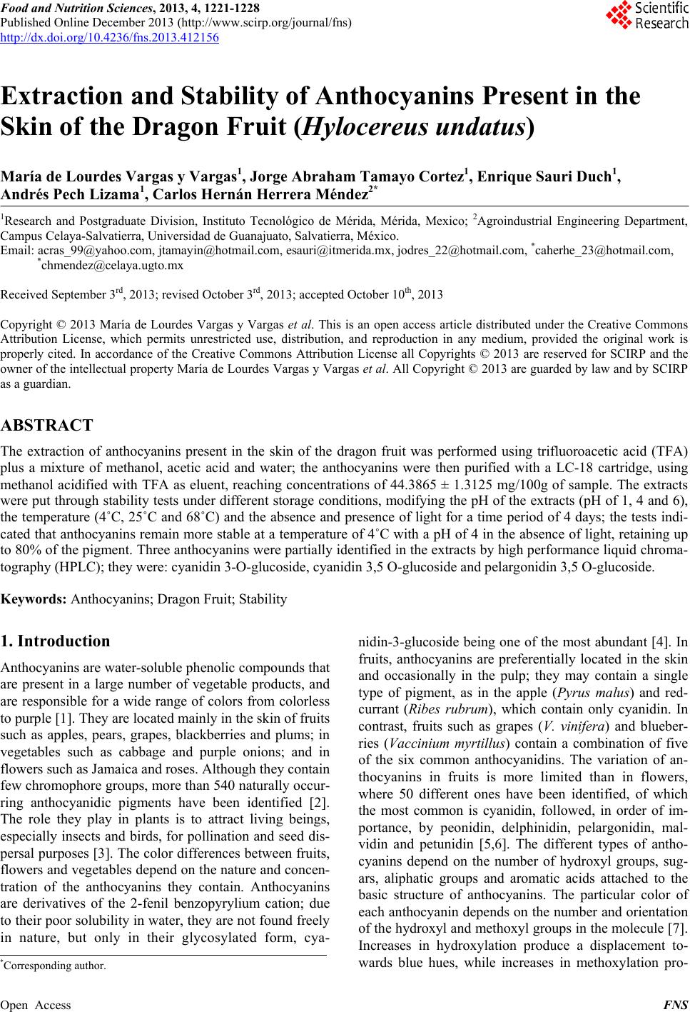
 Food and Nutrition Sciences, 2013, 4, 1221-1228 Published Online December 2013 (http://www.scirp.org/journal/fns) http://dx.doi.org/10.4236/fns.2013.412156 Open Access FNS Extraction and Stability of Anthocyanins Present in the Skin of the Dragon Fruit (Hylocereus undatus) María de Lourdes Vargas y Vargas1, Jorge Abraham Tamayo Cortez1, Enrique Sauri Duch1, Andrés Pech Lizama1, Carlos Hernán Herrera Méndez2* 1Research and Postgraduate Division, Instituto Tecnológico de Mérida, Mérida, Mexico; 2Agroindustrial Engineering Department, Campus Celaya-Salvatierra, Universidad de Guanajuato, Salvatierra, México. Email: acras_99@yahoo.com, jtamayin@hotmail.com, esauri@itmerida.mx, jodres_22@hotmail.com, *caherhe_23@hotmail.com, *chmendez@celaya.ugto.mx Received September 3rd, 2013; revised October 3rd, 2013; accepted October 10th, 2013 Copyright © 2013 María de Lourdes Vargas y Vargas et al. This is an open access article distributed under the Creative Commons Attribution License, which permits unrestricted use, distribution, and reproduction in any medium, provided the original work is properly cited. In accordance of the Creative Commons Attribution License all Copyrights © 2013 are reserved for SCIRP and the owner of the intellectual property María de Lourdes Vargas y Vargas et al. All Copyright © 2013 are guarded by law and by SCIRP as a guardian. ABSTRACT The extraction of anthocyanins present in the skin of the dragon fruit was performed using trifluoroacetic acid (TFA) plus a mixture of methanol, acetic acid and water; the anthocyanins were then purified with a LC-18 cartridge, using methanol acidified with TFA as eluent, reaching concentrations of 44.3865 ± 1.3125 mg/100g of sample. The extracts were put through stability tests under different storage conditions, modifying the pH of the extracts (pH of 1, 4 and 6), the temperature (4˚C, 25˚C and 68˚C) and the absence and presence of light for a time period of 4 days; the tests indi- cated that anthocyanins remain more stable at a temperature of 4˚C with a pH of 4 in the absence of light, retaining up to 80% of the pigment. Three anthocyanins were partially identified in the extracts by high performance liquid chroma- tography (HPLC); they were: cyanidin 3-O-glucoside, cyanidin 3,5 O-glucoside and pelargonidin 3,5 O-glucoside. Keywords: Anthocyanins; Dragon Fruit; Stability 1. Introduction Anthocyanins are water-soluble phenolic compounds that are present in a large number of vegetable products, and are responsible for a wide range of colors from colorless to purple [1]. They are located mainly in the skin of fruits such as apples, pears, grapes, blackberries and plums; in vegetables such as cabbage and purple onions; and in flowers such as Jamaica and roses. Although they contain few chromophore groups, more than 540 naturally occur- ring anthocyanidic pigments have been identified [2]. The role they play in plants is to attract living beings, especially insects and birds, for pollination and seed dis- persal purposes [3]. The color differences between fruits, flowers and vegetables depend on the nature and concen- tration of the anthocyanins they contain. Anthocyanins are derivatives of the 2-fenil benzopyrylium cation; due to their poor solubility in water, they are not found freely in nature, but only in their glycosylated form, cya- nidin-3-glucoside being one of the most abundant [4]. In fruits, anthocyanins are preferentially located in the skin and occasionally in the pulp; they may contain a single type of pigment, as in the apple (Pyrus malus) and red- currant (Ribes rubrum), which contain only cyanidin. In contrast, fruits such as grapes (V. vinifera) and blueber- ries (Vaccinium myrtillus) contain a combination of five of the six common anthocyanidins. The variation of an- thocyanins in fruits is more limited than in flowers, where 50 different ones have been identified, of which the most common is cyanidin, followed, in order of im- portance, by peonidin, delphinidin, pelargonidin, mal- vidin and petunidin [5,6]. The different types of antho- cyanins depend on the number of hydroxyl groups, sug- ars, aliphatic groups and aromatic acids attached to the basic structure of anthocyanins. The particular color of each anthocyanin depends on the number and orientation of the hydroxyl and methoxyl groups in the molecule [7]. Increases in hydroxylation produce a displacement to- wards blue hues, while increases in methoxylation pro- *Corresponding author. 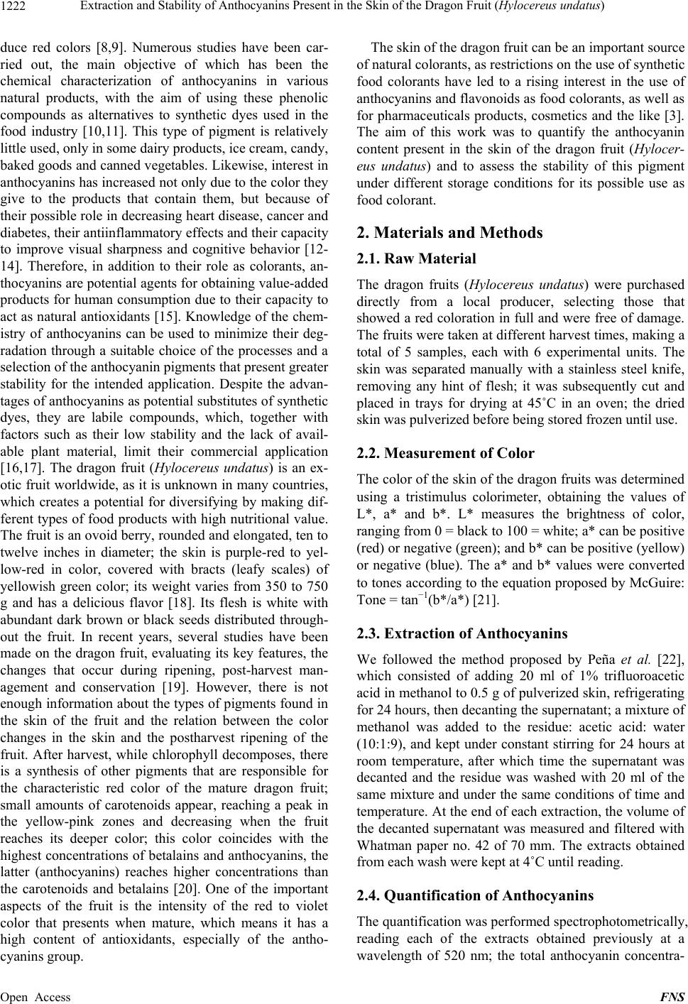 Extraction and Stability of Anthocyanins Present in the Skin of the Dragon Fruit (Hylocereus undatus) 1222 duce red colors [8,9]. Numerous studies have been car- ried out, the main objective of which has been the chemical characterization of anthocyanins in various natural products, with the aim of using these phenolic compounds as alternatives to synthetic dyes used in the food industry [10,11]. This type of pigment is relatively little used, only in some dairy products, ice cream, candy, baked goods and canned vegetables. Likewise, interest in anthocyanins has increased not only due to the color they give to the products that contain them, but because of their possible role in decreasing heart disease, cancer and diabetes, their antiinflammatory effects and their capacity to improve visual sharpness and cognitive behavior [12- 14]. Therefore, in addition to their role as colorants, an- thocyanins are potential agents for obtaining value-added products for human consumption due to their capacity to act as natural antioxidants [15]. Knowledge of the chem- istry of anthocyanins can be used to minimize their deg- radation through a suitable choice of the processes and a selection of the anthocyanin pigments that present greater stability for the intended application. Despite the advan- tages of anthocyanins as potential substitutes of synthetic dyes, they are labile compounds, which, together with factors such as their low stability and the lack of avail- able plant material, limit their commercial application [16,17]. The dragon fruit (Hylocereus undatus) is an ex- otic fruit worldwide, as it is unknown in many countries, which creates a potential for diversifying by making dif- ferent types of food products with high nutritional value. The fruit is an ovoid berry, rounded and elongated, ten to twelve inches in diameter; the skin is purple-red to yel- low-red in color, covered with bracts (leafy scales) of yellowish green color; its weight varies from 350 to 750 g and has a delicious flavor [18]. Its flesh is white with abundant dark brown or black seeds distributed through- out the fruit. In recent years, several studies have been made on the dragon fruit, evaluating its key features, the changes that occur during ripening, post-harvest man- agement and conservation [19]. However, there is not enough information about the types of pigments found in the skin of the fruit and the relation between the color changes in the skin and the postharvest ripening of the fruit. After harvest, while chlorophyll decomposes, there is a synthesis of other pigments that are responsible for the characteristic red color of the mature dragon fruit; small amounts of carotenoids appear, reaching a peak in the yellow-pink zones and decreasing when the fruit reaches its deeper color; this color coincides with the highest concentrations of betalains and anthocyanins, the latter (anthocyanins) reaches higher concentrations than the carotenoids and betalains [20]. One of the important aspects of the fruit is the intensity of the red to violet color that presents when mature, which means it has a high content of antioxidants, especially of the antho- cyanins group. The skin of the dragon fruit can be an important source of natural colorants, as restrictions on the use of synthetic food colorants have led to a rising interest in the use of anthocyanins and flavonoids as food colorants, as well as for pharmaceuticals products, cosmetics and the like [3]. The aim of this work was to quantify the anthocyanin content present in the skin of the dragon fruit (Hylocer- eus undatus) and to assess the stability of this pigment under different storage conditions for its possible use as food colorant. 2. Materials and Methods 2.1. Raw Material The dragon fruits (Hylocereus undatus) were purchased directly from a local producer, selecting those that showed a red coloration in full and were free of damage. The fruits were taken at different harvest times, making a total of 5 samples, each with 6 experimental units. The skin was separated manually with a stainless steel knife, removing any hint of flesh; it was subsequently cut and placed in trays for drying at 45˚C in an oven; the dried skin was pulverized before being stored frozen until use. 2.2. Measurement of Color The color of the skin of the dragon fruits was determined using a tristimulus colorimeter, obtaining the values of L*, a* and b*. L* measures the brightness of color, ranging from 0 = black to 100 = white; a* can be positive (red) or negative (green); and b* can be positive (yellow) or negative (blue). The a* and b* values were converted to tones according to the equation proposed by McGuire: Tone = tan−1(b*/a*) [21]. 2.3. Extraction of Anthocyanins We followed the method proposed by Peña et al. [22], which consisted of adding 20 ml of 1% trifluoroacetic acid in methanol to 0.5 g of pulverized skin, refrigerating for 24 hours, then decanting the supernatant; a mixture of methanol was added to the residue: acetic acid: water (10:1:9), and kept under constant stirring for 24 hours at room temperature, after which time the supernatant was decanted and the residue was washed with 20 ml of the same mixture and under the same conditions of time and temperature. At the end of each extraction, the volume of the decanted supernatant was measured and filtered with Whatman paper no. 42 of 70 mm. The extracts obtained from each wash were kept at 4˚C until reading. 2.4. Quantification of Anthocyanins The quantification was performed spectrophotometrically, reading each of the extracts obtained previously at a wavelength of 520 nm; the total anthocyanin concentra- Open Access FNS 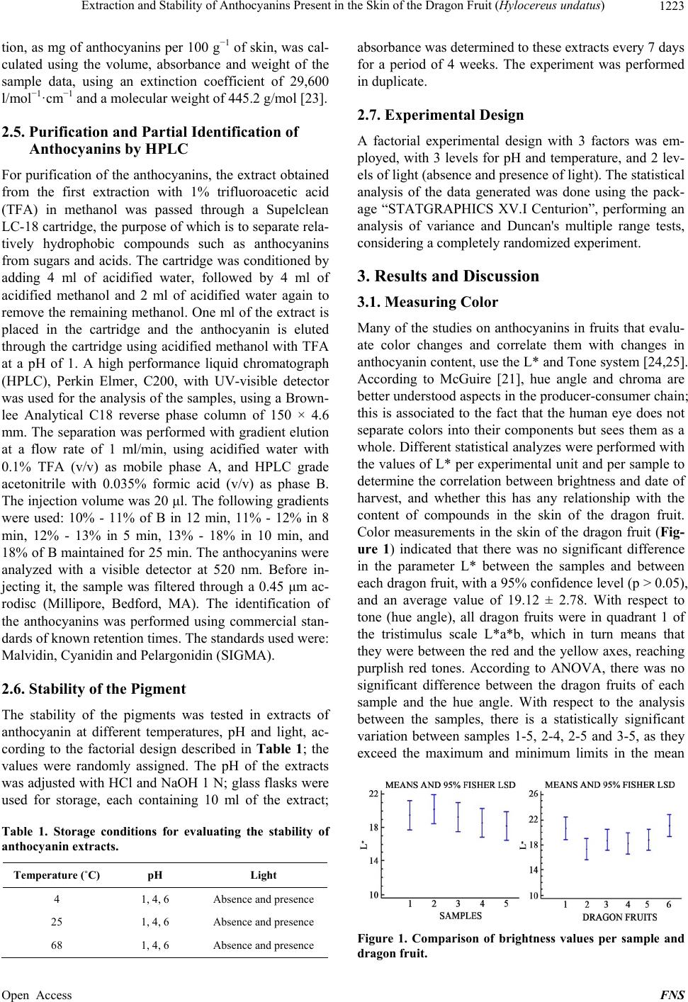 Extraction and Stability of Anthocyanins Present in the Skin of the Dragon Fruit (Hylocereus undatus) 1223 tion, as mg of anthocyanins per 100 g−1 of skin, was cal- culated using the volume, absorbance and weight of the sample data, using an extinction coefficient of 29,600 l/mol−1·cm−1 and a molecular weight of 445.2 g/mol [23]. 2.5. Purification and Partial Identification of Anthocyanins by HPLC For purification of the anthocyanins, the extract obtained from the first extraction with 1% trifluoroacetic acid (TFA) in methanol was passed through a Supelclean LC-18 cartridge, the purpose of which is to separate rela- tively hydrophobic compounds such as anthocyanins from sugars and acids. The cartridge was conditioned by adding 4 ml of acidified water, followed by 4 ml of acidified methanol and 2 ml of acidified water again to remove the remaining methanol. One ml of the extract is placed in the cartridge and the anthocyanin is eluted through the cartridge using acidified methanol with TFA at a pH of 1. A high performance liquid chromatograph (HPLC), Perkin Elmer, C200, with UV-visible detector was used for the analysis of the samples, using a Brown- lee Analytical C18 reverse phase column of 150 × 4.6 mm. The separation was performed with gradient elution at a flow rate of 1 ml/min, using acidified water with 0.1% TFA (v/v) as mobile phase A, and HPLC grade acetonitrile with 0.035% formic acid (v/v) as phase B. The injection volume was 20 μl. The following gradients were used: 10% - 11% of B in 12 min, 11% - 12% in 8 min, 12% - 13% in 5 min, 13% - 18% in 10 min, and 18% of B maintained for 25 min. The anthocyanins were analyzed with a visible detector at 520 nm. Before in- jecting it, the sample was filtered through a 0.45 μm ac- rodisc (Millipore, Bedford, MA). The identification of the anthocyanins was performed using commercial stan- dards of known retention times. The standards used were: Malvidin, Cyanidin and Pelargonidin (SIGMA). 2.6. Stability of the Pigment The stability of the pigments was tested in extracts of anthocyanin at different temperatures, pH and light, ac- cording to the factorial design described in Table 1; the values were randomly assigned. The pH of the extracts was adjusted with HCl and NaOH 1 N; glass flasks were used for storage, each containing 10 ml of the extract; Table 1. Storage conditions for evaluating the stability of anthocyanin extracts. Temperature ( ˚C) pH Light 4 1, 4, 6 Absence and presence 25 1, 4, 6 Absence and presence 68 1, 4, 6 Absence and presence absorbance was determined to these extracts every 7 days for a period of 4 weeks. The experiment was performed in duplicate. 2.7. Experimental Design A factorial experimental design with 3 factors was em- ployed, with 3 levels for pH and temperature, and 2 lev- els of light (absence and presence of light). The statistical analysis of the data generated was done using the pack- age “STATGRAPHICS XV.I Centurion”, performing an analysis of variance and Duncan's multiple range tests, considering a completely randomized experiment. 3. Results and Discussion 3.1. Measuring Color Many of the studies on anthocyanins in fruits that evalu- ate color changes and correlate them with changes in anthocyanin content, use the L* and Tone system [24,25]. According to McGuire [21], hue angle and chroma are better understood aspects in the producer-consumer chain; this is associated to the fact that the human eye does not separate colors into their components but sees them as a whole. Different statistical analyzes were performed with the values of L* per experimental unit and per sample to determine the correlation between brightness and date of harvest, and whether this has any relationship with the content of compounds in the skin of the dragon fruit. Color measurements in the skin of the dragon fruit (Fig- ure 1) indicated that there was no significant difference in the parameter L* between the samples and between each dragon fruit, with a 95% confidence level (p > 0.05), and an average value of 19.12 ± 2.78. With respect to tone (hue angle), all dragon fruits were in quadrant 1 of the tristimulus scale L*a*b, which in turn means that they were between the red and the yellow axes, reaching purplish red tones. According to ANOVA, there was no significant difference between the dragon fruits of each sample and the hue angle. With respect to the analysis between the samples, there is a statistically significant variation between samples 1-5, 2-4, 2-5 and 3-5, as they exceed the maximum and minimum limits in the mean Figure 1. Comparison of brightness values per sample and dragon fruit. Open Access FNS  Extraction and Stability of Anthocyanins Present in the Skin of the Dragon Fruit (Hylocereus undatus) 1224 comparison, which can be seen in Figure 2. The differ- ence presented by these samples is due to the variation in climate, because at the time of the samplings 4 and 5, the rain that fell during the time of harvest was higher, which could have an effect on the color of the skin. 3.2. Extraction and Quantification of Anthocyanins Table 2 shows the average composition with respect to the fractions found in the fruit, both for the edible part (pulp and seeds) and for the skin. Nerd et al. [26] report values of 32.5% for the skin, which resemble the values found for the dragon fruits of this study. However, the average weight of the whole fruit is similar to that re- ported by Centurión et al. [19], who obtained dragon fruits with a weight of 463.7 g, with variations regarding the pulp and the skin, as they mentioned a pulp weight of 368.91 g and a skin weight of 94.8 g, the latter repre- senting a value of 20.4%. This suggests that the fruits of this work had a thicker skin. The average content of an- thocyanins from the skin of dragon fruits harvested in different seasons was 45.15 ± 0.5117 g/100g of dry dra- gon fruit skin. The quantification results of this study are similar to those found in raspberry by Antonne and Kar- jalinen [27], who found values of 19 and 51 mg/100g of fresh fruit; they are also similar to the values obtained by Peña et al. [22] which range from 28.7 to 55.6 mg/100g of fresh fruit. These variations may be due to the sol- vents used or to the methodology used to extract the compound. The analysis of variance showed no signifi- Figure 2. Comparison of means of the hue angle between samples and dragon fruits. Table 2. Average composition of the parts of the dragon fruit (Hylocereus undatus). Average weight of the parts of the dragon fruit Average weight (g) Percentage Whole fruit 463.21 ± 59.74 100 Skin 148.10 ± 31.46 31.97 Pulp and seeds 315.11 ± 54.14 68.027 cant difference in anthocyanin content between the dragon fruits analyzed at different sampling times (Fig- ure 3). The average yield of dragon fruit skin was 319.72 g per kilogram of dragon fruit; anthocyanin content was 45.69 mg/100g of dry skin; therefore, a total of 0.1460 g of anthocyanins was extracted per kilogram of skin. The dragon fruit is thus a suitable source of anthocyanins, since they are obtained solely from the skin, unlike Aguilera et al. [23], who report, for the purple fig of the Mission variety, values 0.295 g per kilogram of whole fruit. The extraction method must be one that allows re- covering the greatest possible amount of anthocyanins with minimum losses due to enzymatic and non-enzy- matic changes. Anthocyanins are generally isolated in acidic media. Among the methods that have been tested for extracting anthocyanins from various natural sources, it was found that using methanol acidified with 1% TFA as an extraction solvent, the amount of pigment extracted increases greatly, while maintaining the stability of the anthocyanins [28]. The efficiency of extraction with TFA is attributed to the low pH that is reached in the system (about 1.3), to the higher solubility of anthocyanins in methanol and to the use of completely pulverized sam- ples, which increases the contact surface of the particles in the solvent. The powdered skin of dragon fruit is sub- jected to extraction in the darkness for 12 hours with overnight stirring in the extraction solvent to allow the diffusion of anthocyanins through the cell membranes and to obtain, during the first extraction, a high concen- tration of anthocyanins; the additional extraction solvent is required to wash the residue of anthocyanins [29]. 3.3. Stability of Anthocyanin Pigments The behavior (Figure 4) of the anthocyanin extracts un- der different storage conditions is to decrease in the first week and then remain more stable from the second week of storage, finding no significant difference (p = 0.05) between the samples stored at 4˚C both in the presence and absence of light, while at 25˚C, in the absence of light, the samples displayed similar values to those stored Figure 3. Average values of total anthocyanins (g of antho- cyanins/100g of skin) present in the skin of dragon fruit, during different harvest times. Open Access FNS 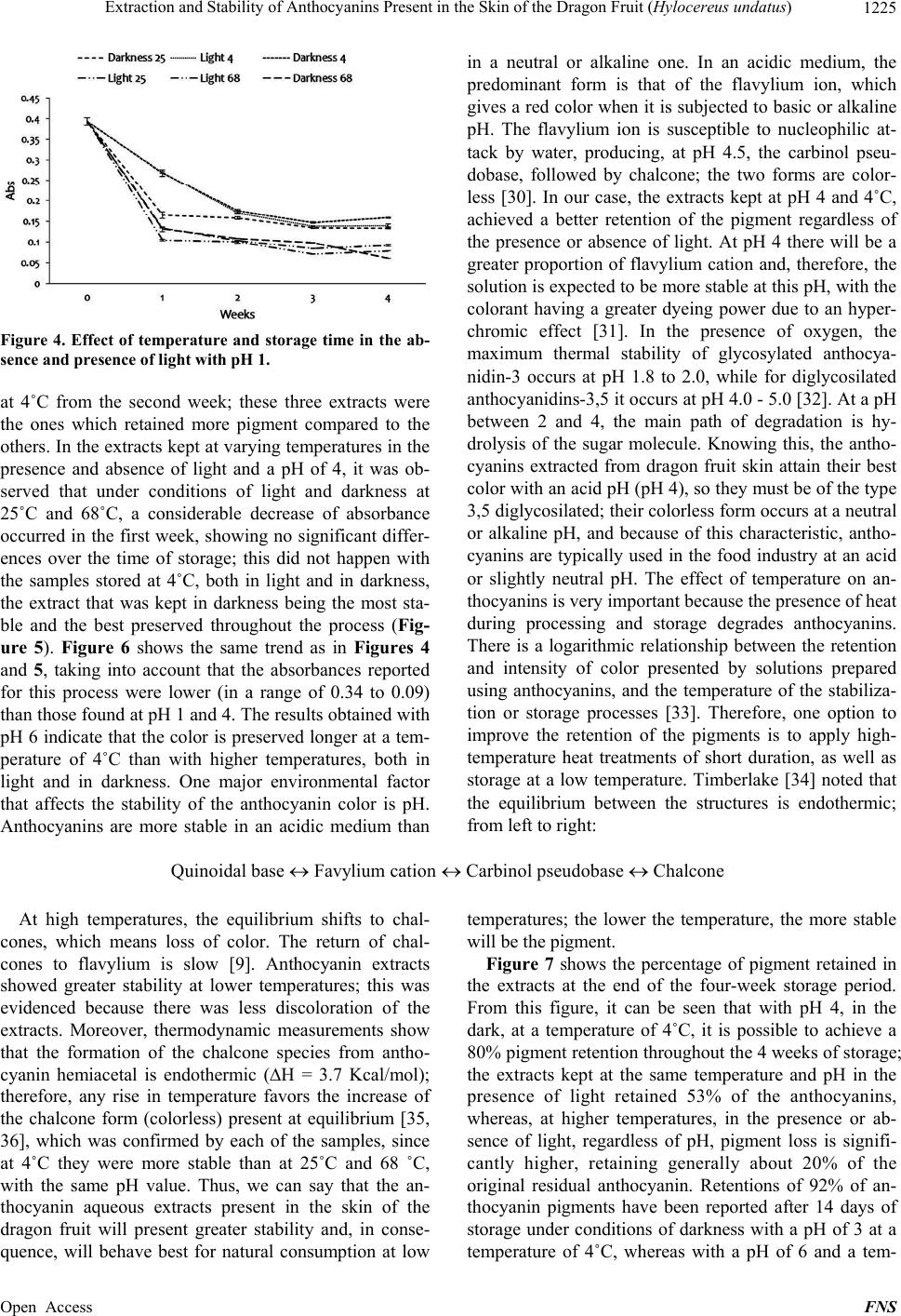 Extraction and Stability of Anthocyanins Present in the Skin of the Dragon Fruit (Hylocereus undatus) Open Access FNS 1225 in a neutral or alkaline one. In an acidic medium, the predominant form is that of the flavylium ion, which gives a red color when it is subjected to basic or alkaline pH. The flavylium ion is susceptible to nucleophilic at- tack by water, producing, at pH 4.5, the carbinol pseu- dobase, followed by chalcone; the two forms are color- less [30]. In our case, the extracts kept at pH 4 and 4˚C, achieved a better retention of the pigment regardless of the presence or absence of light. At pH 4 there will be a greater proportion of flavylium cation and, therefore, the solution is expected to be more stable at this pH, with the colorant having a greater dyeing power due to an hyper- chromic effect [31]. In the presence of oxygen, the maximum thermal stability of glycosylated anthocya- nidin-3 occurs at pH 1.8 to 2.0, while for diglycosilated anthocyanidins-3,5 it occurs at pH 4.0 - 5.0 [32]. At a pH between 2 and 4, the main path of degradation is hy- drolysis of the sugar molecule. Knowing this, the antho- cyanins extracted from dragon fruit skin attain their best color with an acid pH (pH 4), so they must be of the type 3,5 diglycosilated; their colorless form occurs at a neutral or alkaline pH, and because of this characteristic, antho- cyanins are typically used in the food industry at an acid or slightly neutral pH. The effect of temperature on an- thocyanins is very important because the presence of heat during processing and storage degrades anthocyanins. There is a logarithmic relationship between the retention and intensity of color presented by solutions prepared using anthocyanins, and the temperature of the stabiliza- tion or storage processes [33]. Therefore, one option to improve the retention of the pigments is to apply high- temperature heat treatments of short duration, as well as storage at a low temperature. Timberlake [34] noted that the equilibrium between the structures is endothermic; from left to right: Figure 4. Effect of temperature and storage time in the ab- sence and presence of light with pH 1. at 4˚C from the second week; these three extracts were the ones which retained more pigment compared to the others. In the extracts kept at varying temperatures in the presence and absence of light and a pH of 4, it was ob- served that under conditions of light and darkness at 25˚C and 68˚C, a considerable decrease of absorbance occurred in the first week, showing no significant differ- ences over the time of storage; this did not happen with the samples stored at 4˚C, both in light and in darkness, the extract that was kept in darkness being the most sta- ble and the best preserved throughout the process (Fig- ure 5). Figure 6 shows the same trend as in Figures 4 and 5, taking into account that the absorbances reported for this process were lower (in a range of 0.34 to 0.09) than those found at pH 1 and 4. The results obtained with pH 6 indicate that the color is preserved longer at a tem- perature of 4˚C than with higher temperatures, both in light and in darkness. One major environmental factor that affects the stability of the anthocyanin color is pH. Anthocyanins are more stable in an acidic medium than Quinoidal baseFavylium cationCarbinol pseudobaseChalcone At high temperatures, the equilibrium shifts to chal- cones, which means loss of color. The return of chal- cones to flavylium is slow [9]. Anthocyanin extracts showed greater stability at lower temperatures; this was evidenced because there was less discoloration of the extracts. Moreover, thermodynamic measurements show that the formation of the chalcone species from antho- cyanin hemiacetal is endothermic (∆H = 3.7 Kcal/mol); therefore, any rise in temperature favors the increase of the chalcone form (colorless) present at equilibrium [35, 36], which was confirmed by each of the samples, since at 4˚C they were more stable than at 25˚C and 68 ˚C, with the same pH value. Thus, we can say that the an- thocyanin aqueous extracts present in the skin of the dragon fruit will present greater stability and, in conse- quence, will behave best for natural consumption at low temperatures; the lower the temperature, the more stable will be the pigment. Figure 7 shows the percentage of pigment retained in the extracts at the end of the four-week storage period. From this figure, it can be seen that with pH 4, in the dark, at a temperature of 4˚C, it is possible to achieve a 80% pigment retention throughout the 4 weeks of storage; the extracts kept at the same temperature and pH in the presence of light retained 53% of the anthocyanins, whereas, at higher temperatures, in the presence or ab- sence of light, regardless of pH, pigment loss is signifi- cantly higher, retaining generally about 20% of the original residual anthocyanin. Retentions of 92% of an- thocyanin pigments have been reported after 14 days of storage under conditions of darkness with a pH of 3 at a temperature of 4˚C, whereas with a pH of 6 and a tem- 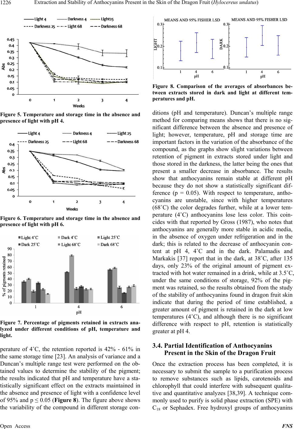 Extraction and Stability of Anthocyanins Present in the Skin of the Dragon Fruit (Hylocereus undatus) 1226 Figure 5. Temperature and storage time in the absence and presence of light with pH 4. Figure 6. Temperature and storage time in the absence and presence of light with pH 6. Figure 7. Percentage of pigments retained in extracts ana- lyzed under different conditions of pH, temperature and light. perature of 4˚C, the retention reported is 42% - 61% in the same storage time [23]. An analysis of variance and a Duncan’s multiple range test were performed on the ob- tained values to determine the stability of the pigment; the results indicated that pH and temperature have a sta- tistically significant effect on the extracts maintained in the absence and presence of light with a confidence level of 95% and p ≤ 0.05 (Figure 8). The figure above shows the variability of the compound in different storage con- Figure 8. Comparison of the averages of absorbances be- tween extracts stored in dark and light at different tem- peratures and pH. ditions (pH and temperature). Duncan’s multiple range method for comparing means shows that there is no sig- nificant difference between the absence and presence of light; however, temperature, pH and storage time are important factors in the variation of the absorbance of the compound, as the graphs show slight variations between retention of pigment in extracts stored under light and those stored in the darkness, the latter being the ones that present a smaller decrease in absorbance. The results show that anthocyanins remain stable at different pH because they do not show a statistically significant dif- ference (p = 0.05). With respect to temperature, antho- cyanins are unstable, since with higher temperatures (68˚C) the color degrades further, while at a lower tem- perature (4˚C) anthocyanins lose less color. This coin- cides with that reported by Gross (1987), who notes that anthocyanins are generally more stable in acidic media, in the absence of oxygen under refrigeration and in the dark; this is related to the decrease of anthocyanin con- tent at pH 4, 4˚C and in the dark. Palamadis and Markakis [37] report that in the dark, at 38˚C, after 135 days, only 23% of the original amount of pigment ex- tracted with hot water remained in a drink, while at 3.5˚C, under the same conditions of storage, 92% of the pig- ment was retained, so the results obtained from the study of the stability of anthocyanins found in dragon fruit skin indicate that during the period of time established, a greater amount of pigment is retained in the dark at low temperatures (4˚C), and although there is no significant difference with respect to pH, retention is statistically greater at pH 4. 3.4. Partial Identification of Anthocyanins Present in the Skin of the Dragon Fruit Once the extraction process has been completed, it is necessary to submit the sample to a purification process to remove substances such as lipids, carotenoids and chlorophyll that could interfere with subsequent qualita- tive and quantitative analyzes [38,39]. A technique com- monly used to purify is solid phase extraction (SPE) with C18 or Sephadex. Free hydroxyl groups of anthocyanins Open Access FNS 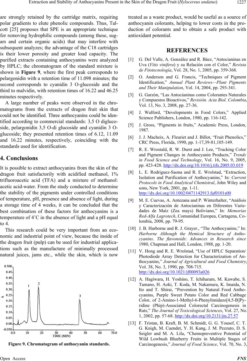 Extraction and Stability of Anthocyanins Present in the Skin of the Dragon Fruit (Hylocereus undatus) 1227 are strongly retained by the cartridge matrix, requiring polar gradients to elute phenolic compounds. Thus, Tal- cott [25] proposes that SPE is an appropriate technique for removing hydrophilic compounds (among these, sug- ars and certain organic acids) that may interfere with subsequent analyzes; the advantage of the C18 cartridges is their lower porosity and greater load capacity. The purified extracts containing anthocyanins were analyzed by HPLC; the chromatogram of the standard mixture is shown in Figure 9, where the first peak corresponds to pelargonidin with a retention time of 11.098 minutes; the second corresponds to cyanidin 3 O-glucoside and the third to malvidin, with retention times of 16.22 and 46.25 minutes respectively. A large number of peaks were observed in the chro- matograms from the extracts of dragon fruit skin that could not be identified. Three anthocyanins could be iden- tified according to commercial standards: 3,5 O digluco- side, pelargonidin 3,5 O-di glucoside and cyanidin 3 O- glucoside; they presented retention times of 6.12, 11.09 and 16.22 minutes, respectively, coinciding with the standards used for identification. 4. Conclusions It is possible to extract anthocyanins from the skin of the dragon fruit satisfactorily with acidified methanol, 1% trifluoroacetic acid (TFA) and a mixture of methanol: acetic acid-water. From the study conducted to determine the stability of the pigments under controlled conditions of temperature, pH, presence and absence of light, during a storage time of 4 weeks, it can be concluded that the best combination of these factors for anthocyanins is a temperature of 4˚C in the absence of light and a pH equal to 4. This research could be very important from an eco- nomic and industrial point of view, because the inside of the dragon fruit (pulp) can be used for industrial applica- tions such as the manufacture of minimally processed natural juices, jams etc., while the skin, which is now Figure 9. Chromatogram of anthocyanin standards. treated as a waste product, would be useful as a source of anthocyanin colorants, helping to lower costs in the pro- duction of colorants and to obtain a safe product with antioxidant potential. REFERENCES [1] G. Del Valle, A. González and R. Báez, “Antocianinas en Uva (Vitis vinifera) y su Relación con el Color,” Revista de Fitotecnología, Vol. 28, No. 1, 2005, pp. 359-368. [2] O. Anderson and G. Francis, “Techniques of Pigment Identification,” Annual Plant Reviews—Plant Pigments and Their Manipulation, Vol. 14, 2004, pp. 293-341. [3] G. Garzón, “Las Antocianinas como Colorantes Naturales y Compuestos Bioactivos,” Revisión. Acta Biol. Colombia, Vol. 13, No. 3, 2008, pp. 27-36. [4] J. Walford, “Developments in Food Colors,” Applied Science Publishers, London, 1980, pp. 116-142. [5] J. Gross, “Pigments in fruits,” Academic Press, London, 1987. [6] J. J. Macheix, A. Fleuriet and J. Billot, “Fruit Phenolics,” CRC Press, Florida, 1990, pp. 1-17,39-81,105-149. [7] R. E. Wrostald, R. W. Durst and J. Lee, “Tracking Color and Pigment Changes in Anthocyanin Products,” Trends in Food Science and Technology, Vol. 16, No. 9, 2005, pp. 423-428. http://dx.doi.org/10.1016/j.tifs.2005.03.019 [8] L. E. Rodriguez-Saona and R. E. Wrolstad, “Extraction, Isolation and Purification of Anthocyanins,” In: Current Protocols in Food Analytical Chemistral, John Wiley and sons, New York, 2001, pp. 1-11. http://dx.doi.org/10.1002/0471142913.faf0101s00 [9] M. E. Cuevas, A. Antezana and P. Winterhalter, “Análisis y Caracterización de Antocianinas en Diferentes Varie- dades de Maíz (Zea mays) Boliviano,” In: Memorias Red-Alfa Lagrotech, Comunidad Europea, Cartagena, Co- lombia, 2008, pp. 79-95. [10] J. B. Harborne and R. J. Grayer., “The Anthocyanins,” In: Harborne Although the Hemical Structure of Antho- cyanins. The flavonoids: Advances in Research since 1980, Chapman and Hall, London, 1988, pp. 1-20. [11] V. Hong and R. E. Wrolstad, “Use of HPLC Separation/ Photodiode Array Detection for Characterization of An- thocyanins,” Journal of Agricultural and Food Chemistry, Vol. 38, No. 3, 1990, pp. 708-715. http://dx.doi.org/10.1021/jf00093a026 [12] A. Hagiwara, H. Yoshino, T. Ichiharam, M. Kawabe, S. Tamano, H. Aoki, T. Koda, M. Nakamura, K. Imaida, N. Ito and T. Shirai, “Prevention by Natural Food Antho- cyanins, Purple Sweet Potato Color and Red Cabbage Color, of 2-Amino-1-Methyl-6-Phenylimidazo[4,5-B]Py- ridine (Phip)-Associated Colorectal Carcinogenesis in Rats,” The Journal of Toxicological Sciences, Vol. 27, No. 1, 2002, pp. 57-68. http://dx.doi.org/10.2131/jts.27.57 [13] F. Tristan, B. Kraft, B. M. Schmidt, G. G. Yousef, C. T. G. Knigh, M. Cuendet, Y. H. Kang, J. M. Pezzuto, D. S. Seigler and M. A. Lila, “Chemopreventive Potential of Wild Lowbush Blueberry Fruits in Multiple Stages of Carcinogenesis,” Journal of Food Science, Vol. 70, No. 3, Open Access FNS 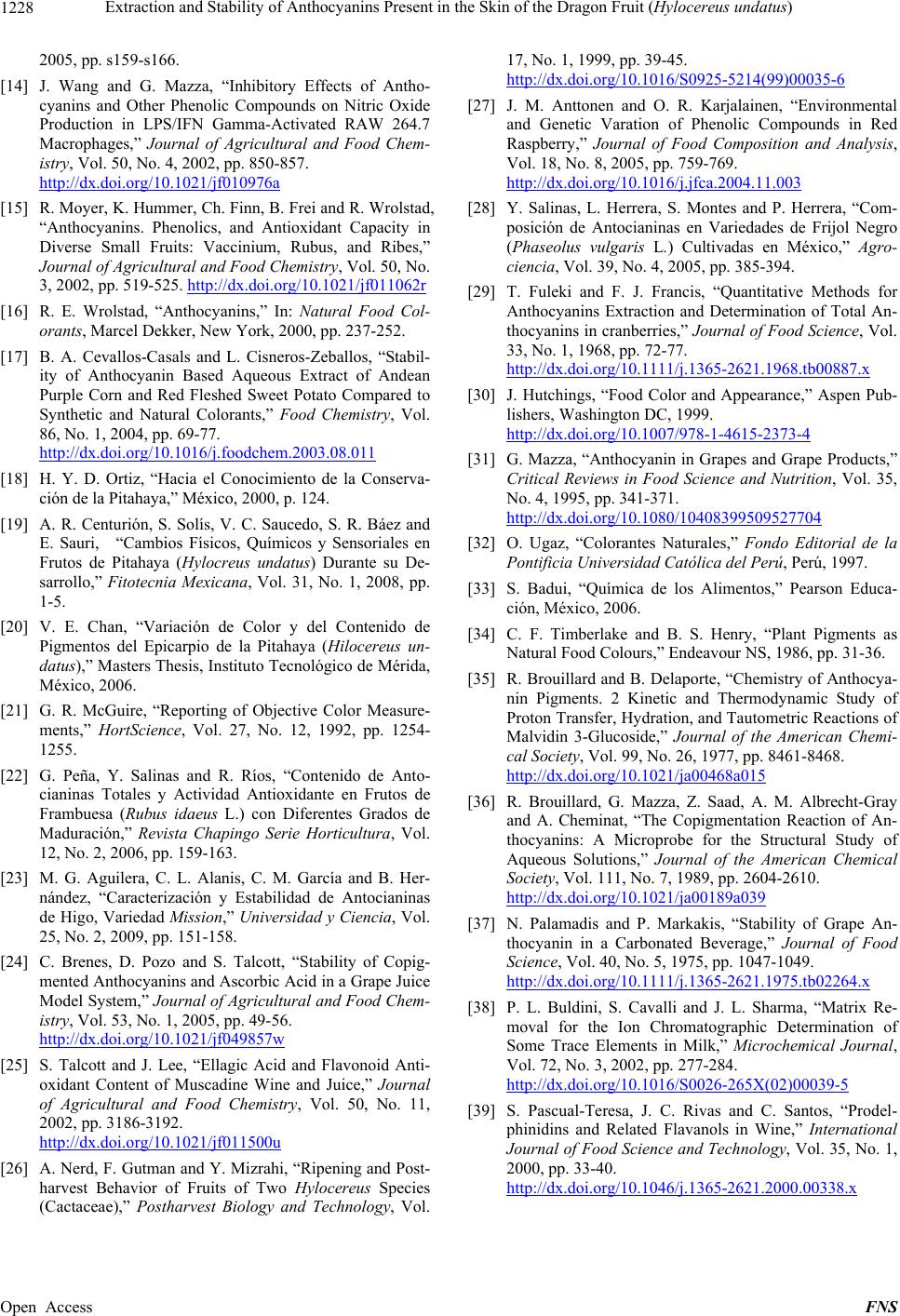 Extraction and Stability of Anthocyanins Present in the Skin of the Dragon Fruit (Hylocereus undatus) Open Access FNS 1228 2005, pp. s159-s166. [14] J. Wang and G. Mazza, “Inhibitory Effects of Antho- cyanins and Other Phenolic Compounds on Nitric Oxide Production in LPS/IFN Gamma-Activated RAW 264.7 Macrophages,” Journal of Agricultural and Food Chem- istry, Vol. 50, No. 4, 2002, pp. 850-857. http://dx.doi.org/10.1021/jf010976a [15] R. Moyer, K. Hummer, Ch. Finn, B. Frei and R. Wrolstad, “Anthocyanins. Phenolics, and Antioxidant Capacity in Diverse Small Fruits: Vaccinium, Rubus, and Ribes,” Journal of Agricultural and Food Chemistry, Vol. 50, No. 3, 2002, pp. 519-525. http://dx.doi.org/10.1021/jf011062r [16] R. E. Wrolstad, “Anthocyanins,” In: Natural Food Col- orants, Marcel Dekker, New York, 2000, pp. 237-252. [17] B. A. Cevallos-Casals and L. Cisneros-Zeballos, “Stabil- ity of Anthocyanin Based Aqueous Extract of Andean Purple Corn and Red Fleshed Sweet Potato Compared to Synthetic and Natural Colorants,” Food Chemistry, Vol. 86, No. 1, 2004, pp. 69-77. http://dx.doi.org/10.1016/j.foodchem.2003.08.011 [18] H. Y. D. Ortiz, “Hacia el Conocimiento de la Conserva- ción de la Pitahaya,” México, 2000, p. 124. [19] A. R. Centurión, S. Solís, V. C. Saucedo, S. R. Báez and E. Sauri, “Cambios Físicos, Químicos y Sensoriales en Frutos de Pitahaya (Hylocreus undatus) Durante su De- sarrollo,” Fitotecnia Mexicana, Vol. 31, No. 1, 2008, pp. 1-5. [20] V. E. Chan, “Variación de Color y del Contenido de Pigmentos del Epicarpio de la Pitahaya (Hilocereus un- datus),” Masters Thesis, Instituto Tecnológico de Mérida, México, 2006. [21] G. R. McGuire, “Reporting of Objective Color Measure- ments,” HortScience, Vol. 27, No. 12, 1992, pp. 1254- 1255. [22] G. Peña, Y. Salinas and R. Ríos, “Contenido de Anto- cianinas Totales y Actividad Antioxidante en Frutos de Frambuesa (Rubus idaeus L.) con Diferentes Grados de Maduración,” Revista Chapingo Serie Horticultura, Vol. 12, No. 2, 2006, pp. 159-163. [23] M. G. Aguilera, C. L. Alanis, C. M. García and B. Her- nández, “Caracterización y Estabilidad de Antocianinas de Higo, Variedad Mission,” Universidad y Ciencia, Vol. 25, No. 2, 2009, pp. 151-158. [24] C. Brenes, D. Pozo and S. Talcott, “Stability of Copig- mented Anthocyanins and Ascorbic Acid in a Grape Juice Model System,” Journal of Agricultural and Food Chem- istry, Vol. 53, No. 1, 2005, pp. 49-56. http://dx.doi.org/10.1021/jf049857w [25] S. Talcott and J. Lee, “Ellagic Acid and Flavonoid Anti- oxidant Content of Muscadine Wine and Juice,” Journal of Agricultural and Food Chemistry, Vol. 50, No. 11, 2002, pp. 3186-3192. http://dx.doi.org/10.1021/jf011500u [26] A. Nerd, F. Gutman and Y. Mizrahi, “Ripening and Post- harvest Behavior of Fruits of Two Hylocereus Species (Cactaceae),” Postharvest Biology and Technology, Vol. 17, No. 1, 1999, pp. 39-45. http://dx.doi.org/10.1016/S0925-5214(99)00035-6 [27] J. M. Anttonen and O. R. Karjalainen, “Environmental and Genetic Varation of Phenolic Compounds in Red Raspberry,” Journal of Food Composition and Analysis, Vol. 18, No. 8, 2005, pp. 759-769. http://dx.doi.org/10.1016/j.jfca.2004.11.003 [28] Y. Salinas, L. Herrera, S. Montes and P. Herrera, “Com- posición de Antocianinas en Variedades de Frijol Negro (Phaseolus vulgaris L.) Cultivadas en México,” Agro- ciencia, Vol. 39, No. 4, 2005, pp. 385-394. [29] T. Fuleki and F. J. Francis, “Quantitative Methods for Anthocyanins Extraction and Determination of Total An- thocyanins in cranberries,” Journal of Food Science, Vol. 33, No. 1, 1968, pp. 72-77. http://dx.doi.org/10.1111/j.1365-2621.1968.tb00887.x [30] J. Hutchings, “Food Color and Appearance,” Aspen Pub- lishers, Washington DC, 1999. http://dx.doi.org/10.1007/978-1-4615-2373-4 [31] G. Mazza, “Anthocyanin in Grapes and Grape Products,” Critical Reviews in Food Science and Nutrition, Vol. 35, No. 4, 1995, pp. 341-371. http://dx.doi.org/10.1080/10408399509527704 [32] O. Ugaz, “Colorantes Naturales,” Fondo Editorial de la Pontificia Universidad Católica del Perú, Perú, 1997. [33] S. Badui, “Química de los Alimentos,” Pearson Educa- ción, México, 2006. [34] C. F. Timberlake and B. S. Henry, “Plant Pigments as Natural Food Colours,” Endeavour NS, 1986, pp. 31-36. [35] R. Brouillard and B. Delaporte, “Chemistry of Anthocya- nin Pigments. 2 Kinetic and Thermodynamic Study of Proton Transfer, Hydration, and Tautometric Reactions of Malvidin 3-Glucoside,” Journal of the American Chemi- cal Society, Vol. 99, No. 26, 1977, pp. 8461-8468. http://dx.doi.org/10.1021/ja00468a015 [36] R. Brouillard, G. Mazza, Z. Saad, A. M. Albrecht-Gray and A. Cheminat, “The Copigmentation Reaction of An- thocyanins: A Microprobe for the Structural Study of Aqueous Solutions,” Journal of the American Chemical Society, Vol. 111, No. 7, 1989, pp. 2604-2610. http://dx.doi.org/10.1021/ja00189a039 [37] N. Palamadis and P. Markakis, “Stability of Grape An- thocyanin in a Carbonated Beverage,” Journal of Food Science, Vol. 40, No. 5, 1975, pp. 1047-1049. http://dx.doi.org/10.1111/j.1365-2621.1975.tb02264.x [38] P. L. Buldini, S. Cavalli and J. L. Sharma, “Matrix Re- moval for the Ion Chromatographic Determination of Some Trace Elements in Milk,” Microchemical Journal, Vol. 72, No. 3, 2002, pp. 277-284. http://dx.doi.org/10.1016/S0026-265X(02)00039-5 [39] S. Pascual-Teresa, J. C. Rivas and C. Santos, “Prodel- phinidins and Related Flavanols in Wine,” International Journal of Food Science and Technology, Vol. 35, No. 1, 2000, pp. 33-40. http://dx.doi.org/10.1046/j.1365-2621.2000.00338.x
|