J. Biomedical Science and Engineering, 2011, 4, 119-121
doi:10.4236/jbise.2011.42017 Published Online February 2011 (http://www.SciRP.org/journal/jbise/
JBiSE
).
Published Online February 2011 in SciRes. http://www.scirp.org/journal/JBiSE
Computer aided modeling and analysis of a new biomedical
and surgical instrument
Zheng L i
University of Bridgeport, Bridgeport, USA.
Email: zhengli@bridgeport.edu
Received 12 November 2010; revised 15 November 2010; accepted 19 November 2010.
ABSTRACT
This paper describes the recent research and devel-
opment of an endo surgical/biomedical instrument in
surgical suture applications for minimally invasive
therapy procedure. The newly developed instruments
can not only protect the wound during the surgical
procedure but also actively help the healing process.
The new mechanism design of the surgical instrument
aids in better ergonomic design, reliable functionality,
and continuous cost reduction in product manufac-
turing. 3-D modeling technique, func tionality an alysis,
kinematical simulation and computer aided solution
have been applied to the instrument design, devel-
opment and future improvement to meet the specific
requirements of minimally invasive surgery proce-
dure. The improved new endo surgical/biomedical
instrument can prevent patient’s vessels and tissues
from being damaging because the distal move of clips
are well controlled without clip drop-off incident.
Plus the operational force to form the clip is lower
than regular surgical/biomedical instruments due to
this special new mechanism design. In addition to the
above, the manufacturing and product cost can be
decreased because the dimensional tolerance of com-
ponents, such as clip channel and jaw guide track,
can be loose due to this new instrument design. The
prototypes of this new endo surgical/biomedical in-
strument design are analyzed through computer
aided modeling and simulation, in order to prove its
feasible functionality, reliable performance, and me-
chanical advantage. All these improved features have
also been tested and verified through the prototypes.
Keywords: Hemostasis; Endoscopic Device;
Computational Simulation; 3-D Modeling; Mechanical
Advantage
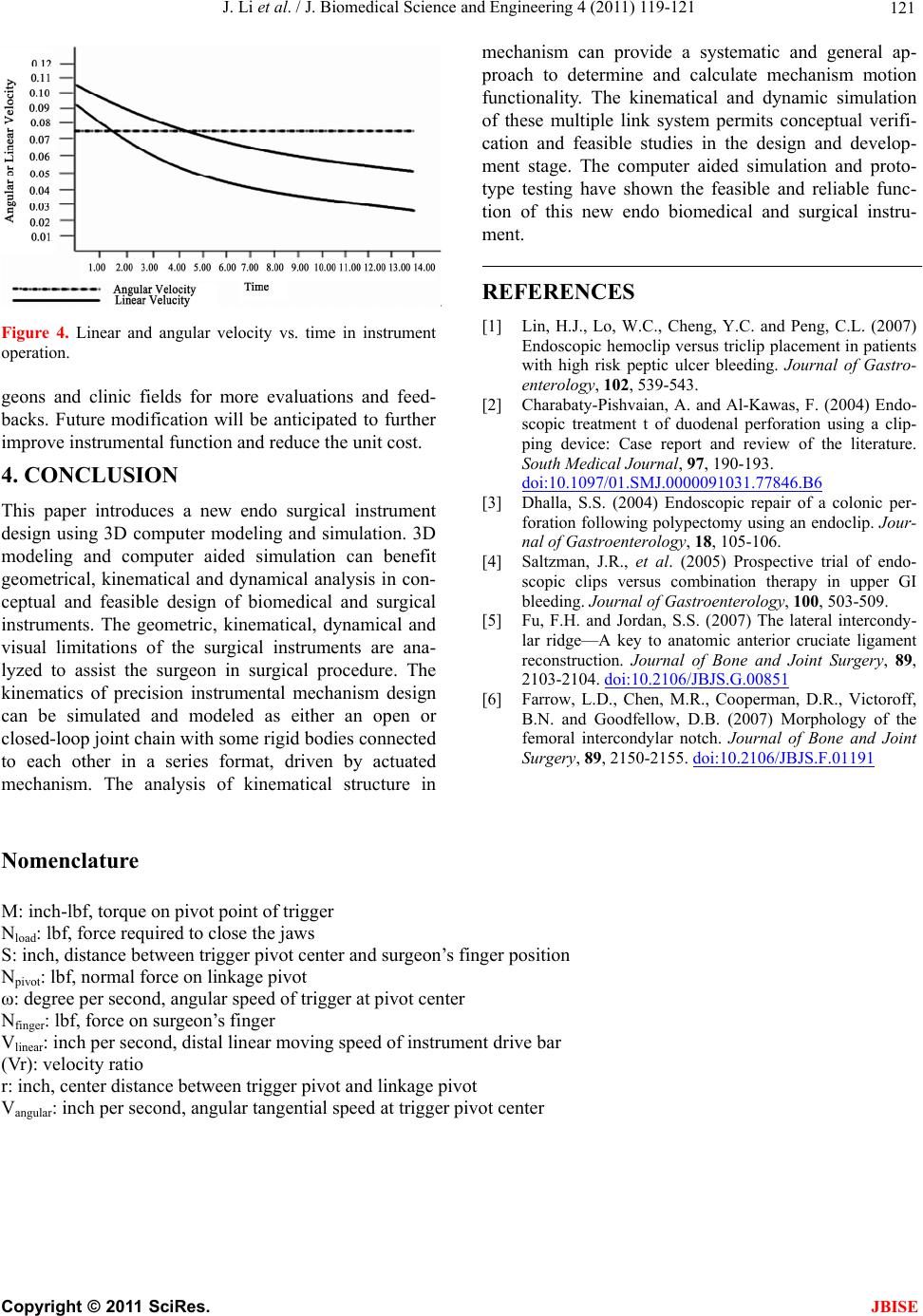
1. INTRODUCTION
The newly developed technologies have directed mini-
mally invasive surgeries [1]. The positive and feasible
changes in surgical instruments have led to the new de-
velopment of surgical techniques [2] and [3]. The bio-
medical and surgical instrument market is always ad-
justed and controlled for its functionality, performance,
feasibility, quality, safety, and manufacturing cost. The
surgical instrument market is very competitive, price
sensitive and depicted by advanced technologies [4] and
[5]. Biomedical and surgical instrument is technology
based product and normally advanced techniques are
especially required to develop special technology to
compete the products in today’s challenging market [6].
The applications include the closure of tissue defects,
perforations, and anastomotic leakage in the esophagus
and stomach. The endo surgical instrument has also been
used to prevent post-polypectomy bleeding, placement
of enteral feeding tubes. The recent studies show the
versatility of endo surgical clips in therapeutic and en-
doscopic applications.
This endo surgical instrument is the innovative product
that will allow for greater ease of use for surgeons and
help to improve patient outcomes. Based on the field and
clinical feedback, the new technology that simultaneously
opens and aligns the jaws has been implemented, allow-
ing well controlled surgical clip feeding and closure. This
new surgical instrument design can provide more con-
sistent and reliable mechanism to protect the clip from
external and unanticipated disturbance while the surgical
clip sits in the jaw track.
Endo surgical instrument has been widely used in
hemostasis during endoscopy of the upper and lower
gastrointestinal tract in which the bleeding lesions can be
successfully clipped. The alternatives to endoscopic
clipping of peptic ulcers are thermal therapy (such as
electrocautery to burn the vessel causing the bleeding),
or injection of epinephrine to constrict the blood vessel.
Comparative studies between endo surgical clips and
thermal therapy verify that endo surgical clips cause less
trauma to the mucosa around the ulcer than electrocau-
tery.