 Advances in Bioscience and Biotechnology, 2013, 4, 999-1006 ABB http://dx.doi.org/10.4236/abb.2013.411133 Published Online November 2013 (http://www.scirp.org/journal/abb/) Evaluation of antimicrobial activity of a lectin isolated and purified from Indigofera heterantha Sakeena Qadir, Ishfak Hussain Wani, Shaista Rafiq, Showkat Ahmad Ganie, Akbar Masood, Rabia Hamid* Department of Biochemistry, University of Kashmir, Srinagar, India Email: *rabeyams@yahoo.co.in Received 13 August 2013; revised 26 September 2013; accepted 12 October 2013 Copyright © 2013 Sakeena Qadir et al. This is an open access article distributed under the Creative Commons Attribution License, which permits unrestricted use, distribution, and reproduction in any medium, provided the original work is properly cited. ABSTRACT Indigofera heterantha commonly called indigo bush is a member of leguminoseae family found in the Hima- layan region of Kashmir. A lectin has been isolated from the seeds of Indigofera heterantha by the purifi- cation procedure involving anion exchange chroma- tography on DEAE-cellulose followed by gel filtra tion chromatography on Sephadex G 100. Molecular cha- racterization of the lectin was done by gel filtration and SDS-PAGE. Activity of the lectin was checked by hemagglutination assay and the sugar specificity by sugar inhibition tests. The antimicrobial activity of the purified lectin was carried out by Agar disc diffu- sion using appropriate standards. On the ion ex- change column , the boun d protein when eluted with 0 - 0.5 M NaCl gradient emerged as three peaks—peak I, peak II and peak III out of which only peak II showed the hemagglutinating activity. The lectin fur- ther resolved into two peaks G1 and G2 on gel filtra- tion, with the lectin activity residing in G1, corre- sponding to a molecular weight of 70 KDa. The puri- fied lectin named as Indigofera heterantha Lectin (IHL) produced a single band on SDS PAGE (18 KDa), revealing the tetrameric nature of the lectin. It agglutinated human erythrocytes (A, B, AB, and O). Hemagglutination was inhibited by D-galactose, D- mannose and D-arabinose. The lectin is reasonably thermostable showing full activity within a tempera- ture range of 30˚C to 90˚C. pH stability of the lectin falls in the range of 2 - 9. IHL demonstrated a re- markable antibacterial activity against the pathogenic bacteria Klebsiella pneumoniae, Staphylococcus au- reus, Escherichia coli, and Bacillus subtilis. IHL also inhibited the growth of phytopathogenic fungi Asper- gillus niger, Aspergillus oryzae and Fusarium oxys- porum. Keywords: Indigofera heterantha Lectin; Hemagglutination; Antimicrobial 1. INTRODUCTION Lectins are proteins of non-immune origin that bind to carbohydrates and sugar containing substances in a spe- cific and reversible way or precipitate glycoconjugates [1]. The main properties of lectins are based on their ability to interact with carbohydrates and thus combine with glycocomponents on the cell surface, leading to their biological properties [2]. Lectins are able to tightly bind to and cause the precipitation of specific polysac- charides and glycoproteins because they are polyvalent. They vary, however, in molecular size, amino acid com- position, metal ion requirements, and three-dimensional structure [3]. Lectins can recognize mono-, oligo- or polysaccharides, as well as glycoconjugates and thus recognize glycoproteins or glycolipids, e.g. on the sur- face of cells [4]. Generally, lectins are glycoproteins con- sisting of subunits ranging in molecular mass from 25 to 35 kDa, arranged as dimers or tetramers, and existing as multiple isoforms sharing similar biochemical properties [5]. The subunits are usually identical or very similar, made up of single polypeptide chains that are encoded by different genes or by a family of closely related genes [6]. In addition to their carbohydrate binding activity, some of the lectins such as those of hemagglutinin, galactosi- dase, polynucleotide adenosine glucosidase, or ribosome inactivating proteins, also show other biological activi- ties [7]. Lectins in higher plants defend against patho- genic bacteria and fungi by recognizing and immobiliz- ing the infecting microorganisms via binding, thereby preventing their subsequent growth and multiplication. They act as a sort of immune system for plants by “sticking” themselves to the structural carbohydrates (sugars) of invaders. Recently, lectin biology has applied these tools in the biomedical field and also in the treat- *Corresponding author. OPEN ACCESS  S. Qadir et al. / Advances in Bioscience and Biotechnology 4 (2013) 999-1006 1000 ment of diseases, including cancer [8]. Another applica- tion reported in the literature involving lectins is their antimicrobial activity where lectins may act against mi- croorganisms by interfering with their growth and play- ing a role in defense systems [9]. Antibacterial activity on Gram-positive and Gram-negative bacteria occurs through the interaction of lectin with components of the bacterial cell wall including techoic and teichuronic acids, peptidoglycans and lipopolysaccharides; study revealed that the isolectin I from Lathyrus ochrus seeds binds to muramic acid and muramyl dipeptide through hydrogen bonds between ring hydroxyl oxygen atoms of sugar and carbohydrate binding site of lectin and hydrophobic in- teractions with the side chains of residues Tyr100 and Trp128 of isolectin I [10]. The inhibition of fungal growth can occur through lectin binding to hyphas re- sulting in poor absorption of nutrients as well as by in- terference on spore germination process [11]. The poly- saccharide chitin is constituent of fungi cell wall and chitin-binding lectins showing antifungal activity; im- pairment of synthesis and/or deposition of chitin in cell wall may be the reasons of antifungal action [12]. Proba- bly the carbohydrate-binding property of lectin is in- volved in the antifungal mechanisms and lectins of dif- ferent specificities can promote distinct effects. In this report, we describe the purification and characterization of a lectin from Indigofera heterantha seeds and also the evaluation of its potential antimicrobial activity. 2. MATERIALS AND METHODS The plants (Indigofera heterantha) were collected from the premises of the Department of Botany of Kashmir University and were authenticated from Centre of Plant Taxonomy of the same department. DEAE-cellulose and Sephadex G-100 were purchased from Sigma Aldrich Company, USA. Other chemicals were of highest purity grade. 2.1. Isolation and Purification of Lectins from Indigofera heterantha Seeds Twenty five grams of dry Indigofera heterantha seeds were allowed to swell in distilled water at 4˚C overnight. The seeds were then ground in a mortar and pestle and proteins extracted by suspending the ground seed meal in 0.15 M NaCl (10% w/v) for 1 hour at room temperature and subjecting the mixture to homogenization in a Remi auto mix blender for 10 min. The homogenate thus pro- duced was stirred for 2 h at 4˚C and then filtered through double layered cheesecloth. The filtrate was centrifuged at 5000 × g for 20 min at 4˚C. The pellet was discarded. The storage proteins present in the supernatant were pre- cipitated by slowly adding 1 M acetic acid to the stirred solution until pH 5.0 was reached. After an additional hour of stirring at 4˚C, the suspension was again centri- fuged at 5000 × g for 20 min. The pellet was again dis- carded and the supernatant readjusted to pH 8.0 by the addition of 0.1 M NaOH. The supernatant thus obtained was dialyzed against 20 mM phosphate buffer, pH 7.2 for 24 hr. The viscous extract thus obtained was the crude extract and was stored at 4˚C. To purify the lectin further, the crude extract was sub- jected to ion exchange chromatography on DEAE cellu- lose column in 50 mM Tris-HCL buffer, pH 8. The pro- tein was eluted using linear sodium chloride gradient from 0 to 0.5 M. The active fractions were pooled and dialyzed against Tris-HCL buffer, pH 8. To check the size homogeneity, the active fractions eluted from ion-exchange column were chromatographed on Sephadex G 100 column in 0.05 M Tris-HCl, pH 8.0. The charge homogeneity of the fractions eluted from gel filtration column was checked by polyacrylamide gel electrophoresis. Estimation of the subunit molecular weight of the purified lectin was done by SDS-PAGE. 2.1.1. Pr otein Esti m a t ion Protein concentration was determined by the method of Lowry et al. [13] using BSA as the standard protein. 2.1.2. Polyacrylamide Gel Electrophoresis Polyacrylamide gel electrophoresis in presence and ab- sence of SDS was performed at pH 8.3 on 10% and 12% gels respectively with a discontinuous buffer system. The gels were stained with Coomassie Brilliant Blue R-250. 2.2. Hemagglutination Assay and Inhibition of Hemagglutination The hemagglutination activity of the lectin was deter- mined by a slight variation of the method devised by Peumans et al. [14], using human erythrocytes bearing blood groups A, B, O, AB and chick erythrocytes. The human blood was collected from SKIMS, Srinagar and chick blood was obtained from a local veterinary outlet. The erythrocytes were separated by centrifugation of the blood at 2500 g (5000 rpm) for 10 min, and the R.B.Cs washed thrice with normal saline. Finally the erythro- cytes were suspended in normal saline to obtain a final concentration of 3% erythrocyte suspension. The assay of agglutination was carried out on glass slides by mix- ing the erythrocytes with the test solution. 50 µl erythro- cyte suspensions (A, B, O, AB and Chick erythrocytes) were taken on a slide. To this, 50 µl of the test solution was added. After an incubation period of 15 min at room temperature, agglutination was monitored unaided on the slides. Also the control slide, using the buffer instead of the lectin solution was run simultaneously. The lectin activity has been expressed as EU (Erythroagglutinating Unit). One erythroagglutinating unit (EU) is defined as Copyright © 2013 SciRes. OPEN ACCESS  S. Qadir et al. / Advances in Bioscience and Biotechnology 4 (2013) 999-1006 1001 the minimum amount of the lectin per ml required to give positive agglutination of 1 ml of a 5% human ery- throcyte suspension. EU is expressed as μg of lectin per ml of the protein solution. The carbohydrate specificity was investigated by ob- serving the inhibition of the lectin induced hemaggluti- nation by various sugars namely D-glucose, D-galactose, D-mannose, fructose, lactose, maltose, sucrose, ribose and sugar derivatives like N-acetyl galactosamine and N-acetyl glucosamine. The inhibition assay was per- formed on glass slides. Different dilutions of the above sugars (final volume 20 µl) were added to glass slides on which agglutination was performed. To each dilution, 20 µl of purified lectin was added. The mixture was incu- bated at room temperature for 1 hr after which 80 µl of 3% suspension of erythrocytes was added to each slide. The minimum concentrations of each sugar capable of fully inhibiting agglutination after 1 hr at room tempera- ture were noted. 2.3. Effect of pH and Temperature on the Stability of the Lectin The pH dependence of the lectin was determined by in- cubating 50 μg of IHL with buffers in different pH: 0.1 M glycine/HCl (pH 2 - 3), 0.05 sodium acetate/acetic acid (pH 4 - 5), 0.05 M potassium phosphate (pH 6 - 7), 0.05 M Tris-HCl (pH 8 - 9) and 0.1 M glycine-NaOH (pH 10 - 11) for 5 hrs at 25˚C and pH adjusted to 7.2 just prior to hemagglutination assay. The effect of tempera- ture on the agglutinating activity of the IHL lectin was determined by carrying out assay at different tempera- tures according to the method described by Ngai and Ng [15]. The purified lectin was incubated in a water bath for 30 min at various temperatures: 10˚C, 20˚C, 30˚C, 40˚C, 50˚C, 60˚C, 70˚C, 80˚C, 90˚C and 100˚C, and then cooled to 20˚C in each case. Hemagglutination assay was carried out as previously described. The assay was per- formed in duplicate [16]. 2.4. Molecular Weight Determination The subunit molecular mass of the lectin was estimated by discontinuous SDS-PAGE according to method of Laemmli [17]. Native molecular mass was determined by gel filtration on Sephadex G-100 column using the fol- lowing protein markers: BSA (66 kDa), Ovalbumin (45 kDa), Pepsin (34.7 kDA) and cytochrome c (12.4 kDa). The gel filtration data was treated according to the rela- tion given by Andrew [18]. 2.5. Evaluation of Antimicrobial Activity For the evaluation of antibacterial activity of Indigofera heterantha lectin, firstly all the glassware was sterilized in an autoclave. After sterilization, the subsequent steps were the preparation of media, selection of the test or- ganisms and sensitivity tests of antibacterial activity. Purified IHL was used for analysis. Ciprofloxacin 5 µg/ml was used as standard. Human pathogenic bacteria Pseudomonas spp. Escherichia coli, Shigella bodyi, Klebsiella pneumonea, Streptococci, Salmonella typhi and Staphylococcus aureus obtained from Department of Microbiology, Sheri Kashmir Institute of Medical Sci- ences Srinagar were used to check the antibacterial activ- ity. Antifungal activity was checked against Aspergillus niger, Aspergillus Oryzae and Fusarium oxysporum and were obtained from Sheri Kashmir University of Agri- cultural Science and Technology, Shalimar. The Agar disc diffusion method was used to determine the anti- microbial activity. This test was done according to the method of Edward [19] and Lansing [20]. The microbial (bacterial/fungal) colony was picked by the inoculating wire and the media plates were subsequently inoculated with specific microbial strains and labeled accordingly. The inoculated plates were left to dry for at least 5 - 10 minutes, after which Whatman filter paper discs 6 mm (previously saturated with crude extract and purified lectin) were placed on the plates with the help of a steril- ized forcep. Standard antibiotic discs of ciprofloxacin (positive control) were also impregnated on plates at previously labeled positions. Loaded plates were kept as such for some time under Laminar hood and then in- cubated at 37˚C for 24 hours in case of bacterial strains and 48 hours in case of fungal strains in the incubator. A transparent ring around the paper disc signified antim- icrobial activity. Zone diameters (mm) around each of the discs were measured to the nearest mm. 3. RESULTS 3.1. Isolation and Purification A lectin was isolated and purified from the Indigofera heterantha seeds by ion exchange chromatography on DEAE-Cellulose followed by gel filtration on Sephadex G-100. The crude extract obtained after homogenization of the finely ground seed meal in 0.15 M NaCl was as- sayed for hemagglutinating activity using human eryth- rocytes of blood group types A, B, AB and O. This was then applied on to a DEAE-Cellulose column equili- brated with 50 mM Tris-HCl buffer, pH 8 (containing 0.02% NaN3). Major portion of the protein in crude ex- tract passed unbound through the column. This fraction did not carry any hemagglutinating activity. The bound protein when eluted with 0 - 0.5 M NaCl gradient emerged as three peaks—peak I, peak II and peak III out of which only peak II showed the hemagglutinating ac- tivity. Typical profile is shown in Figure 1. Fractions under the active peak were pooled together and concen- trated, and then subjected to gel filtration on Sephadex Copyright © 2013 SciRes. OPEN ACCESS  S. Qadir et al. / Advances in Bioscience and Biotechnology 4 (2013) 999-1006 1002 Figure 1. Ion exchange chromatography of the crude extract on DEAE-cellulose column (1.6 × 10 cm). About 60 mg of protein was applied on DEAE-cellulose column pre-equilibrated with 50 mM Tris-HCL buffer, pH-8. The protein was eluted using 0 - 0.5 M NaCl gradient in 5 ml fractions at a flow rate of 30 ml/hr. G-100 column (2.6 × 40 cm) where it resolved into two peaks, G1 and G2 (Figure 2). Lectin activity resided majorly in G1. The fractions in the peak (G1) were again pooled together, concentrated and stored at 4˚C in 0.05 M Tris-HCl, pH 8.0 containing 0.02% NaN3. This prepa- ration was called the Indigof era heterantha lectin or IHL, and was used for physicochemical and biological char- acterization. Data on the purification of Indigofera het- erantha lectin is summarized in Table 1. The yield of the lectin was about 0.46% and about a 5.2 fold purification of the lectin was achieved. 3.2. Electrophoretic Analysis The charge homogeneity of the lectin was established by subjecting the lectin to polyacrylamide gel electrophore- sis under native conditions. The electrophoretogram shows that the lectin moved as a single band (Figure 3). In 12% SDS-PAGE, the lectin again moved as a single band, establishing that the lectin is composed of similar type of subunits (Figure 4). 3.3. Characterization 3.3.1. Molecular Weight Determination Molecular weight of the Indigofera heterantha lectin as determined by gel filtration on a Sephadex G-200 col- umn (2.6 × 40 cm) equilibrated in 0.05 M Tris-HCl buffer, pH 8 was about 70 KDa (Figure 2). In SDS- PAGE under reducing conditions, the lectin moved as a single protein band with molecular weight corresponding to 18 KDa. The results of SDS-PAGE (Figure 4) to- gether with gel filtration data revealed that IHL (Indigo- fera heterantha lectin) is a homotetramer. 3.3.2. Hemagg l uti nation The Indigofera heterantha lectin (IHL) does not show any marked blood group specificity (Table 2 ). As is evi- dent, the lectin agglutinated human erythrocytes of all blood groups, being somewhat more specific towards blood group A erythrocytes. Extent of hemagglutination was found to be same, when human erythrocytes of blood group A, B, AB and O were incubated with puri- fied IHL for overnight at 7˚C and for 6 hr at 27˚C & 37˚C. 3.3.3. Carbohydrate Specificity In order to determine the sugar specificity of the lectin, inhibition and reversal of inhibition by a number of sug- ars and sugar derivatives was studied. In each case the ability of the sugar to inhibit agglutination was measured. Results on such specificity studies are shown in Table 3. It is clear from the table that arabinose is the most potent inhibitor of the Indigofera heterantha lectin mediated hemagglutination followed by the disaccharide sucrose. Galactose also inhibits agglutination though at very high concentrations. 3.4. Effect of pH and Temperature The examination of IHL activity in different pH (pH 2 - 13) values showed that the lectin was stable in the pH range of 2 - 9 (Figure 5) indicating that the amino acid residues involved in carbohydrate binding are not af- fected by changes in pH in this range. The activity how- ever falls of rapidly thereafter, with essentially all activ- ity lost after pH 12. The purified lectin sample was found to be heat stable up to 90˚C, at incubation temperatures of 30 to 90˚C (Figure 6). Even heating at 100˚C for 30 minutes caused a loss of only 25% of its original activity. However the hemagglutinating activity of the lectin was completely lost when exposed to 110˚C. 3.5. Antimicrobial Activity 3.5.1. Antibacte ri a l Act i vity Purified lectin obtained from ion exchange chromatog- raphy and gel filtration chromatography as well as the crude extract were tested against different bacterial strains and compared to that of antibacterial antibiotic, ciprofloxacin. The results of the sensitivity test are shown in Figure 7. Purified IHL (500 μg/ml) exhibited a significant antibacterial effect on four strains namely Klebsiella pnuemoniae, Staphylococcus aureus, Escheri- chia coli, and Bacillus subtilis. The diameters of the zones of inhibition by the addition of IHL were 10 mm, 6 mm, 4 mm and 4 mm respectively. The diameters of the zones of inhibition with the standard drug used were 22 mm, 20 mm, 22 mm and 13 mm for the four strains re- spectively. Results with the crude extract were not sig- ificant. n Copyright © 2013 SciRes. OPEN ACCESS  S. Qadir et al. / Advances in Bioscience and Biotechnology 4 (2013) 999-1006 Copyright © 2013 SciRes. 1003 Table 1. Purification profile of Indigofera heterantha lectin. OPEN ACCESS Vo l u m e (ml) Purification step Protein (mg/ml) Total protein (mg) Activity (EU)* Total activity Specific activity (E.U*/mg) Fold purification Yeild % Crude extract 500 9 4500 81 40,500 9 1 100 Ion exchange Chromatography 10 1.7 17 52.02 520.2 30.6 3.4 1.28 Sephadex gel chromatography 4 1 4 46.80 187.2 46.8 5.2 0.46 *One erythroagglutinating unit (EU) is defined as the minimum amount of the lectin per ml required to give positive agglutination of 1 ml of a 5% erythrocyte suspension. EU is expressed in microgram of lectin/ml of the protein solution. Table 2. Blood group specificity of purified Indigofera hete- rantha lectin. Blood group Specific activity (EU*/mg) A 46.8 B 35.3 O 31.2 *One erythroagglutinating unit (EU) is defined as the minimum amount of the lectin per ml required to give positive agglutination of 1 ml of a 5% erythrocyte suspension. EU is expressed in μg of lectin/ml of the protein solution. Table 3. Carbohydrate Inhibition of Indigofera heterantha lec- tin mediated hemagglutination. Figure 2. Gel filtration of fraction PII from ion exchange col- umn on Sephadex G-100 column (2.6 cm × 40 cm). About 20 milligram of protein was applied on the column pre-equili- brated with 50 mM Tris-HCL buffer, pH 8.0. The protein was eluted using the same buffer in 3 ml fractions at a flow rate of 30 ml/hr. Sugar Minimal inhibitory concentration (mM)* Arabinose 0.039 Sucrose 0.188 D-galactose 0.275 Ribose No inhibition N-acetyl galactosamine No inhibition Raffinose No inhibition Lactose No inhibition Mannose No inhibition Glucose No inhibition *Minimum sugar concentration necessary for complete inhibition of agglu- tination of human erythrocytes by Indigofera heterantha lectin solution of 4 µg/ml. 3.5.2. An ti fungal Ac tivity In vitro antifungal susceptibility by IHL was determined against three phytopathogenic fungi Aspergillus oryzie, Aspergillus niger and Fusarium oxysporum with cipro- floxacin as positive control. The lectin showed some, though not significant inhibition of growth against all the three strains with a zone inhibition diameter of 9 mm, 3 mm and 6 mm respectively in each case (Table 4). Figure 3. Polyacrylamide Gel Electrophoresis (PAGE) pattern of Indigofera heterantha lectin under native conditions. About 40 μg of the purified IHL was applied on 10% polyacrylamide gel. PAGE was performed in Tris-glycine buffer, pH 8.0. Current applied was 25 mA for 2 hrs at room temperature. 4. DISCUSSION Legume lectins represent the largest and most thoroughly studied family of the simple lectins. The members of this  S. Qadir et al. / Advances in Bioscience and Biotechnology 4 (2013) 999-1006 1004 Figure 4. SDS-Polyacrylamide gel electrophoresis of Indigofera heterantha lectin. About 60 μg of lectin was electrophoresed on 12% polyacrylamide gel at a current of 3.0 mA/well in presence of 0.1% SDS. Tris-glycine buffer pH 8.0 was used. The staining reagent used was Coomassie brilliant blue G-250. Lane 1: molecular weight markers (66 KDa, 45 KDa, 35 KDa, 25 KDa, 18 KDa and 14 KDa). Lane 2: Purified Indigofera heterantha lectin. Figure 5. Effect of pH variations on IHL activity. The pH de- pendence of the lectin was determined by incubating 50 μg of IHL with buffers in different pH: 0.1 M glycine/HCl (pH 2 - 3), 0.05 sodium acetate/aceticacid (pH 4 - 5), 0.05 M potassium phosphate (pH 6 - 7), 0.05 M Tris-HCl (pH 8 - 9) and 0.1 M glycine-NaOH (pH 10 - 11) for 5 hrs at 25˚C and pH was ad- justed to 7.2 just prior to hemagglutination assay. protein family consists of two or four subunits (protom- ers), either identical or slightly different each with a sin- gle small carbohydrate combining site with the same specificity. A lectin was isolated from seeds of Indigo- fera heterantha using a combination of ion exchange and gel filtration chromatographies. The lectin is a tetramer and exhibits a molecular weight of 70 KDa with a sub- unit Mr of 18 KDa. The tetrameric nature of IHL is similar to the lectin obtained from Phaseolus acutifolius Figure 6. Effect of temperature on IHL activity. The tempera- ture dependence of the lectin was determined by incubating the agglutination mixture (50 μg of IHL in Tris-HCl buffer, pH 8) at different temperatures for 30, cooled to 20˚C and determin- ing the hemagglutination activity. (a) (b) (c) (d) Figure 7. Antibacterial assay of Indigofera heter- antha lectin with four different bacterial strains (a) Klebsiella pnuemoniae; (b) Staphylococcus aureus; (c) Escherichia coli; and (d) Bacillus subtilis. The agar disc diffusion method was used to determine antibacterial activity. Ciprofloxacin (5 μg/ml) was used as standard. Copyright © 2013 SciRes. OPEN ACCESS 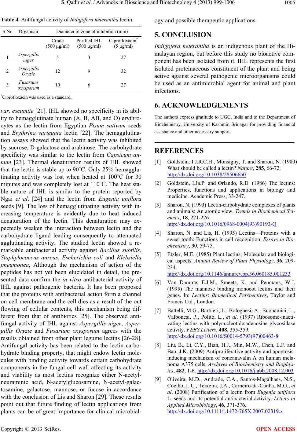 S. Qadir et al. / Advances in Bioscience and Biotechnology 4 (2013) 999-1006 1005 Table 4. Antifungal activity of Indigofera heterantha lectin. S.No Organism Diameter of zone of inhibition (mm) Crude (500 µg/ml) Purified IHL (500 µg/ml) Ciprofloxacin* (5 µg/ml) 1 Aspergillis niger 5 3 27 2 Aspergillis Oryzie 12 9 32 3 Fusarium oxysporum 10 6 27 *Ciprofloxacin was used as a standard. var. escumite [21]. IHL showed no specificity in its abil- ity to hemagglutinate human (A, B, AB, and O) erythro- cytes as the lectin from Egyptian Pisum sativum seeds and Erythrina variegata lectin [22]. The hemagglutina- tion assays showed that the lectin activity was inhibited by sucrose, D-galactose and arabinose. The carbohydrate specificity was similar to the lectin from Capsicum an- num [23]. Thermal denaturation results of IHL showed that the lectin is stable up to 90˚C. Only 25% hemagglu- tinating activity was lost when heated at 100˚C for 30 minutes and was completely lost at 110˚C. The heat sta- ble nature of IHL is similar to the protein reported by Ngai et al. [24] and the lectin from Eugenia uniflora seeds [9]. The loss of hemagglutinating activity with in- creasing temperature is evidently due to heat induced denaturation of the lectin. This denaturation may ex- pectedly weaken the interaction between lectin and the carbohydrate ligand leading consequently to attenuated agglutinating activity. The studied lectin showed a re- markable antibacterial activity against Bacillus subtilis, Staphylococcus aureus, Escherichia coli and Klebsiella pneumon ea, Although the mechanism of action of the peptides has not yet been elucidated in detail, the pre- sented data confirm the in vitro antibacterial activity of IHL against pathogenic bacteria. It has been proposed that the proteins with antibacterial action form a channel on cell membrane and the cell dies as a result of the out flowing of cellular contents, this mechanism being dif- ferent from that of antibiotics [25]. The observed anti- fungal activity of IHL against Aspergillis niger, Asper- gillis Oryzie and Fusarium oxysporum agrees with the results obtained from other plant legume lectins [26-28]. Antifungal activity has been related to the lectin carbo- hydrate binding property, that might endow lectin mole- cules with binding activity towards certain carbohydrate components in the fungal cell wall affecting its activity and viability as most lectins recognize either N-acetyl- neuraminic acid, N-acetylglucosamine, N-acetyl-galac- tosamine, galactose, mannose, or fucose in accordance with the conclusion of Lis and Sharon [29]. These results point out that future finding of lectin applications from plants can be of great importance for clinical microbial- ogy and possible therapeutic applications. 5. CONCLUSION Indigofera heterantha is an indigenous plant of the Hi- malayan region, but before this study no bioactive com- ponent has been isolated from it. IHL represents the first isolated proteinaceous constituent of the plant and being active against several pathogenic microorganisms could be used as an antimicrobial agent for animal and plant infections. 6. ACKNOWLEDGEMENTS The authors express gratitude to UGC, India and to the Department of Biochemistry, University of Kashmir, Srinagar for providing financial assistance and other necessary support. REFERENCES [1] Goldstein. I.J.R.C.H., Monsigny, T. and Sharon, N. (1980) What should be called a lectin? Nature, 285, 66-72. http://dx.doi.org/10.1038/285066b0 [2] Goldstein, I.Ja.P. and Orlando, R.D. (1986) The lectins: Properties, functions and applications in biology and medicine. Academic Press, 33-247. [3] Sharon, N. (1993) Lectin-carbohydrate complexes of plants and animals: An atomic view. Trends in Biochemical Sci- ences, 18, 221-226. http://dx.doi.org/10.1016/0968-0004(93)90193-Q [4] Sharon, N. and Lis, H. (1995) Lectins—Proteins with a sweet tooth: Functions in cell recognition. Essays in Bio- chemistry, 30, 59-75. [5] Etzler, M.E. (1985) Plant lectins: Molecular and biologi- cal aspects. Annual Review of Plant Physiology, 36, 209- 234. http://dx.doi.org/10.1146/annurev.pp.36.060185.001233 [6] Van Damme, E.J.M., Smeets, K. and Peumans, W.J. (1995) The mannose binding monocot lectins and their genes. In: Lectins: Biomedical Perspectives, Taylor and Francis Ltd., London. [7] Battelli, M.G., Barbieri, L., Bolognesi, A., Buonamici, L., Valbonesi, P., Polito, L., et al. (1997) Ribosome-inacti- vating lectins with polynucleotide:adenosine glycosidase activity. FEBS Letters, 408, 355-359. http://dx.doi.org/10.1016/S0014-5793(97)00463-8 [8] Liu, B., Li, C.Y., Bian, H.J., Min, M.W., Chen, L.F. and Bao, J.K. (2009) Antiproliferative activity and apoptosis- inducing mechanism of concanavalin A on human mela- noma A375 cells. Archives of Biochemistry and Biophys- ics, 482, 1-6. http://dx.doi.org/10.1016/j.abb.2008.12.003 [9] Oliveira, M.D., Andrade, C.A., Santos-Magalhaes, N.S., Coelho, L.C., Teixeira, J.A., Carneiro-da-Cunha, M.G., et al. (2008) Purification of a lectin from Eugenia uniflora L. seeds and its potential antibacterial activity. Letters in Applied Microbiology, 46, 371-376. http://dx.doi.org/10.1111/j.1472-765X.2007.02319.x Copyright © 2013 SciRes. OPEN ACCESS  S. Qadir et al. / Advances in Bioscience and Biotechnology 4 (2013) 999-1006 Copyright © 2013 SciRes. 1006 OPEN ACCESS [10] Sun, J., Wang, L., Wang, B., Guo, Z., Liu, M., Jiang, K., et al. (2008) Purification and characterization of a natural lectin from the plasma of the shrimp Fenneropenaeus chinensis. Fish and Shellfish Immunology, 25, 290-297. http://dx.doi.org/10.1016/j.fsi.2008.06.001 [11] Bourne, Y., Ayouba, A., Rouge, P. and Cambillau, C. (1994) Interaction of a legume lectin with two compo- nents of the bacterial cell wall. A crystallographic study. The Journal of Biological Chemistry, 269, 9429-9435. [12] Lis, H. and Sharon, N. (1981) Lectins in higher plants. In: Marcus, A., Ed., The Biochemistry of Plants, Academic Press, New York, 371-447. [13] Lowry, O.H., Rosebrough, N.J., Farr, A.L. and Randall, R.J. (1951) Protein measurement with the Folin phenol reagent. The Journal of Biological Chemistry, 193, 265- 275. [14] Cammue, B.P., Peeters, B. and Peumans, W.J. (1985) Iso- lation and partial characterization of an N-acetylgalacto- samine-specific lectin from winter-aconite (Eranthis hy- emalis) root tubers. The Biochemical Journal, 227, 949- 955. [15] Ngai, P.H. and Ng, T.B. (2007) A mannose-specific tetra- meric lectin with mitogenic and antibacterial activities from the ovary of a teleost, the cobia (Rachycentron ca- nadum). Applied Microbiology and Biotechnology, 74, 433-438. http://dx.doi.org/10.1007/s00253-006-0649-2 [16] Wang, H., Ye, X.Y. and Ng, T.B. (2001) Purification of chrysancorin, a novel antifungal protein with mitogenic activity from garland chrysanthemum seeds. Biological Chemistry, 382, 947-951. http://dx.doi.org/10.1515/BC.2001.118 [17] Laemmli, U.K. (1970) Cleavage of structural proteins during the assembly of the head of bacteriophage T4. Nature, 227, 680-685. http://dx.doi.org/10.1038/227680a0 [18] Andrews, P. (1970) Estimation of molecular size and molecular weights of biological compounds by gel filtra- tion. Methods of Biochemical Analysis, 18, 1-53. http://dx.doi.org/10.1002/9780470110362.ch1 [19] Edward, A. (2001) Microbiology. Jones and Bartlett Pub- lishers, Massacheustte. [20] Lansing, M., Prescott, J.P.H. and Donald, A.K. (2006) Microbiology. 6th Edition, McGraw Hill, New York. [21] Castillo-Villanueva, A., Caballero-Ortega, H., Abdullaev- Jafarova, F., Garfias, Y., del Carmen Jimenez-Martinez, M., Bouquelet, S., et al. (2007) Lectin from Phaseolus acutifolius var. escumite: Chemical characterization, sugar specificity, and effect on human T-lymphocytes. Journal of Agricultural and Food Chemistry, 55, 5781-5787. http://dx.doi.org/10.1021/jf063644k [22] Datta, T.K. and Basu, P.S. (1983) Human erythrocyte specific lectin from the seeds of Indian coral tree, Ery- thrina variegata Linn, var. orientali Linn, Merrill. Jour- nal of Bioscience, 5, 25-30. [23] Adenike, K.O.O., Kemi, A.M.A. and Toyin, O. (2009) Purification of a mannose/glucose-specific lectin with an- tifungal activity from pepper seeds (Capsicum annuum). African Journal of Biochemistry Research, 3, 272-278. [24] Ngai, P.H. and Ng, T.B. (2004) A mushroom (Ganoder- ma capense) lectin with spectacular thermostability, po- tent mitogenic activity on splenocytes, and antiprolifera- tive activity toward tumor cells. Biochemical and Bio- physical Research Communications, 314, 988-993. http://dx.doi.org/10.1016/j.bbrc.2003.12.196 [25] Talas-Ogras, T., Ipekci, Z., Bajrovic, K. and Gozukirmizi, N. (2005) Antibacterial activity of seed proteins of Ro- binia pseudoacacia. Fitoterapia, 76, 67-72. http://dx.doi.org/10.1016/j.fitote.2004.10.020 [26] Yan, Q., Jiang, Z., Yang, S., Deng, W. and Han, L. (2005) A novel homodimeric lectin from Astragalus mong- holicus with antifungal activity. Archives of Biochemistry and Biophysics, 442, 72-81. http://dx.doi.org/10.1016/j.abb.2005.07.019 [27] Ciopraga, J., Gozia, O., Tudor, R., Brezuica, L. and Doyle, R.J. (1999) Fusarium sp. growth inhibition by wheat germ agglutinin. Biochimica et Biophysica Acta, 1428, 424- 432. http://dx.doi.org/10.1016/S0304-4165(99)00085-9 [28] Ye, X.Y., Ng, T.B., Tsang, P.W. and Wang, J. (2001) Isolation of a homodimeric lectin with antifungal and an- tiviral activities from red kidney bean (Phaseolus vul- garis) seeds. Journal of Protein Chemistry, 20, 367-375. http://dx.doi.org/10.1023/A:1012276619686 [29] Lis, H. and Sharon, N. (1998) Lectins: Carbohydrate-Spe- cific proteins that mediate cellular recognition. Chemical Reviews, 98, 637-674. http://dx.doi.org/10.1021/cr940413g
|