Journal of Biomedical Science and Engineering
Vol.08 No.02(2015), Article ID:54096,5 pages
10.4236/jbise.2015.82010
Computer-Aided Design and Fabrication of Finger Prosthesis
Takeshi Murayama1*, Kosei Oono1, Mitsunori Tada2, Toru Eguchi3, Misuzu Nagami4, Mitsuhiro Tamamoto1
1Graduate School of Biomedical and Health Sciences, Hiroshima University, Hiroshima, Japan
2Digital Human Research Center, Agency for Industrial Science and Technology (AIST), Tokyo, Japan
3Graduate School of Engineering, Hiroshima University, Hiroshima, Japan
4Hello Tomorrow Japan Co. Ltd., Tokyo, Japan
Email: *murayatk@hiroshima-u.ac.jp
Copyright © 2015 by authors and Scientific Research Publishing Inc.
This work is licensed under the Creative Commons Attribution International License (CC BY).
http://creativecommons.org/licenses/by/4.0/


Received 24 January 2015; accepted 10 February 2015; published 15 February 2015
ABSTRACT
Custom-made esthetic finger prostheses, which are used for rehabilitation of patients with missing or impaired fingers, have been fabricated manually. However, such fabrication is time-consuming and requires manual skill. Here we propose a computer-aided method for fabricating finger prostheses to save time and allow fabrications that do not require considerable manual skill. In this method, the dimensions of a patient’s healthy finger on the contralateral hand are first measured using a caliper. Using these dimensions, a three-dimensional model is constructed for fabricating a prosthesis for the patient’s impaired finger. Using the 3D model, a mold is designed using 3D modeling tools and a computer-aided design system. The resulting mold is then fabricated using a 3D printer. A finger prosthesis is fabricated by pouring silicone resin into the mold. A finger prosthesis for a volunteer was experimentally fabricated according to the proposed method. To evaluate the size and shape of the finger prosthesis, the difference between the finger prosthesis and the original finger of the volunteer was analyzed. Because the average difference between them was 0.25 mm, it was concluded that the proposed method could be used to fabricate a finger prosthesis of adequate size and shape.
Keywords:
Finger Prostheses, Esthetic Prostheses, Computer-Aided Design, 3D Printer, Additive Manufacturing

1. Introduction
Esthetic finger prostheses are used for rehabilitation of patients with missing or impaired fingers. Because the characters (size, shape, color, etc.) of fingers are different from person to person and also differ with the types of fingers, the finger prostheses differ with patients. Therefore the finger prostheses must be custom-made. The custom-made finger prostheses have been fabricated manually by referring the shapes of patient’s fingers [1] . However, such fabrication is time-consuming and requires manual skill.
Computer-aided techniques have been proposed by several researchers to enable fabrication of finger prostheses using methods that would both save time and require less manual skill [2] [3] . In these techniques, a healthy finger is scanned using a laser scanner (e.g., if the index finger of a patient’s right hand is absent, the healthy index finger of the patient’s left hand is scanned) to produce a three-dimensional (3D) model of the healthy finger. Next, a 3D model of the finger prosthesis for the patient’s impaired finger is produced by mirroring the 3D model of the healthy finger. Using a 3D printer, a mold is manufactured, into which silicone resin is poured to fabricate the finger prosthesis. Similar techniques have also been applied to facial prostheses [4] - [11] .
Although this technique enables workers with less manual skills to fabricate finger prostheses, it has some disadvantages. To fabricate a complete 3D model of the healthy finger, a healthy finger needs to be scanned several times from several different directions; moreover, the multiple 3D models obtained by the laser scanner need to be aligned and combined. Furthermore, the surface abnormalities need to be eliminated and the surface gaps on the combined 3D model need to be filled. These tasks are troublesome and time-consuming. Therefore, from our experience [3] , we consider that these techniques do not necessarily reduce the time required for fabricating finger prostheses.
To avoid these troublesome tasks, we propose a fabricating method that enables us to fabricate a complete 3D model of a finger using the method [12] developed in the Digital Human Research Center.
2. Methods
A custom-made esthetic finger prosthesis requires the following characteristics:
1) The external shape of the finger prosthesis should resemble the patient’s healthy fingers.
2) The internal shape of the finger prosthesis should fit the impaired part of the patient’s hand or finger.
In this paper, we focus on the first characteristic; the second characteristic will be considered in our future study.
2.1. Synthesizing a 3D Model of the Finger
We use the method proposed by Kimura et al. [12] to make a 3D model. The external shape of this model closely resembles the individual’s healthy fingers. The method does not require the troublesome tasks mentioned in the Introduction section.
First, eight representative dimensions of a healthy finger are measured using a caliper, as shown in Figure 1. If a finger on the right hand is impaired or missing, the dimensions of its counterpart on the left hand are measured and vice versa.
Using the dimensions and Geometric Database [12] , a 3D model of the finger is synthesized automatically. The Geometric Database was constructed in the Digital Human Research Center by computing the difference between individuals from their MRI images and analyzing these differences statistically to obtain the principal


Figure 1. Measurement of the eight representative dimensions of a healthy finger [12] .
features of the geometry. The MRI images of 50 people were used for the construction and Principal Component Analysis (PCA) was used for the statistic analysis. We can synthesize possible variation of fingers by computing weighted summation of the principal features. To synthesize a 3D model of a target individual, an optimization method is used for minimizing the errors in the representative dimensions between the synthesized model and the target individual. The details on the database and synthesizing method can be found in a previous report [12] .
Because the database has data for only right-hand index fingers, 3D models of these fingers can be readily synthesized. However, the dimensions of either the right or left hand can be used as inputs because the differences between the dimensions of both hands are minimal. If a patient has an impaired right index finger, the dimensions on the left healthy index finger are measured and a 3D model of the right index finger is synthesized. This model can be used as a 3D model of the finger prosthesis for the impaired right hand. If a patient has an impaired left index finger, the dimensions on the right healthy index finger are measured and a 3D model of the right index finger is synthesized. In this case, a 3D model of the finger prosthesis for the impaired left finger is made by mirroring the synthesized model, in the manner proposed by previous studies [2] [3] . To deal with the other types of fingers as well as index fingers, the database needs to be extended to include data for all fingers.
In this study, we experimentally fabricated 3D model and finger prosthesis for a healthy volunteer who is not a patient and has no impaired fingers. In this experiment, we assumed that the left index finger was impaired. Figure 2 shows the synthesized 3D model for the volunteer. Figure 3 shows the mirroring of the synthesized model to obtain the finger model for the left hand.
2.2. Design and Manufacturing of the Mold
We use the 3D modeling tools (Rapid Form, INUS Technology & Free Form, Sens Able Technologies) and Computer-aided Design (CAD) system (Solid Works, Dassault Systemes) to design a mold that is used for fabricating the finger prosthesis.
First, a cuboid is made by using Solid Works, and the 3D model of the finger is subtracted from the cuboid by using Rapid Form. Next, the cuboid is separated into upper and lower parts by using Free Form. Figure 4 shows the 3D model of the mold, which was designed using the steps mentioned above. The hemisphere in the bottom of the mold shown in Figure 4 is the stand for the finger prosthesis, and the two hemispheres in the right and left
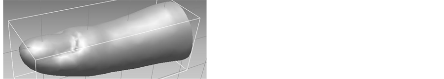
Figure 2. Synthesized 3D model of the index finger.

Figure 3.Mirroring the finger model of the right hand to obtain the finger model of the left hand.
parts of the mold are intended to join the upper and lower parts without forming a gap. These hemispheres are created by using Solid Works and are subtracted from/added to the mold model by using Free Form.
Mold manufacturing is performed using a 3D printer (Z-Printer 450, 3D Systems) that continuously fabricates thin layers of plaster until the entire mold is completed. Figure 5 shows the mold fabricated by the 3D printer.
A finger prosthesis is fabricated by pouring silicone resin into the mold. Figure 6 shows the fabricated finger prosthesis.
3. Evaluation and Results
To evaluate the size and shape of the finger prosthesis, we analyzed the differences between the finger prosthesis and the original finger of the volunteer using the procedure described below.
First, the left index finger of the volunteer was duplicated using the dental impression technique and consequently a plaster cast model of the index finger was made. Next, the plaster cast model and the finger prosthesis were scanned by the laser scanner (VIVID9i, Konica Minolta, Inc.), which allowed construction of the 3D models. Then, taking the two 3D models as inputs, Rapid Form analyzed the differences between the two 3D models. Figure 7 shows the results obtained using Rapid Form. As the figure indicates, the average difference was 0.25 mm and the largest difference was less than 0.87 mm. We concluded that the finger prosthesis had adequate size and shape.
4. Conclusion
We proposed a computer-aided method of designing and fabricating a finger prosthesis. We used the Geometric Database to synthesize a 3D model of a finger prosthesis automatically. The use of the synthesized model is the most distinctive feature of our method. This method enables fabrication of finger prostheses more easily than

Figure 4. The 3D model of the mold.

Figure 5. The mold fabricated using the 3D printer.

Figure 6. The fabricated finger prosthesis.

Figure 7. The differences between the finger prosthesis and the original finger.
previously proposed methods. We also used the 3D modeling tools, CAD, and 3D printer to design and fabricate a mold that was used for fabricating the finger prosthesis. We fabricated the finger prosthesis for a healthy volunteer and analyzed the differences between the finger prosthesis and the original finger of the volunteer. As a result, we found that the size and shape of the finger prosthesis were adequate.
Because the Geometric Database contains data only for index fingers, our current approach can be used to fabricate index fingers alone. However, our method can also be used to fabricate other types of fingers once the database is extended to include all of the types of fingers.
Cite this paper
TakeshiMurayama,KoseiOono,MitsunoriTada,ToruEguchi,MisuzuNagami,MitsuhiroTamamoto, (2015) Computer-Aided Design and Fabrication of Finger Prosthesis. Journal of Biomedical Science and Engineering,08,98-103. doi: 10.4236/jbise.2015.82010
References
- 1. Shanmuganathan, N., Uma Maheswari, M., Anandkumar, V., Padmanabhan, T.V., Swarup, S. and Jibran, A.H. (2011) Aesthetic Finger Prosthesis. The Journal of Indian Prosthodontic Society, 11, 232-237.
http://dx.doi.org/10.1007/s13191-011-0074-9 - 2. Botolin, L., Gazroda, S., Maver, T. and Ganter, G. (2007) Use of Rapid Manufacturing Technology in Comprehensive Rehabilitation of a Patient with Physical Body Disorders.
http://digicen.si/clanki/nasi_clanki/ICIT_07_784_botolin.pdf - 3. Fujita, S., Murayama, T., Tamamoto, M., Shimoe, S., Makihira, S., Sasahara, K., Kawahara, K., Satoda, T., Nikawa, H., Niitani, Y., Hara, K., Matsumoto, A., Takemoto, T. and Amano, H. (2009) Design and Fabrication of the Mold of Finger Prostheses by Using CAD and 3d-Printer. 93rd Regular Meeting of Hiroshima University Dental Society, Hiroshima, 24-25 October 2009, 120. (In Japanese)
- 4. De Crescenzio, F., Fantini, M., Ciocca, L., Persiani, F. and Scotti, R. (2011) Design and Manufacturing of Ear Prosthesis by Means of Rapid Prototyping Technology. Proceedings of the Institution of Mechanical Engineers, Part H, 225, 296-302.
- 5. Ciocca, L., Mingucci, R., Gassino, G. and Scotti, R. (2007) CAD/CAM Ear Model and Virtual Construction of the Mold. The Journal of Prosthetic Dentistry, 98, 339-343.
http://dx.doi.org/10.1016/S0022-3913(07)60116-4 - 6. Jiao, T., Zhang, F., Huang, X. and Wang, C. (2004) Design and Fabrication of Auricular Prostheses by CAD/CAM System. The International Journal of Prosthodontics, 17, 460-463.
- 7. Karayazgan-Saracoglu, B., Gunay, Y. and Atay, A. (2009) Fabrication of an Auricular Prosthesis Using Computed Tomography and Rapid Prototyping Technique. Journal of Craniofacial Surgery, 20, 1169-1172.
http://dx.doi.org/10.1097/SCS.0b013e3181acdb95 - 8. Liacouras, P., Garnes, J., Roman, N., Petrich, A. and Grant, G.T. (2011) Designing and Manufacturing an Auricular Prosthesis Using Computed Tomography, 3-Dimensional Photographic Imaging. The Journal of Prosthetic Dentistry, 105, 78-82.
http://dx.doi.org/10.1016/S0022-3913(11)60002-4 - 9. Turgut, G., Sacak, B., Kiran, K. and Bas, L. (2009) Use of Rapid Prototyping in Prosthetic Auricular Restoration. Journal of Craniofacial Surgery, 20, 321-325.
http://dx.doi.org/10.1097/SCS.0b013e3181992266 - 10. Subburaj, K., Nair, C., Rajesh, S., Meshram, S.M. and Ravi, B. (2007) Rapid Development of Auricular Prosthesis Using CAD and Rapid Prototyping Technologies. International Journal of Oral and Maxillofacial Surgery, 36, 938-943.
http://dx.doi.org/10.1016/j.ijom.2007.07.013 - 11. Murayama, T., Ogasawara, M., Eguchi, T., Morishita, Y. and Tamamoto, M. (2013) Computer-Aided Technique for the Design and Manufacturing of Auricular Prostheses. IFMBE Proceedings, 43, 593-596.
http://dx.doi.org/10.1007/978-3-319-02913-9_151 - 12. Kimura, K., Tada, M., Terabayashi, K. and Umeda, K. (2011) Subject-Specific Finger Model from Geometric Database. Proceedings of ISB2011, Brussels, 3-7 July 2011, 155.
NOTES

*Corresponding author.