Journal of Cancer Therapy
Vol.5 No.8(2014), Article
ID:47877,28
pages
DOI:10.4236/jct.2014.58086
Gastrointestinal Stromal Tumors in the 21st Century
Rani Kanthan1, Jenna-Lynn Senger1, Shahid Ahmed2, Selliah Chandra Kanthan3
1Department of Pathology & Laboratory Medicine, University of Saskatchewan, Saskatoon, Canada
2Divison of Medical Oncology, Department of Medicine, University of Saskatchewan, Saskatoon, Canada
3Department of Surgery, University of Saskatchewan, Saskatoon, Canada
Email: rani.kanthan@saskatoonhealthregion.ca
Copyright © 2014 by authors and Scientific Research Publishing Inc.
This work is licensed under the Creative Commons Attribution International License (CC BY).
http://creativecommons.org/licenses/by/4.0/


Received 12 May 2014; revised 10 June 2014; accepted 3 July 2014
ABSTRACT
Gastrointestinal stromal tumours (GISTs) are rare mesenchymal lesions accounting for only 0.2% of all gastrointestinal neoplasms. These tumors arise from the interstitial cells of Cajal, with mutations described in proto-oncogenes such as KIT, PDGFRA, DOG-1, and SDH. The majority of these lesions are asymptomatic, thus the true incidence remains unknown. While patients typically undergo initial endoscopy, CT scan and/or MRI, findings are often nonspecific and require a biopsy to identify the tumor. As such, immunohistochemical evaluation is the gold-standard for the accurate diagnosis of GIST. Though surgical excision remains the gold-standard for curative management, the discovery of imatinib, a tyrosine kinase inhibitor (TKI), has revolutionized the treatment of GIST in the 21st century as a “prototype” of molecular targeted therapy for solid tumors. Risk assessment for recurrence divides these tumors into low and high-risk categories. In the latter, a role for adjuvant therapy with TKI confers a significantly better prognosis than previously observed. However, secondary mutations conferring drug resistance remain an ongoing challenge for management, as few alternative treatment options are available for patients intolerant/refractory to TKI therapy. In this review, we summarize the epidemiology, molecular pathogenesis, clinical presentation, diagnosis, pathology features, management options, and prognostic features of GISTs.
Keywords:Gastrointestinal Stromal Tumor, Targeted Therapy, Drug Resistance, Histopathology, Surgery

1. History & Evolution of the Terminology “GIST”
Gastrointestinal stromal tumours, though rare, are the most common primary mesenchymal tumor of the gastrointestinal tract. In the past, there has been considerable discord regarding its nomenclature, cellular origin, diagnosis, treatment, and prognosis [1] . These lesions were originally thought to be smooth muscle/spindle cell neoplasms and were thus labelled as leiomyomas, leiomyoblastomas, leiomyosarcomas, or schwannomas [1] [2] . These tumors have also been referred to as “gastrointestinal autonomic nerve tumors” (GANT) [3] . In the 1960s, with the advent of electron microscopy, some of these gastric tumors demonstrated a lack of the typical smooth muscle differentiation [4] .
The term “GIST” was first introduced in 1983 by Mazur and Clark to describe tumors lacking smooth muscle differentiation or immunohistochemical features of Schwann cells [4] . In 1998, Kindblom et al. described GISTs as originating from pluripotent mesenchymal stem cells programmed to differentiate into the interstitial cells of Cajal which are considered to act as the gastrointestinal pacemaker cell [5] . It is now recognized that these tumors arise from the cells of Cajal in the muscular layer of the gastrointestinal tract, anywhere from the foregut to the hindgut [6] . In the same year, Hirota et al. discovered a functional mutation in the c-kit proto-oncogene [5] [7] . KIT is a transmembrane receptor tyrosine kinase (TK) protein that is a product of the KIT proto-oncogene. The CD117 molecule is part of the KIT (c-kit) receptors and most GISTs stain positive for CD117. The use of molecular targeted therapy against c-kit with agents such as imatinib mesylate, a tyrosine kinase inhibitor (TKI), has revolutionized the management of GIST in the 21st century. Detection of this proto-oncogene protein by immunohistochemistry is thus a pivotal diagnostic and therapeutic target. Thus, gastrointestinal stromal tumor (GIST), as defined by their molecular signature, is the accepted nomenclature of this family of tumors.
The following is a comprehensive review that provides a discussion on the epidemiology, molecular pathogenesis, clinical presentation, diagnosis, pathology, management, and prognostic features of GIST.
2. Methodology
A systematic review of the published medical literature for English-language articles using PubMed and Medline was carried out using the search terms: “gastrointestinal stromal tumor” and “GIST” with a special emphasis on review articles. Secondary references obtained from these publications were also reviewed as appropriate. Selected relevant abstracts from key oncology meetings were also reviewed. We limited our search to reports published since January 1, 2000 as GIST was not a generally recognized tumor entity prior to this time.
3. Epidemiology
GISTs are rare, accounting for only 1% to 3% of all malignant gastrointestinal tumors and only 0.2% of all GI tract neoplasms [8] [9] . The annual incidence of GIST is 11 - 14/million annually and the median age of diagnosis is 60 years (range 51 - 60) [1] [10] . These tumors are very rare in the pediatric population, with <1% occurring prior to age 21 [11] . This number may be an underestimation, as it is likely that many smaller GISTs go undetected as they are asymptomatic [12] . The diagnosis of GIST has increased 25-fold in the past 2 decades which is likely related to incidental discoveries with increased use of many advanced imaging modalities and reclassification of many GI smooth muscle tumors as GISTs [13] . While some articles have reported no gender predisposition, others have indicated a distinct male preponderance [12] . GISTs are typically solitary tumors; however, there are published reports of synchronous GISTs in the stomach and jejunum. Familial GISTs however are a notable exception to this observation, wherein multiple GISTs are more frequently encountered [14] .
3.1. Anatomical Location: GIST vs. EGIST
GISTs may arise anywhere along the gastrointestinal tract, with the most common sites including wherever the interstitial cells of Cajal are present including the stomach (60%), small bowel (30%), duodenum (5%), colorectum (4%) and esophagus [1] [15] . While rare, extra-gastrointestinal GIST (eGIST) are most common (80%) in the omentum and mesentery; however, these have also been reported in the pleura, pancreas, abdominal wall, liver, rectovaginal septum, gallbladder, and urinary bladder. eGISTs are morphologically and immunophenotypically similar to gastrointestinal GISTs; however, the pathogenesis and their exact cell of origin remains controversial. It is postulated that these lesions likely arise from multipotential mesenchymal stem cells, or possibly represent extramural outgrowths of GISTs that have lost contact with the muscular wall of the GI tract [15] [16] .
3.2. Biological Behavior
The biological behavior of GISTs is not clearly defined. GISTs usually present as a localized lesion; intraabdominal spread is common in tumors with potentially aggressive biological behavior. However, lymph node dissemination is extremely uncommon in adults. Up to 30% of all GISTs will exhibit malignant behavior including metastasis and infiltration [1] . Whether the benign vs. malignant GIST represents two distinct entities or a continuing spectrum remains undetermined; however, it is suggested clonal expansion may be responsible for this transformation. This theory is supported by a case report that showed two GISTs in a single stomach, one of low malignant potential and the other with high malignancy [17] . GISTs have also been reported to arise synchronously with adenocarcinomas of the jejunum, colon and stomach [14] [18] [19] .
4. Molecular Pathogenesis
4.1. KIT-Mutant GIST
In 1986 the oncogene v-KIT was identified encoded in the viral genome of HZ4-FeSV (Hardy-Zuckerman 4 feline sarcoma virus). V-KIT was subsequently found to encode a transmembrane tyrosine kinase receptor, KIT, and it was determined that the KIT receptor is expressed by the interstitial cells of Cajal (ICCs), the pacemaker cells of the gastrointestinal tract found in the myenteric plexus [1] [2] [20] . ICCs are able to transdifferentiate into smooth muscle cells and thus have stem-cell-like characteristics [11] . It is suggested that hyperplasia of the ICCs may be a precursor lesion to the development of GIST [21] . It has been shown that microscopic foci of KIT-positive spindle hyperplasia is common in patients with KIT/PDGFRA mutations; however, <1% will progress to a clinically significant GIST [22] .
Normally KIT is autoinhibited, unless bound by a stem cell factor (SCF), which causes KIT receptor homodimerization. This subsequently leads to activation of KIT tyrosine kinase activity, affecting intracellular signal transduction [1] . Downstream pathways including mitogen-activated protein kinase and phosphatidylinositol 3-kinase (PI3K) are activated [23] . Additionally, the RAS-RAF-MAPK pathway is also implicated [24] . It is recognized that products of RAS are downstream of KIT and PDGFRA and thus likely play a role as an oncogenic driver mutation in GISTs. This signal transduction is involved in the regulation of cellular proliferation, differentiation, and anti-apoptotic signaling [1] .
KIT mutations have been identified in GISTs, wherein they play a central role in its pathogenesis [1] . This is thought to be an early event in the tumorigenesis of GIST, particularly involving exon 11 [25] . KIT mutations release the autoinhibition such that the receptor remains in a constitutively active state [26] . Mutations may be further classified into primary vs. secondary mutations, the latter developing through exposure to tyrosine kinase inhibitors leading to drug resistance [4] . Primary mutations include in-frame deletions or insertions, point mutations, and duplications [4] [20] . The majority of these mutations occur in exon 11, and cause disruption of the juxtamembrane scaffolding, thus preventing the kinase activation loop from alternating into an active configuration [13] [24] . In-frame deletions are the most common (60% - 70%) mutation at this site followed by missense point mutation (20% - 30%) [24] . Less commonly exons 9 (extracellular domain), 13, and 17 may be involved [6] . Mutations of the extracellular domain of KIT, predominately exon 9, and in kinase I and II domains (exons 13 and 17) may also be present [20] .
In the presence of a mutation, when c-KIT is autophosphorylated, ligand-independent tyrosine kinase activity leads to uncontrolled cellular proliferation. As SCF-KIT interactions are involved in the normal development of melanocytes, erythrocytes, germ cells, mast cells, and ICCs, mutations cause defects in melanogenesis, hematopoiesis, gametogenesis, and in the interstitial cells of Cajal [1] . Secondary mutations occur in KIT kinase domains during or after imatinib treatment, leading to drug resistance [4] .
4.2. PDGFRA-Mutant GIST
In KIT-negative GISTs, a variety of other mutations have been identified. Three-to-five percent of GISTs have a mutation of platelet-derived growth factor receptor alpha (PDGFR-alpha) [2] . PDGFRA is a transmembrane tyrosine kinase homolog of KIT [13] . These tumours are associated with mutations in exons 12, 14, and 18 of the PDGFRA gene which is also on chromosome 4 [6] [22] [27] . Mutation-type depends on the exon involved: the juxtamembrane domain (exon 12), the ATP binding side (exon 14), or the activation loop (exon 18) [13] . Of note, exon 18 mutations confer resistance to tyrosine kinase inhibition [13] . PDGFRA-mutant GISTs arise most commonly in the stomach, mesentery, and omentum [28] . These tumors typically have a low malignant potential and confer a better prognosis than KIT-mutant tumors [29] . KIT and PDGFRA mutations are typically mutually exclusive [22] [29] .
4.3. Wild-Type GIST
Approximately 15% of GISTs have no KIT or PDGFRA mutations and are thus classified as “wild-type tumors”. Nevertheless, these tumors stain positively for KIT on immunohistochemistry [13] . Many of these wild-type tumors have BRAF V600E mutations at exon 15 [22] . This mutation is thought to arise in early-stage incidental GISTs, and confer a low risk for malignant potential. Tumors with BRAF mutations most commonly arise in the small bowel [30] . Mutations of NF1, BRAF, NRAS, and KRAS have also been described [6] as well as variants in CYP1B1, RAD23B, GSTM1, and ERCC2 [31] . A germline alteration of the BAX gene, which encodes a BCL2- associated protein that is directly activated by p53, has also been reported in GISTs [32] .
A subset of KIT/PDGFRA wild-type GISTs are characterized by germline/somatic mutations of succinate dehydrogenase (SDH) subunits A, B, C, D (SDHA, SDHB, SDHC, SDHD). Only 7.5% of GISTs are driven by SDH deficiency rather than the typical KIT/PDGFRA mutation, of which half are due to mutations of the SDHsubunit genes and half are attributable to epigenetic silencing. SDH deficient cells accumulate succinate which leads to overexpression of aberrant transcription factors resulting in hypoxia-associated tumorigenesis and angiogenesis, referred to as “hypoxic drive” or “pseudohypoxia signaling”. SDH-deficient GISTs are also associated with over expression of Insulin-Like-Growth Factor-1 Receptor (IGF1) possibly by gene amplification. Thus, high expression of IGF-1 protein detected immunohistochemically is a strong surrogate marker for the diagnosis of SDH-deficient GISTs in the appropriate clinical scenario. SDH-mutated GISTs are more frequent in a younger population, encompassing the majority of pediatric GISTs as well as GISTs associated with CarneyStratakis syndrome, with a greater prevalence in females, and arise exclusively in the stomach, typically in a multinodular pattern. On histopathological examination, cells typically have an epithelioid morphology with eosinophilic cytoplasm, contrasting the typical spindle-cells and pale cytoplasm of KIT-mutant GISTs. These GISTs express KIT equally strong as KIT-mutant GISTs and are also uniformly positive for DOG1/Anoctamin-1 by immunohistochemistry. SDHA mutation has an excellent correlation with immunohistochemical loss of SDHA expression and is thus an excellent screening tool and surrogate marker for SDHA mutation analysis. Up to 50% of patients present with lymphovascular invasion. Despite the presence of node positivity or liver metastases, SDH-mutated GISTs usually have an indolent course with patients living many years after identification of metastases. A minority of SDH-silenced GISTs are fatal within a few years of diagnosis (up to 15%), while others will metastasize >10 years after the initial diagnosis. Delayed recurrences up to 42 years have also been described [33] [34] .
4.4. Chromosomal Alterations
Typical chromosomal alterations in GISTs include losses at 1p, 14q, 15q, and 22q [4] . Loss of 9p is associated with an aggressive behavior, corresponding to loss of CDKN2A. Similarly, aggressive/metastatic behavior is associated with gains in chromosomes 5p, 20q, 8q, and 17q [22] .
4.5. Epigenetics
While KIT or PDGFRA mutations are present in the majority of cases, it is speculated that epigenetic alterations may also contribute to the malignant transformation. Hypomethylation of LINE1 correlating with the aggressiveness of GISTs, is proportional to the total number of chromosomal aberrations, and may be a marker for risk assessment. Hypomethylation of SATA and NBL2 additionally are associated with high-risk GIST. Hypermethylation has been detected in several genes, the most common being MGMT (47%), P16/INK4A (45%), RASSFIA (40%), E-Cadherin (37%), HMLH1 (34%), and APC (31%) [6] . GISTs may also have dior tri-methylation of lysine 4 on histone H3. The active histone H3K4me3 marks multiple genes including HOTAIR in malignant GISTs.
4.6. MicroRNA
MicroRNA has been implicated in the pathogenesis and tumorigenesis of GISTs. The most well-studied microRNA (mRNA) in GISTs is miR-221/222, which target KIT and ETV1. Over expression of miR-222 as well as -17 and -20a inhibits cell proliferation, affects cell cycle progression, induces apoptosis, and downregulates proteins. Upregulation of these microRNAs is associated with downregulation of ETV1 protein [35] . Other microRNAs that have been implicated include miR-494, -132, -504, -134/170, -196a, and 133b [6] . miR-494 is reported to target downstream KIT, thus inhibiting GIST cell growth while inducing apoptosis [35] .
5. Clinical Presentation
Only 70% of patients with GISTs are symptomatic, with 20% being asymptomatic incidentalomas and 10% being detected at autopsy. Presenting signs and symptoms depend on the site of involvement; however, abdominal discomfort is the most common symptom (60% - 70%) followed by gastrointestinal bleeding due to erosion into the lumen (30% - 40%). If bleeding is intraperitoneal, patients may present as an acute abdomen whereas hemorrhage into the lumen may present more chronically with hematemesis, melena, or anemia [1] . Anemia is present in ~50% of patients with GIST [36] . Bleeding is more common as a clinical presentation in duodenal GISTs (75%) compared with gastric (54%) or ileojejunal (28%) [37] . Many patients may have vague complaints including nausea, vomiting, weight loss, or early satiety [1] [2] . Bowel obstruction is uncommon in GISTs as it, like other sarcomas, displaces rather than invades adjacent structures [2] . More rarely, symptoms such as pelvic pain, pleuritic chest pain, and dysuria may be present [24] . Site-specific symptoms may include dysphagia (esophagus), biliary obstruction (ampulla of Vater), or intussusception (small bowel) [1] [38] . Rare presentations include perforation, and hypoglycemia secondary to paraneoplastic production of insulin-like-growthfactor-II [8] .
When <1.0 cm, these tumors are commonly referred to as “microGIST” and are most often incidental findings. These lesions are present in 10% - 22.5% of stomachs in middle-aged to elderly patients [20] . This high prevalence suggests that the majority of KIT-positive microGISTs do not progress to malignant behavior [39] . Such microGISTS are most typically found at the GE junction or proximal stomach. MicroGISTs generally are immunoreactive for KIT and contain KIT or PDGFRA mutations [40] .
5.1. Familial GIST
Familial GISTs are the result of an autosomal dominant mutation of KIT exon 8, 11, 13, or 17. Such individuals are at risk of developing multiple GISTs as early as age 18. This germline mutation has 100% penetrance [41] . These patients may present with pigmented macules on the perineum, axilla, hands, and face as well as urticaria pigmentosa [20] . Morphologically, these GISTs are indistinguishable from sporadic tumors. This patient population is thought to confer a high risk of metastatic spread irrespective of tumor size and mitotic activity [22] . The worse prognosis in this patient population is, however, primarily due to local complications such as hemorrhage, perforation, or surgical complications due to the large number of multiple GISTs [41] .
5.2. Carney’s Triad
Carney’s triad is a rare non-heritable syndrome mainly in young females that includes gastric GIST, paraganglioma, and pulmonary chondroma. These lesions tend to be of the epithelioid subtype and have a higher risk of metastatic spread including lymphadenopathy. Tumors usually do not have KIT or PDGFRA mutations, with some occurring in the context of SDH germline mutation (SDH-deficient GIST) [34] . These GISTs are thought to be sporadic, non-familial, growing more slowly with frequent metastases, and have minimal or equivocal response to imatinib therapy [22] .
5.3. Carney-Stratakis Syndrome
Carney-Stratakis syndrome occurs secondary to germline mutation of succinate dehydrogenase (SDH) that predisposes individuals to multifocal GISTs, paragangliomas, and pheochromocytomas [22] . Recently esophageal leiomyoma and adrenal cortical adenoma have been added as potential components to this syndrome [42] . These tumors are negative for KIT or PDGFRA mutations yet have loss of expression of succinate dehydrogenase subunit B. This syndrome is most commonly seen in young females, with >80% presenting prior to the age of 30. Unlike sporadic GISTs, these tumors more commonly have lymph node metastases [43] .
5.4. Type I Neurofibromatosis
Patients with type 1 neurofibromatosis (NF1) often develop small bowel GISTs [20] . These tumours occur in a younger population than sporadic GISTs [44] . One autopsy series revealed up to one-third of patients with NF1 had undiagnosed GISTs [11] . These tumors are typically small, multifocal, and are usually KIT and PDGFRAnegative [43] [44] .
5.5. Other Associations
Additionally, GISTs are thought to have a relationship with desmoid tumors. It is not yet understood whether the presence of a GIST predisposes to the development of a desmoid, or vice versa [43] .
5.6. Pediatric GISTs
GISTs in children are rare, accounting for ~1% of these tumors [43] . Pediatric GISTs are most common in the stomach of females in the 2nd decade of life, and are often multifocal with a multinodular pattern of growth [42] [43] . These lesions are distinct in their pathogenesis and clinical behavior and are thus considered separate from their adult counterpart. Many of the clinical features of pediatric GIST are similar to tumors associated with Carney’s triad [22] . Children most often present with symptoms of anemia, pallor, fatigue, vertigo, vomiting, abdominal pain, distension, and/or intestinal obstruction (intussusception) [38] . Up to 20% of pediatric patients will present with metastases at diagnosis, typically to the liver, lymph nodes and peritoneum [3] . They uniformly express KIT and Dog1, though rarely have detectable mutations in KIT or PDGFR. Generally, pediatric GISTs follow an indolent course and some argue that treatment with imatinib therapy should be reserved only for patients with metastatic disease or restricted to patients in whom the tumor is unresectable [22] .
6. Diagnosis
The majority of GISTs are detected by imaging. The radiologic appearance of GISTs is highly variable, complicating their accurate detection and diagnosis. Tumors may have intraluminal, intramural, or external components with/without pedunculated extramural or cystic appearance [11] . These tumors arise within the muscularis propria and appear as endophytic or exophytic nodules with a mean size of 3 - 5 cm in diameter and are usually highly vascular and friable lesions [45] .
6.1. Endoscopy
Upper GI endoscopy is normally the first investigation chosen, which typically reveals a smooth lesion protruding into the bowel lumen with normal-appearing overlying mucosa associated with or without superimposed ulceration [45] . Upper endoscopy with biopsy has a low diagnostic yield of only 17% - 42% [46] . Additionally, GI endoscopy is unable to determine the accurate size of the lesion due to the submucosal location of GISTs [45] .
Endoscopic ultrasound (EUS) may be used to assess the depth of invasion and to obtain a tissue sample for histopathological confirmation. Features of a GIST on EUS include irregular extraluminal borders, heterogeneous echo patterns, and the presence of cystic spaces and echogenic foci [1] [39] [47] . Lesions are typically in the fourth sonographic wall layer corresponding to the muscularis propria. This technique is limited by inter-observer variability resulting in a diagnostic accuracy as low as 43%. By contrast, other studies have found a 100% positive predictive value when EUS features include a) irregular extraluminal margins, b) cystic spaces, and c) lymph nodes with malignant pattern [46] .
6.2. CT Scan
Contrast-enhanced computed tomography (CECT) of the abdomen and pelvis is the mainstay imaging technique to detect and characterize the lesion, assess its size, plan surgical treatments, determine the presence/absence of metastases, and monitor for therapy response or recurrence [1] [48] . GIST classically appears as a hyperdense, enhancing mass closely associated with the stomach or bowel [2] . Larger tumors (>6 cm) may be heterogenous with regions of necrosis, hemorrhage, and/or cystic changes [48] . Cysts may be so large as to obscure full visualization of the tumor itself [49] . Pitfalls in the accurate diagnosis of GIST using CT include: a) when the tumor is connected to the GIT wall, contrast may inhibit visualization, b) lack of characteristic features pathognomonic for GISTs, c) presence of necrosis or cystic degeneration reminiscent of intraabdominal abscess, inflamed intestinal loops, or pancreatic lesions, or d) difficulty in defining the epicenter of origin in large lesions [50] . Additionally, CT scanning may not be able to accurately identify peritoneal seeding [51] .
6.3. MRI
Magnetic resonance imaging (MRI) has a comparable diagnostic yield to CT scans and is the preferred choice for the evaluation of GISTs in specific locations such as the rectum or liver [8] . It is additionally indicated when CT scanning is inconclusive or contraindicated [45] . GISTs appear as hypointense lesions on T1-weighted imaging (T1WI) and hyperintense on T2-weighted imaging (T2WI) [48] .
6.4. Biopsy
Biopsies are not routinely recommended for GISTs due to fear of intraabdominal seeding; however, it is a useful technique in the diagnosis of an unidentified abdominal neoplasm [39] . Percutaneous biopsy is not recommended due to a high risk of inducing tumor rupture, and endoscopic modalities have supplanted this option [3] . Endoscopic biopsy specimens are, however, often unable to obtain sufficient tissue to make a diagnosis, as they are only successful in 20% - 30% of cases [11] . As GISTs are submucosal, routine forceps biopsy technique is not reliable [3] . Core-needle biopsy with a 19gauge Tru-cut needle may improve yield rates; however, this technique is limited to tumors in the fundus, antrum, and duodenal bulb [39] . Lesions not amenable to endoscopic biopsy may require a laparoscopic or open biopsy [46] . The choice of which biopsy technique to pursue depends on the anatomic location of the lesion, the patient’s physiological status, and the equipment and resources available [3] .
Recently, endoscopic ultrasound-guided fine needle aspiration (EUS-FNA) has been reported to have an accuracy rate of 80% - 85% and is thus the procedure of choice to obtain a tissue diagnosis when indicated [1] . However, EUS-FNA fails to yield an adequate tissue sample in up to one-third of specimens [39] . Factors that affect the diagnostic yield include: site, size and characteristics of the tissue; technical and procedural factors; endosonographer and cytopathologist expertise [52] . Larger tumors have a higher diagnostic accuracy, with reported rates for tumours of 2 cm, 2 - 4 cm, and 4+ cm at 71%, 86%, and 100% respectively [53] . A similar procedure, endoscopic ultrasound-guided tru-cut biopsy (EUS-TCB) which uses a stiffer device, has additionally been described as rapid and safe; however, no standards for its use have been determined [52] .
6.5. PET Scan
Positron emission tomography (PET) can be used to detect GISTs because the receptor tyrosine kinase increases glucose transport protein signaling. This imaging modality is used to identify small metastases not detectable on CECT and to differentiate active tumor vs. necrotic/scar tissue [1] . The reported sensitivity of PET scanning is 86% - 100% [8] . PET scans may also be used to assess early response to tyrosine kinase inhibitor therapy, as this treatment creates changes at the metabolic level prior to detectable anatomic changes which are then detectable by CECT [1] .
6.6. Miscellaneous
Video capsule endoscope (VCE) and double balloon enteroscopy (DBE) have both been used successfully in the detection of GISTs, with detection rates of 80% and 60% respectively [36] .
The use of fluoroscopy has been previously described in the detection of GISTs. A recent study has successfully used fluorescent anti-KIT antibodies to label GIST as an in vivo imaging modality in transgenic mice. Fluorophore-conjugated antibodies against KIT are effective in labeling KIT-positive GISTs for the detection of primary and metastatic disease. Thus similar to CT scans, fluoroscopy may be used in the initial diagnosis and treatment planning as well as monitoring disease response to therapy [51] .
7. Pathology
7.1. Gross Features
GISTs range in size from a few millimeters to >30 - 40 cm (median 5 - 8 cm) [1] [54] . On gross examination, they appear as gray/white, well-circumscribed growths often within a pseudocapsule [1] . Lesions may grow into the peritoneum exophytically or into the bowel’s lumen endophytically [55] . Small gastric GISTs typically appear as serosal/intramural nodules, and large tumors may have intraluminal, intramural, and/or external components [56] . Hemorrhagic foci, central cystic degeneration, and/or necrosis may also be seen [40] .
7.2. Cytopathology
Cytopathological findings obtained in FNA specimens suggestive of a GIST may include spindle shaped cells with scant cytoplasm and elongated-to-oval nuclei [52] . Cellularity is typically moderate to high. The presence of cohesive and tight fascicular bundles with minimal pleomorphism is suggestive of this neoplasm. Chromatin is typically fine, and nuclei are most often indistinct. The diagnosis of GIST can, however, only be suspected on cytology and confirmatory immunostaining is necessary for definitive diagnosis [57] .
7.3. Histopathology
Three distinct histopathological subtypes have been described: spindle, epithelioid, and mixed [8] [20] . The spindle-type is the most common phenotype seen in 70% of GISTs, and fascicles of uniform spindled neoplastic cells with minimal nuclear atypia and perinuclear vacuoles can be visualized [23] . Extracellular dense collagen deposits (skeinoid fibres) may be seen [40] . Individual cells may have ill-defined cell borders with ovoid nuclei, fine nuclear chromatin, and inconspicuous nucleoli. The cytoplasm is often pale, eosinophilic, with a fibrillary quality. Nuclear palisading reminiscent of Antoni A regions of schwannoma may be observed. The epithelioidtype is characterized by the presence of round cells in sheets or nests [22] . Cytoplasm may be clear, rhabdoid, plasmacytoid retracted spider-like, or vacuolated [58] . The mixed-type contains elements of both the spindle and epithelioid morphology in varying proportions.
Certain morphological patterns have a predilection in relation to the anatomical location. On histopathology, common features of gastric GISTs include cells with a sclerosing matrix, perinuclear vacuolization, and nuclear palisading, with an epithelioid or sarcomatoid, mitotically active morphology [11] . Gastric GISTs appear as solid/nested tumors with an epithelioid morphology and a stroma showing myxoid change whereas small bowel tumors are spindle-type with a paragangliomatous architectural pattern [1] . Small bowel GISTs are more homogenous than gastric GISTs, and contain extracellular collagen globules and Verocay bodies or neuropil-like material [11] [56] . Colonic GISTs typically mirror their small bowel counterpart, and histologic features of rectal GISTs include features of both gastric and small bowel [56] .
7.4. Immunohistochemistry
The immunohistochemistry of GISTs has been well researched and remains the only true diagnostic test for its accurate and definitive diagnosis. The vast majority (>95%) of GISTs are positive for CD117, with diffuse strong expression in the spindle sub-type and focal expression in the epithelioid-type [8] . KIT expression is the most specific marker for these tumors; however, the absence of CD117 (KIT) expression does not rule out the diagnosis [47] . Though staining by CD117 is sensitive, it is not specific for GISTs as other tumors such as metastatic melanoma, pulmonary small cell carcinoma, other types of carcinomas, angiosarcoma, Ewing’s sarcoma, and seminoma can also be KIT positive [4] .
Additionally, the majority (60% - 70%) of GISTs stain positive for CD34 (60% - 70%), smooth muscle actin (30% - 40%), S100 (5%), vimentin and rarely desmin [2] [59] . Immunostaining with PDGFRA is paranuclear in the majority of cases [58] . Caldesmon immunoreactivity is present in >66% of GISTs [40] .
Accurate identification of CD117-negative GISTs remains a challenge. Protein kinase C-θ (PKC-θ) has been reported to be a useful biomarker for such tumors, as it is a downstream effector in the kit signaling pathway [24] [60] . DOG-1 (Discovered on GIST-1), also known as anoctamin 1, can assist in identifying some of these lesions as it holds a high specificity, possibly even higher than that of CD117, for the accurate diagnosis of GIST [8] [56] [60] [61] . Other tumors that may express DOG-1 include leiomyomas and certain GI carcinomas, especially squamous cell [56] . DOG-1 is a chloride channel composed of eight transmembrane domains that is activated by calcium. This channel is also referred to as FLJ10261, TMEM16A, and ANO1. Sensitivity of DOG-1 staining in GISTs is 75% - 100% [11] . The presence or absence of DOG1 expression has not been correlated with any clinicopathological characteristics [61] . Additionally, histopathology features do not reliably relate to the immunophenotype or the molecular genetics of the lesion [8] ie: there is no strong genotype/phenotype correlation.
8. Management Principles
Small (<2 cm) GISTs merit surgical resection if symptomatic; however, if asymptomatic with no high-risk EUS features endoscopic resection or a conservative management of surveillance q6-12 months is appropriate. Endoscopic mucosal resection is more challenging for tumors >2 cm or those arising from the muscularis propria [1] . This conclusion is supported by the observation of Lim et al. who reported an increase in size of only 8/252 gastric tumors over 59 months [39] .
8.1. Surgical Management
8.1.1. General Principles
Whenever possible, surgical management is indicated as it is the only potentially curative option and the most important factor with regard to overall survival (OS) regardless of the approach chosen or the technique employed [62] . Surgical resection with negative margins is the gold-standard treatment for all resectable GISTs [13] . Surgery may be positioned as the initial step in management, following neoadjuvant therapy or as a debulking procedure for symptomatic relief in advanced metastatic disease [27] . Ideally, the pseudocapsule should remain intact [1] . The surgical procedure chosen must minimize the risk of tumor rupture, as it is a highly unfavorable prognostic factor that may lead to peritoneal seeding [62] [63] . Excessive tumor manipulation increases the risk of bleeding and intraperitoneal dissemination [2] .
Four classes of surgical approaches have been described for the treatment of GISTs [45] :
1) Endoscopy (risk of positive margins)2) Laparoscopy (risk of technical challenges with large tumors)3) Laparoendoscopy (evidence limited to case reports)4) Laparotomy (more invasive).
Segmental or wedge resection aimed at histologically negative margins is ideal. Wide excision with multivisceral and radical en bloc excisions should be avoided [1] as GISTs do not typically spread intramurally though a clear margin of 1 - 3 cm has been recommended by some authors [9] whereas others suggest that microscopically free millimeter margin is sufficient in gastric GISTs [13] . Incomplete tumor excision is, however, associated with a decreased 5-year survival of 8% - 9% (compared with 42% in complete resection) [5] .
8.1.2. Laparoscopic Surgery Principles
Laparoscopic treatment is reported to confer decreased recurrence rates, shorter hospital stays, and a lower morbidity. This treatment strategy may be indicated in certain anatomic locations including the anterior wall of the stomach, jejunum, and ileum [1] . Typically GISTs do not invade adjacent organs and as such the tumor can be lifted away, making laparoscopic surgery a viable preferred modality [4] . The reported success rates are excellent, from 92% - 96% [11] . There are, however, concerns about the oncologic safety and technical feasibility of laparoscopic resection, predominately related to tumor size, risk of tumor rupture, and adequacy of the resection margins. The National Comprehensive Cancer Network Clinical Practice Guidelines for Optimal Management of Patients with GIST states that a laparoscopic approach should be reserved for tumors <2 cm. However, studies report this technique being used for tumors 2.7 - 6.0 cm with low recurrence rates and margin negativity suggests that a large tumor size should not be an absolute contraindication to laparoscopic resection [64] . For GISTs > 5 cm, the use of an endobag may be used to protect the exophytic lesion from lacerations [65] . Specially designed techniques such as a “non-touch lesion-lifting” method have been described in the treatment of GISTs [66] .
The subsequent steps in the event of a microscopically positive margin post-resection are controversial. If the original margin can be located, re-excision is a possibility with low associated morbidity [13] .
8.1.3. Localized GISTs
1) Gastric GISTs In gastric GISTs, wedge resection is often possible, particularly for tumors of the greater curvature, with segmental or total gastrectomy reserved for large tumors [13] . GISTs at the gastroesophageal junction or pylorus may not be amenable to wedge resection and require a proximal/distal gastrectomy. If the tumor infiltrates the surrounding organs, a complete en bloc multivisceral resection including the adjacent liver, spleen, pancreas and/or colon should be considered [39] . Lymph node involvement is rare and therefore routine lymphadenectomy is not indicated unless locoregional nodes are enlarged. Additionally, as GIST does not typically exhibit an intramural mode of spread, wide margins of resection are deemed unnecessary [2] .
A recent meta-analysis reviewed laparoscopic vs. gastric resections for the treatment of 765 patients with gastric GISTs [64] . As would be expected, a higher proportion of high-risk tumors and gastrectomies were performed by open technique in comparison with laparoscopic gastric resection. The precise laparoscopic surgical techniques for GISTs located at different locations within the stomach have been previously reported [39] ; however, novel techniques continue to emerge. Gasless laparoscopic surgery has recently been described to decrease the postoperative hospital stay, wound length, white blood cell count on post-op day 1, and peak daily body temperatures when compared with open surgery [67] . Single-incision laparoscopic surgery (SILS) is another new safe and feasible procedure for treating gastric GISTs. In the hands of an experienced laparoscopic surgeon, SILS allows for improved direct visualization and better control of surgical margins [68] . In general, laparoscopic surgery was found to confer a lower risk of intraoperative blood loss, less minor complications, a shorter postoperative hospital stay, and less time in resuming oral intake. Operation time, major complication rate, margin positivity, local recurrence, and recurrence-free survival were however found to be comparable between laparoscopic vs. open procedures. These results were influenced by selection/publication bias as technically more complex cases were chosen for an open approach whereas the simpler cases were completed laparoscopically [69] .
Laparoscopic and endoscopic cooperative surgery (LECS) is a procedure in which the tumor is resected with minimal surgical margins [1] . This procedure allows for intraoperative definition of the tumor size and additional information required to inform operative decision making [21] . This technique is particularly useful for tumors at the esophagogastric junction [66] .
2) Duodenal GISTs GISTs of the small intestine are typically resected [20] . Surgical treatment of duodenal GISTs can be categorized as a limited resection (LR, wedge and segmental resection) or extended resection (ER, pancreaticoduodenectomy) [70] . Factors influencing choice of surgical procedure include the size, location and proximity of the GIST to the duodenal papilla [59] [71] . Wedge resection is recommended for GISTs up to 1cm located over 2 cm from the ampulla of Vater while segmental duodenectomy is the preferred choice for larger tumors not involving the ampulla and extended resection is reserved for tumors involving the ampulla. Whether limited duodenal resection is a better option for tumors close to the ampulla of Vater remains controversial [55] . Segmental duodenectomy typically has good oncologic outcomes and is associated with a lower morbidity and mortality. Limited resections do, however, carry a higher risk of anastomotic leakage, stenosis and/or recurrence [59] . Larger tumors involving the antimesenteric border of the second and third part of the duodenum should be treated with a partial duodenectomy with Roux-en-Y duodenojejunostomy [37] .
When the tumor specifically involves the papilla of Vater, extensive resection such as a Whipple’s is the standard treatment of choice [37] [71] [72] . Limited resection, such as a pancreas-sparing duodenectomy with reimplantation of the major papilla may be an option; however, this technique is only viable if the tumor does not invade adjacent organs/lymph nodes and if the papilla itself is preservable [73] . A study by Colombo et al. compared patients who underwent pancreaticoduodenectomy vs. limited resection and found no difference in overall outcome [74] .
3) Colorectal GISTs The traditional treatment of colonic GIST is segmental colectomy as skip metastases is extremely rare [75] . Regarding rectal GISTs, there is debate whether conservative therapy is equal to abdominoperineal resection, as survival and the rate of distant metastasis is reported to be similar [76] . Other authors suggest that all rectovaginal GISTs require a biopsy with excision regardless of the size due to the risk of increased mortality [45] . In the case of small tumors, transanal or trans-sphincteric methods may be attempted [13] . Rectal GISTs < 3 cm may be treated with local excision providing clear margins are possible [75] .
8.1.4. Risk Assessment of Recurrence
Surgery is the mainstay treatment for localized GIST as previously discussed. However, a significant number of patients develop disease recurrence within 5 years of surgery. The median time to recurrence following surgery of the primary tumor ranges from 18 to 24 months [77] . A population based study involving 2560 patients demonstrated that 5- and 15-year recurrence-free survival (RFS) rates for patients with GISTs treated with surgery alone were 70.5% and 59.9%, respectively [78] .
A number of risk-stratification schemes have been created to estimate the risk of postoperative recurrence. Through the years, the number of prognostic factors examined to estimate recurrence risk has increased with each scheme. The first proposed schematic was developed by Fletcher et al. of the US National Institutes of Health (NIH) in 2002 which used tumor size and mitotic rate to group GISTs into very low, low, intermediate, or high-risk tumors; however, this criterion was criticized for not including tumor site [79] [80] . The Armed Forces Institute of Pathology led by Miettinen in 2006 responded by the creation of their own criteria that included mitoses, size, and location to create eight prognostic groups. A second NIH classification was then created to include these three factors as well as tumor rupture. The prognostic accuracy is, however, thought to be equal in all three schemes [4] [79] . Using these same parameters, a “rule of 5s” has been proposed to determine low vs. higher risk that incorporates the three main risk factors of tumor size, proliferation, and location. In this “rule”, intermediate-to high risk gastric GISTs are both >5 cm and have >5 mitoses/50HPF. Non-gastric GISTs are considered high-grade if they are either >5 cm in size or >5 mitoses/50HPF [81] .
Two prognostic normograms have been created by Gold et al. and Rossi et al., both aimed at predicting the likelihood of recurrence. Gold’s normogram estimates outcome at 2 years and 6 years post-resection based on a continuous spectrum of tumor sizes and categorical mitotic count [22] [79] . By contrast the Rossi normogram measures tumor size and mitotic count both as continuous variables; however, this normogram is only applicable to patients <65 years and is predictive for 10-year overall survival [79] .
Prognostic heat maps are a risk stratification tool that uses tumor size, tumor site, mitotic count, and rupture status to depict recurrence risk with different colors. These maps are reported to be more accurate than NIH or AFIP criteria [79] .
Moving away from these traditional prognostic parameters, the CINSARC (Complexity Index in SARComas) evaluates 67 genes involved in maintaining chromosome integrity and mitotic control to differentiate GISTs into lowand high-risk groups. The Genomic Index (GI), calculated by determining the ratio of the square of the total number of genomic alterations vs. the number of chromosomes involved, is used to segregate intermediate-risk GISTs into two prognostic groups. The true prognostic value of genomic profiling is, however, unestablished to date [79] .
8.2. Advanced GIST
Surgery for advanced GIST, defined as locally invasive or metastatic disease, may have a role to play in a) alleviation of symptoms for acute surgical complications such as bowel obstruction, tumor rupture, or GI hemorrhage [20] , or b) debulking or curative resection. Cytoreductive surgery can be used in a) patients with stable disease or who respond to TKI therapy, b) resecting clones of disease with drug resistance, and c) surgical emergencies such as perforation or abscess [47] . The true survival benefit of cytoreductive surgery for recurrences or metastases however, is largely undetermined [82] .
Often recurrent GIST is multifocal within the peritoneal cavity and surgery alone may thus not be a viable curative option. Such patients may undergo downsizing of the tumor with TKI in order to increase the likelihood of surgical success as part of a multidisciplinary approach. In these instances, the optimal time for surgical resection is within a 6 - 12 month drug treatment interval, and the drug should be continued postoperatively. If, however, the multi-site tumor is not responsive to drug therapy, there is generally no benefit observed from surgery [13] . In the presence of peritoneal and/or liver metastases, resection should be considered for all surgical candidates in order to eliminate the drug-resistant clones such that imatinib-therapy may be continued [1] . Each case must, however, be considered individually.
8.2.1. Targeted Therapy in GIST
Conventional chemotherapy and radiation therapy have limited value in GIST. Hence, until the advent of targeted therapy, there was no effective treatment available for advanced GISTs. Over the past decade there has been rapid progress in the management of GIST. The discovery of oncogenic kinase mutations in the vast majority of these tumors and the introduction of specific targeted molecular therapies that inhibit the molecular defect have revolutionized the management and prognosis of GIST. These specific agents block signaling of KIT or PDGFRA by binding to the ATP-binding pocket essential for phosphorylation and activation of the receptor. Figure 1 summarizes the role of systemic therapy in the management of resectable, unresectable/borderline resectable and metastatic GISTs.
8.2.2. First Line Treatment of Advanced GISTs
1) Imatinib Imatinib (Gleevec) was developed as a prototype tyrosine kinase receptor inhibitor which was shown to inhi-
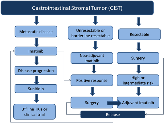
Figure 1. Framework of management of gastrointestinal stromal tumors (GIST). This flowchart provides a summarized framework for the management of resectable, unresectable/borderline resectable, and metastatic gastrointestinal stromal tumors.
bit the intracellular kinases ABL and BCR-ABL fusion protein in chronic myeloid leukemia (CML) cells. It was subsequently found that imatinib can induce dramatic and rapid clinical benefit in GIST as well [83] . A formal phase I study involving 40 patients demonstrated clinical benefit in 91% patients (54% partial response and 37% stable disease) [84] . Subsequently, several phase 2 trials assessed efficacy of imatinib at varying doses from 400 to 800 mg per day in patients with GIST and confirmed the benefit reported in the phase 1 trial [85] -[87] . For example in a phase 2 study, 147 patients were randomly assigned to receive 400 mg or 600 mg of imatinib daily. Overall, 54% patients had a partial response within six months of treatment. Early resistance to imatinib was noted in 14% patients. Therapy was well tolerated, although mild-to-moderate edema, diarrhea, and fatigue were common side effects [85] . In a long-term follow-up of a cohort of 56 patients who continued to take imatinib beyond three years, 26 (18% of the initial cohort) remained on imatinib at a median follow-up period of 9.4 years [88] .
The median time to objective response is about 12 - 14 weeks; however, some patients have a dramatic improvement in symptoms and signs within days of therapy initiation. Duration of imatinib therapy, in advanced GISTs, should be as long as possible, providing that the patient is still responding to the treatment. Following the introduction of imatinib, the median survival of patients with advanced GIST increased from an average of 18 to 57 months in an extended follow-up trial. Nearly 50% of patients with advanced GIST who were treated with imatinib mesylate survived for more than 5 years regardless of imatinib dose (400 mg/d vs. 600 mg/d) [89] .
There have been two large randomized controlled phase III trials assessing the optimum dosage of imatinib [86] [90] . In the European trial 946 patients were randomly assigned to imatinib 400 mg or 800 mg daily. Patients who were assigned to 400 mg, on progression were offered the option of crossover. Overall, 5% patients achieved a complete response, 47% a partial response, and 32% stable disease, with no difference in response rate between the groups. However, at median follow up period of 760 days, 56% of patients who received imatinib 400 mg daily developed disease progression compared with 50% of patients who were assigned 800 mg (HR: 0.82 [95% CI 0.69 - 0.]">98]). Overall survival was 85% at 1 year and 69% at 2 years for the 400 mg group and 86% at 1 year and 74% at 2 years for the 800 mg group [86] .
In the US intergroup phase III study, 746 patients with advanced GIST were assigned to imatinib 400 mg or 800 mg daily. With a median follow-up of 4.5 years there were no statistically significant differences in objective response rates, progression-free survival, or overall survival. For instance median progression-free survival was 18 months for patients on imatinib 400 mg compared with 20 months for those receiving high-dose imatinib. Median overall survival was 55 and 51 months, respectively. Of note, 33% of patients who crossed over to the high-dose imatinib regimen, on progression, achieved either an objective response or stable disease [90] .
The meta-analysis of two large randomized studies revealed that after a median follow up of 45 months, a small but significant progression free survival advantage was seen in the high dose arm. Overall survival was identical in the two arms (median survival 4.08 years in the high dose group versus 4.05 years in the standard dose group, HR 1.00). The presence of a KIT exon 9 mutation was the predictive marker for benefit from higher doses. Patients with an exon 9 mutation had a significantly better response rate (47 versus 21%) and progression free survival [PFS] (HR 0.58, 95% CI 0.38 - 0.91) with high-dose therapy. However, no difference was observed between the treatment arms in the absence of such mutations [91] .
2) Continuous vs. Interrupted Imatinib A small phase III French trial assessed optimal schedule of imatinib and compared continuous with interrupted imatinib therapy beyond 1 year of treatment in patients with advanced GIST. Of 58 eligible patients, documented progression was seen in 8 of 26 patients in the continuous group and 26 of 32 patients in the interrupted group (p < 0.001). Although there were no differences in overall survival, imatinib resistance or quality of life between the two groups, due to rapid progression after treatment interruption, this study supports the use of continuous imatinib [92] .
3) Imatinib Dose Escalation The standard dose of imatinib is 400 mg daily. Clinical data from the EORTC and North American Intergroup studies, where patients were allowed to cross over to high-dose imatinib upon progression, revealed that approximately one third of these patients were able to regain disease control [86] [89] . Hence, imatinib dose escalation can occasionally control advanced GISTs that progress on imatinib [93] . The standard approach as recommended by the European Society of Medical Oncology (ESMO) Clinical Practice Guidelines in the case of tumor progression on 400 mg is to increase the imatinib dose to 800 mg daily. Dose escalation is particularly useful in the case of a KIT exon 9 mutated GIST [94] [95] .
4) Combination Strategy Combination strategies have focused on targeting upregulated signaling pathways in imatinib-resistant patients or on reducing the stability of activated signaling proteins [93] . Agents such as everolimus targeting the PI3K/Akt/mTOR signaling pathway and heat shock protein 90 and histone deacetylase inhibitors in combination with imatinib have been studied [96] -[]">98] . However, disappointing efficacy or toxicity results have precluded their use in routine clinical practice.
8.2.3. Response Evaluation
Measurement of lesion density is important for response assessment. Both tumor size and tumor density on CT scan, or consistent changes on MRI, should be considered as criteria for tumor response [94] [95] . An FDG-PET scan has proved to be highly sensitive in early assessment of tumor response and may be useful in doubtful cases, or when early prediction of the response is highly useful [99] . In one study, more than 10% reduction in lesion size on CT, or a greater than 15% reduction in density had 97% sensitivity and 100% specificity for identification of patients who showed a response on PET scan [100] .
The measure of tumor response can be classified into four categories based on tumor size, degree, extent of enhancement, and the presence/absence of intra-tumoral solid nodules. The categories are as follows [41] :
• Category 1—Complete response: the original lesion previously identified by imaging is no longer present• Category 2—Partial response: two subcategories include:
a) Continuous regression throughout treatmentb) Initial regression followed by stabilization without further change• Category 3—Stable disease: tumor size and enhancement remains unchanged• Category 4—Progressive disease: two subcategories include:
a) Initial regression followed by development of new lesions and increased size/enhancementb) Continuous progression of increasing tumor size/enhancement throughout treatment.
The Choi criterion is an alternative set of response evaluation criteria utilizing tumor size and density and predicts response to therapy better than other criteria. A 10% decrease in unidimensional tumor size or a 15% decrease in tumor density on contrast-enhanced CT scans correlates well with PET scan findings [100] . However, research continues in determining additional such criteria for accurate response evaluation.
8.2.4. Preoperative (Neoadjuvant) Treatment
Preoperative imatinib for locally advanced GIST may facilitate resection and permits organ-preserving surgery
[101] . Although level I evidence is lacking, non-randomized studies have demonstrated that preoperative imatinib can reduce tumor bulk in GIST, and permit staged resection of initially unresectable or borderline resectable disease [102] -[106] . For instance, in a retrospective study of 46 patients, 11 with a locally advanced primary tumor, and 35 with recurrent or metastatic disease, all patients were treated with imatinib. Eleven patients who were treated for a locally advanced disease underwent surgical resection. At a median follow-up of 19.5 months, 10 patients were recurrence free and all were alive. Among the 35 patients who were treated for metastatic disease, 11 patients were able to undergo a compete resection. Of 11 patients 6 developed recurrences at median follow up period of 15.1 months [103] .
In a prospective phase II trial, 63 patients with KIT-positive GIST, with either a resectable primary greater than 5 cm, or resectable recurrent disease were treated with preoperative imatinib 600 mg daily for 8 to 12 weeks. Following surgery, all patients received at least two additional years of postoperative imatinib. At a median follow-up of 5.1 years, the estimated five-year progression-free and disease-specific survival rates for patients presenting with localized primary disease were 57% and 77%, respectively [107] .
Consensus guidelines for preoperative or neoadjuvant therapy includes 1) unresectable or borderline resectable primary tumor, 2) potentially resectable tumor that requires extensive surgery, 3) local recurrence of locally advanced disease, or 4) selected patients with limited potentially resectable metastatic disease. The ESMO and National Comprehensive Cancer Network (NCCN) guidelines support the use of preoperative imatinib in cases of limited disease if it would facilitate less extensive surgery with organ sparing [94] [95] . Imatinib is usually administered for 2 - 6 months before surgery. Tumor mutation analysis and longitudinal imaging should be done to identify patients who do not benefit from preoperative imatinib. The most appropriate method to assess response to targeted therapies is controversial. PET scan using fluorodeoxyglucose (FDG-PET) is highly sensitive for disease with a high glucose metabolism [108] . Evidence suggests that radiographic response and tumor cell apoptosis occur within the first week of imatinib therapy.
8.2.5. Adjuvant Treatment
The high risk of recurrence following surgery has led to exploration of the role of adjuvant imatinib in patients with completely resected GIST. Several single-arm and phase II studies assessed adjuvant imatinib for 12 - 26 months duration and suggested benefit [109] .Three major phase III trials have evaluated the benefit of adjuvant imatinib in GIST after surgery [110] -[112] . The American College of Surgeons Oncology Group Z9000 pivotal trial revealed that 1 year of adjuvant imatinib therapy provides significantly superior RFS in patients with GIST after surgical resection, when compared to placebo. In this trial, 713 patients with a completely resected 3+ cm GIST were randomly assigned to one year of adjuvant imatinib (400 mg daily) versus placebo. At median follow-up of 19.7 months 30 (8%) patients in the imatinib group and 70 (20%) in the placebo group developed recurrence or died. The one-year RFS rate was 98% in imatinib group versus 83% in the placebo group (Hazard ratio [HR]: 0.35, 95% CI 0.22 to 0.53) [109] [110] [113] . In an unplanned subset analysis, tumor size, particularly over 10 cm, was an independent predictor of benefit from adjuvant imatinib.
The Scandinavian Sarcoma Group compared 36 versus 12 months of adjuvant imatinib in 400 patients with high-risk GIST with at least one of the following: tumor size greater than 10 cm, mitotic count greater than10 per 50 high-power fields (hpf), tumor size greater than 5 cm with mitotic rate greater than 5 per hpf, or tumor rupture. The median follow-up time was 54 months. Patients assigned for 36 months of imatinib had longer RFS compared with those assigned for 12 months (HR, 0.46; 95% CI, 0.32 - 0.65). Patients who received 36 months of therapy had a significantly better 5-year RFS (65.6% vs. 47.9%) and overall survival (HR, 0.45; 95% CI, 0.22 - 0.89) with 5-year survival of 92% compared with 81.7% in group treated with 12 months of imatinib [111] .
The Intergroup European Organization for Research and Treatment of Cancer (EORTC) 62024 trial randomly assigned 908 patients with intermediate or high risk GIST to two years of imatinib versus observation alone. The preliminary report revealed that at a median follow-up of 4.7 years, five-year imatinib free survival (IFS) was 87% in the imatinib arm compared with 84% percent in the control arm (HR 0.80, 95% CI 0.51 - 1.26), and three-year RFS was 84 versus 66% [112] .
Specific gene mutations result in differential imatinib sensitivity. The most common is a substitution mutation in PDGFRA exon 18 seen in up to 10% of patients with high-risk GIST [113] -[115] . It is not known if patients with localized GISTs carrying KITexon 9 mutations can benefit from a dose higher than 400 mg daily on the basis of observations made in the treatment of advanced GIST [116] .
8.2.6. Surveillance
Although most recurrences are noted within 2 years of curative surgery, late relapses and delayed recurrences up to 42 years [34] are not uncommon for intermediate and high risk GISTs thereby necessitating lifelong surveillance. The availability of effective targeted therapy and feasibility of salvage surgery in selected patients makes it important to detect early recurrences. The goals of post-treatment surveillance are 1) early recognition and treatment of potentially curable disease recurrences, 2) recognition of therapy-related complications, and 3) detection of symptoms consistent with metastatic disease. In patients treated with curative intent, follow up includes a careful history and physical examination with periodic postoperative radiologic surveillance for recurrent disease [94] [95] [117] . Details explaining the best-practice post-operative follow-up remain ill-defined. It is however recommended that patients at highand intermediate-risk perhaps should undergo a CT or MRI q 3 - 6 months while undergoing adjuvant therapy, then every 3 months after cessation of therapy for the following 2 years, then 2 times per year for the next 5 years. For low-risk patients, 1 - 2 scans/year for 5 years is generally acknowledged to be acceptable though its usefulness remains unknown [39] . Recently Plumb et al. further defined follow-up in GIST patients into high-risk (CT every six months ×3 years then annually ×2 years), intermediate risk (CT at 9 months then annually ×4 years), low-risk (CT at 1 year post-op) and very low risk (no further imaging). This being said, the clinical relevance of early detection remains unclear, as there is no evidence that earlier detection with treatment initiation leads to improved outcomes [118] . However, as 20% - 35% of GISTs recur during the first 2 years after completion of adjuvant imatinib, more frequent imaging at intervals of 3 - 4 months during this period is not unreasonable [78] .
Site specific GISTs may need alternative surveillance regimens due to their natural history, i.e.: duodenal GISTs have been reported to recur even ten years after the initial excision, and as such longer follow-up may be necessary in these cases [119] . Additionally, as delayed recurrences of GIST up to 30 years have also been reported in the literature, some authors recommend lifelong clinical follow-up.
8.3. Recurrence & Metastases
Approximately 40% of GISTs localized at detection will develop metastases, and 10% - 20% of patients will present initially with metastatic disease. The most common sites of spread include the liver, omentum, and peritoneum; extraabdominal metastases are uncommon [20] . Spread to the lung, bone, and other soft tissue sites generally only occurs in late-stage disease [45] . GISTs have been reported to metastasize to the lung, pleura, bone, subcutaneous tissues, and central nervous system [42] . The rectovaginal space, urinary bladder, pharynx, and gallbladder have additionally been reported to be involved [4] . Lymph node metastases are more common with epithelioid GISTs compared with the spindle type [42] . Two main routes of metastases have been reported: intraperitoneal dissemination and hematogenous spread predominately to the liver [120] . Delayed metastases may be seen over five years after the initial surgery [40] .
8.3.1. Liver Metastases
Post-resection, approximately half of patients will have recurrences, of which two-thirds will have liver metastases and half will have peritoneal disease [45] . Within this population, 25% of patients with liver metastases will be candidates for metastasectomy. While traditionally the only treatment option available for metastatic or recurrent GISTs was TKI therapy, emerging data suggests TKI combined with metastasectomy may increase progression-free and overall survival in specific patients [121] . With clear margins at metastasectomy, the presence of extrahepatic disease does not affect overall survival; however, residual disease is associated with a worse prognosis. Appropriate timing for hepatic resection remains controversial, with three predominating paradigms: 1) “wait for progression” 2) “plateau approach” in which surgery should take place once the maximal therapeutic response has occurred, or 3) when patients are offered resection after a predetermined period of six-months post-TKI therapy [122] . The survival benefits of this procedure are drastically increased with the addition of imatinib therapy, with a 2-year progression-free survival and overall increased survival from 0% and 36% to 61% and 100% respectively [45] . Surgery is an important consideration in addition to the traditional TKI therapy for advanced GIST as the majority of patients who respond to imatinib will develop resistance in a median time of 2 years. It is suggested surgical debulking may delay the time to this development [121] . The role of hepatectomy for liver metastases is controversial with a reported mortality risk of <1% in non-cirrhotics [123] . Alternative strategies such as radiofrequency ablation for liver metastases have been successful and safe in past experiences [1] . While hepatic artery embolization and debulking surgery have been attempted, results were not promising [2] . Enucleation or shellout procedures are not recommended due to the risk of peritoneal dissemination secondary to disruption of the pseduocapsule and/or tumor rupture [39] [63] .
8.3.2. Non-Hepatic Metastases
Surgery for metastatic disease often requires multivisceral resections as many of the recurrences are diffuse. In well selected patients with stable or responsive disease on imatinib, a benefit has been reported from such extensive surgical resections which may include cytoreductive surgery ± multivisceral resection. Omentectomy and peritoneal stripping are often required as, in contrast to primary tumors, metastatic deposits often adhere to surrounding structures and can seldom be peeled off with negative margins. Though formal lymphadenectomy is not required, all enlarged nodes encountered during surgery should be removed. In summary, elective surgery can be considered as part of the multidisciplinary approach in the management of carefully selected patients with metastatic disease; while, emergency surgeries may be required in situations such as perforation, highgrade obstruction, or refractory bleeding [121] .
8.4. Treatment of GIST after First Line Therapy
8.4.1. Resistance to Imatinib
Despite the effectiveness of imatinib in advanced GISTs, complete responses are uncommon (less than 10 percent). Ten to fifteen percent of patients are primarily resistant to treatment with imatinib and most patients who initially respond to imatinib eventually develop resistance via additional mutations in KIT. Resistance to imatinib can develop due to a new mutation in the target protein, gene amplification, target modulation and functional resistance [124] [125] (Table 1). Moreover, retrospective data suggest that suboptimal plasma levels of imatinib are associated with a worse outcome [95] .
Two types of resistance to imatinib have been described; primary and secondary. Primary resistance, occurring in 10% patients with GIST, is defined as progression within the first six months of treatment and incidence is related to the causational mutation. The D842V mutation of PDGFRA consistently has a low response rate to imatinib. By contrast, secondary resistance occurs after an initial benefit of >6 months and typically occurs due to acquired mutations in KIT or PDGFRA [26] . Thus, different molecular mechanisms are responsible for primary and secondary imatinibresistance in GISTs.
8.4.2. Imatinib Toxicity
Typically imatinib toxicities are controllable, including edema, rash, diarrhea, nausea, abdominal pain, and fatigue [77] . Additionally, patient intolerance to imatinib is observed. “Cystic-like” transformation of some hepatic metastases has been noted as a response to imatinib therapy and is a diagnostic challenge to distinguish relapse and regrowth vs. true response/toxicity [99] . Hepatotoxicty has been described as a risk of imatinib therapy, occurring in 2% - 5% of treated patients. Though the majority of these present with elevated transaminases within the first 2 - 3 months of therapy which resolve with dose reduction, histopathological findings of periportal necrosis and massive hepatic necrosis has been reported, as well as five deaths from liver failure [126] . Corticosteroids may contribute to reversing hepatotoxicity.
8.4.3. Sunitinib
Sunitinib (Sutent) is a multi-targeted tyrosine kinase that has demonstrated activity in imatinib-refractory or intolerant patients [127] -[131] . In a phase III trial, 312 patients who were resistant to, or intolerant of, previous

Table 1 . Resistance to targeted therapy (imatinib)—type and mechanism.
treatment with imatinib were randomized to receive sunitinib (50 mg a day, orally, in 6-week cycles, 4 weeks on and 2 weeks off) or placebo [127] . Median time to tumor progression was 27.3 weeks in patients treated with sunitinib compared with 6.4 weeks in the placebo group (hazard ratio 0.33; p < 0.0001). Therapy was reasonably well tolerated and the most common treatment-related adverse events were fatigue, diarrhea, skin discoloration, and nausea. Due to cross-over design there was no statistical difference in overall survival (median 72.7 in sunitinib arm vs. 64.9 weeks in the placebo arm) [128] .
Similar to imatinib, the efficacy of sunitinib is influenced by both primary and secondary mutations in the predominant pathogenic kinases. In a phase I/II trial of sunitinib in 97 patients with metastatic, imatinib-resistant/intolerant GIST, clinical benefit with sunitinib was observed for the three most common primary GIST genotypes: KIT exon 9 (58%), KIT exon 11 (34%), and wild-type KIT/PDGFRA (56%) [130] . Progression-free survival was significantly longer for patients with primary KIT exon 9 mutations or with a wild-type genotype than for those with KIT exon 11 mutations. Following progression on imatinib, patients with KIT exon 9 mutation or a PDGFRA mutation had a median time to progression of 19 months, compared with 5 months for patients with exon 11 mutations. The same pattern was observed for overall survival. Patients with secondary KIT exon 13 or 14 mutations had longer PFS and overall survival compared with those with exon 17 or 18 mutations.
8.4.4. Regorafenib
Regorafenib is a structurally unique inhibitor of multiple cancer-associated kinases, including KIT and PDGFR, with broad-spectrum anticancer activity. Because KIT and PDGFRA remain oncogenic drivers of GIST after resistance to imatinib and sunitinib, regorafenib was evaluated in patients who developed resistant to them. In a prospective phase II trial 34 patients received regorafenib, 160 mg daily, on days 1 to 21 of a 28-day cycle. Clinical benefit rate was 79% (95% CI, 61% to 91%) with overall median progression-free survival was 10.0 months [132] .
Efficacy of regorafenib was confirmed in a phase III trial in which 199 patients were randomized to regorafenib or placebo [133] . Median PFS was 4.8 months for regorafenib compared with 0.9 months for placebo (HR: 0.27, 95% CI 0.19 - 0.39). After progression, 56 patients (85%) assigned placebo crossed over to regorafenib. Drug-related adverse events were reported in 98% of patients assigned to regorafenib and 68% of patients assigned to the placebo arm. The most common serious adverse effects were hypertension, hand-foot-skin reaction and diarrhea.
8.4.5. Nilotinib & Other Tyrosine Kinase Inhibitors
Nilotinib is a potent second-generation TKI that targets c-KIT and PDGFR and has demonstrated efficacy in patients with imatinib-refractory GISTs [134] -[137] . In an open phase III trial 248 patients with advanced GIST following prior imatinib and sunitinib failure were randomized to nilotinib or best supportive care [137] . Median PFS, based on blinded central radiology review was similar between the groups (109 days versus 111 days; P = 0.56). Local investigator-based intent-to-treat analysis, however, showed a significantly longer median PFS with nilotinib (119 versus 70 days; P = 0.0007). A trend in longer median overall survival was noted with nilotinib (332 versus 280 days; P = 0.29).
Limited data are available on the efficacy of other TKIs in patients with imatinib and sunitinib-refractory GISTs. Sorafenib is a multi-TKI that has demonstrated efficacy in such patients [138] -[140] . Retrospective data demonstrated median PFS of 6.4months and median overall survival of 13.5 months in patients treated with sorafenib as thirdor fourth-line treatment [140] . A phase II trial revealed disease control rate of 68%, and median PFS of 5.2 months. In addition, there are published reports of efficacy of motesanib, dasatinib, and vatalanib in refectory GISTs previously treated with sunitinib and or imatinib [141] -[143] . Masitinib mesylate has shown promising results in phase I and II clinical trials. Currently it is being compared in a first-line setting with imatinib in a randomized phase III trial [144] .
Future directions include strategies to maximize the effect of imatinib and delay the appearance of resistant clones by the development of next-generation TKIs for KIT inhibition through irreversible binding of epidermal growth factor receptor with drugs such as afatinib. Such steps would have a potential impact on both the adjuvant and metastatic settings. Next generation TKIs, notably ponatinib, is currently being studied in a phase II clinical trial in patients with GIST. Additionally, dual inhibition of KIT downstream PI3K/AKT and RAS/RAF/ MAPK pathways is currently being investigated in ongoing clinical trials. Crenolanib, a potent inhibitor of imatinib resistance PDGFRA mutants in GISTs, is being studied in a phase II clinical trial in a selected GIST subpopulation. Recent development of a monoclonal antibody against KIT studied in vitro and in vivo, a) shows a decrease in proliferation, b) induces KIT downregulation, and c) enhances cell-mediated tumor clearance and thus may be the future drug for the 22nd century [144] .
9. Prognostic Features
The three most important prognostic factors predictive of postoperative GIST recurrence are the tumor proliferation rate, tumor size and tumor location [79] . Additional prognostic factors have been identified that include the surgical/medical treatment undertaken, tumor location, and other tumor characteristics on gross and microscopic evaluation.
9.1. Surgical Resection
Surgical resection alone is associated with a recurrence rate of 50% and a 5-year survival rate also of 50% [2] [13] . According to some authors, tumor rupture automatically makes a GIST high-risk [63] [79] . The median time for post-resection recurrence is 19 - 25 months. These most commonly occur in the abdomen, involving the peritoneum and/or liver, and are generally non-resectable [13] . Though recurrence is uncommon ten years postoperatively, delayed recurrences up to 25 - 30 years after initial diagnosis have also been reported [79] .
9.2. Medical Treatment
GISTs are known to be refractory to conventional chemotherapy and radiation. Since the advent of imatinib in the 21st century, the response rate has increased from 5 to 80%, with an associated increase in median survival from 15 months to 5 years [2] . Survival in advanced GIST has also improved dramatically with the advent of imatinib, from 10 - 20 months to 51 - 57 months [20] . GISTs with an exon 11 mutation which increases the likelihood of responding to imatinib confers a better prognosis [23] . Though PDGFRA mutations appear to confer resistance to imatinib therapy, the American College of Surgeons Oncology Group Z9000 clinical trial showed this population to have the best outcome, with 90% recurrence-free survival at 3 years [12] .
9.3. Tumor Location
Comparable tumors of the small bowel, colon, rectum, or mesentery confer a poorer outcome than if located in the stomach. Half of all duodenal GISTs are malignant of which just under 50% are metastatic at presentation [37] . Characteristics of colonic GIST are similar to rectal GIST and receive a high-risk categorization in Miettinen and Lasota system [5] . Among colonic GISTs, small tumors <2 cm with low mitotic rate (<5 mitoses/ 50HPF) do not show evidence of clinical progression. Larger tumors or tumors with a higher mitotic rate (>5/50 HPF) results in a metastatic rate of >50% [4] . Approximately 75% of recurrences are asymptomatic [118] .
9.4. Gross Characteristics
The malignant potential of GIST is highly variable, as even those <2 cm with bland histologic features may have malignant potential [8] . The presence of tumor ulceration or necrosis confers a shorter overall survival; however, this relationship has not been shown to be statistically significant [145] .
9.5. Histopathology
Three characteristics on histopathology have been reported to predict GIST behavior: size, mitotic rate, and tumor location [9] . Mitotic index is the main predictor of recurrence, with tumors at least 5 mitoses/50HPF being 15-fold more likely to recur than those with <5 mitoses/50HPF. Higher recurrence rates are also associated with larger tumors with a diameter >10cm. Tumors originating in the small intestine have a worse prognosis in comparison with gastric GISTs [2] . As such, gastric GISTs with ≤5 mitoses/50HPF and a size ≤10 cm have a <5% risk of distant metastases; when the size is 2 - 5 cm and >5 mitoses/50HPF show progression in 10% - 15%. On the other hand in duodenal GISTs, tumors 2 - 5 cm have a high risk of clinical progression even if fewer mitoses are present [4] . Other studies have found no predictive value in mitotic count, tumor size, or Fletcher risk score [10] .
9.6. Immunohistochemistry
Other factors that have been identified as high-risk behavior include down-regulation of p16, loss of Raf kinase inhibitory protein, ezrin overexpression, bcl-2, and predominately fetal-expressed tetramerization domain expression [22] . CD34 immunoexpression is reported to be associated with larger tumors and higher mitotic activity. Overexpression of S100 and desmin did not correlate with worse prognosis. Ki67 > 30% conferred a shorter overall and disease-free survival [145] . CD9 expression has also been reported to be associated with recurrence and/or metastasis of GIST [146] . While the GIST-specific transcription factor E twenty-six variant 1 (ETV1) has been reported to have no prognostic potential, expression of its downstream gene KCTD10 confers a favorable prognosis [147] .
10. Conclusion
Gastrointestinal stromal tumors though rare, are the commonest primary mesenchymal neoplasm of the gastrointestinal tract. These tumors are heterogeneous with respect to patient demographics affected, clinical presentation, tumor size, mitotic count, histologic subtypes, and malignant potential. The recognition that GIST is a disease driven by an oncogenic kinase mechanism and the development of novel targeted tyrosine kinase inhibitors such as imatinib mesylate (Gleevec), has revolutionized the clinical understanding and management of GIST in the 21st century. Accurate recognition of GIST by the expression of CD117 (c-kit) and/or DOG-1 now clearly defines this family of tumors. There is a growing body of evidence to suggest that additionally, complex epigenetic alterations occur during GIST tumorigenesis, which correlates with tumor aggressiveness. The decoding of this molecular signature may help in predicting clinical behavior/susceptibility to standard treatment. Mediators of the epigenetic mechanisms and the recognition of other specific molecular abnormalities may serve as potential therapeutic targets in the future.
11. Future Directions
Surgical resection is the gold standard treatment for GISTs. Technical advances in imaging and minimally invasive surgical techniques hold great promise for future improved patient outcomes. Improved techniques maintaining patient safety and oncological integrity encourages future explorations of minimally invasive options such as robotic and fluorescent-guided imaging surgery. Novel surgical-decision-making based on tumor genetics and response to targeted therapy is expected to evolve in the future. The combined use of surgery with peri-/ post-operative imatinib makes it critical for GIST to be coordinated by an expertise of a multidisciplinary team to ensure maximum clinical benefits over the entire course of this disease. No longstanding evidence-based standard therapeutic guidelines are currently available.
Despite an understanding of the pathogenetic mechanisms involved in GIST, the exact cause of GIST is still unknown and therefore little has been done for prevention or screening of these tumors. Prevention of GIST is challenging as there are no identifiable risk factors for sporadic GISTs. Future research on genetic and environmental causes of these rare and understudied tumors is warranted as early identification and accurate diagnosis of GIST is extremely important to guide selection of effective appropriate interventional therapy.
New challenges are emerging as primary and secondary-drug resistance develops, necessitating further insights into the molecular basis of resistance to guide pharmacologic innovations. The mechanism of side effects and its correlation with pharmacogenetic data during first/second/third line TKI therapy and other drugs need further study. Future research will focus on investigating alternative molecular mechanisms that contribute to tumor progression with the fervent hope of providing additional therapeutic modalities to reduce recurrence and prolong survival in patients diagnosed with GIST. Application of newer technologies in the functional assessment of tumor response accompanied by detailed mutational analysis of the underlying tumor will hopefully lead to a more personalized treatment approach for the individual patients in this population. In conclusion, as accurate identification and treatment is often challenging, optimal management of GIST remains a dynamic process that requires the committed multidisciplinary care of the gastroenterologist, radiologist, pathologist, surgeon, medical oncologist, and oncology nurses for successful outcomes.
References
- Rammohan, A., Sathyanesan, J., Rajendran, K., Pitchaimuthu, A., Perumal, S.K., Srinivasan, U., Ramasamy, R., Palaniappan, R. and Govindan, M. (2013) A Gist of Gastrointestinal Stromal Tumors: A Review. World Journal of Gastrointestinal Oncology, 5, 102-112. http://dx.doi.org/10.4251/wjgo.v5.i6.102
- Chaudhry, U.I. and Dematteo, R.P. (2011) Advances in the Surgical Management of Gastrointestinal Stromal Tumor. Advances in Surgery, 45, 197-209. http://dx.doi.org/10.1016/j.yasu.2011.03.018
- Janeway, K.A. and Weldon, C.B. (2012) Pediatric Gastrointestinal Stromal Tumor. Seminars in Pediatric Surgery, 21, 31-43. http://dx.doi.org/10.1053/j.sempedsurg.2011.10.003
- Beham, A.W., Schaefer, I.M., SchüLer, P., Cameron, S. and Ghadimi, B.M. (2012) Gastrointestinal Stromal Tumors. International Journal of Colorectal Disease, 27, 689-700. http://dx.doi.org/10.1007/s00384-011-1353-y
- Kapoor, R., Khosla, D., Kumar, P., Kumar, N. and Bera, A. (2013) Five-Year Follow up of Patients with Gastrointestinal Stromal Tumor: Recurrence-Free Survival by Risk Group. Asia-Pacific Journal of Clinical Oncology, 9, 40-46. http://dx.doi.org/10.1111/j.1743-7563.2011.01494.x
- Sioulas, A.D., Vasilatou, D., Pappa, V., Dimitriadis, G. and Triantafyllou, K. (2013) Epigenetics in Gastrointestinal Stromal Tumors: Clinical Implications and Potential Therapeutic Perspectives. Digestive Diseases and Sciences, 58, 3094-3102. http://dx.doi.org/10.1007/s10620-013-2785-8
- Hirota, S., Isozaki, K., Moriyama, Y., Hashimoto, K., Nishida, T., Ishiguro, S., et al. (1998) Gain-of-Function Mutations of C-Kit in Human Gastrointestinal Stromal Tumors. Science, 279, 577-580. http://dx.doi.org/10.1126/science.279.5350.577
- Lamba, G., Gupta, R., Lee, B., Ambrale, S. and Liu, D. (2012) Current Management and Prognostic Features for Gastrointestinal Stromal Tumor (GIST). Experimental Hematology & Oncology, 1, 14. http://dx.doi.org/10.1186/2162-3619-1-14
- Atmatzidis, S., Chatzimavroudis, G., Ananiadis, A., Kapoulas, S. and Atmatzidis, K. (2013) Giant GIST of the Small Intestine in a Young Man. Journal of Gastrointestinal Surgery, 17, 1536-1537. http://dx.doi.org/10.1007/s11605-013-2268-4
- Bhalgami, R., Manish, K., Patil, P., Mehta, S. and Mohandas, K.M. (2013) Clinicopathological Study of 113 Gastrointestinal Stromal Tumors. Indian Journal of Gastroenterology, 32, 22-27. http://dx.doi.org/10.1007/s12664-012-0273-2
- Miettinen, M. and Lasota, J. (2013) Gastrointestinal Stromal Tumors. Gastroenterology Clinics of North America, 42, 399-415. http://dx.doi.org/10.1016/j.gtc.2013.01.001
- Shaheen, S. and Guddati, A. (2013) Gastrointestinal Stromal Tumor: A Rare Abdominal Tumor. Case Reports in Oncology, 6, 148-153. http://dx.doi.org/10.1159/000350061
- Eisenberg, B.L. and Pipas, J.M. (2012) Gastrointestinal Stromal Tumor—Background, Pathology, Treatment. Hematology/Oncology Clinics of North America, 26, 1239-1259. http://dx.doi.org/10.1016/j.hoc.2012.08.003
- Nakayama, Y., Kadowaki, K., Higure, A., Hisaoka, M. and Yamaguchi, K. (2013) Synchronous Sporadic Gastrointestinal Stromal Tumors in the Stomach and Jejunum: Report of a Case. Case Reports in Gastroenterology, 7, 69-74. http://dx.doi.org/10.1159/000348754
- Skandalos, I.K., Hotzoglou, N.F., Matsi, K.C., Pitta, X.A. and Kamas, A.I. (2013) Giant Extra Gastrointestinal Stromal Tumor of the Lesser Omentum Obscuring the Diagnosis of a Choloperitoneum. International Journal of Surgery Case Reports, 4, 818-821. http://dx.doi.org/10.1016/j.ijscr.2013.07.006
- Louis, A.R., Singh, S., Gupta, S.K. and Sharma, A. (2013) Primary GIST of the Liver Masquerading as Primary Intra-Abdominal Tumour: A Rare Extra-Gastrointestinal Stromal Tumour (EGIST) of the Liver. Journal of Gastrointestinal Cancer.
- Nakajima, T., Ushijima, T., Kihara, A., Murata, K., Sugiyama, T., Tsuneyama, K., Imura, J., Fukushima, J. and Horiuchi, H. (2012) A Gastrointestinal Stromal Tumor of the Stomach Demonstrating A Stepwise Progression From Lowto High-Grade Malignancy. Case Reports in Gastrointestinal Medicine, 2012, Article ID: 606832.
- Seshadri, R.A., Singh, S.S. and Ratnagiri, R. (2012) Synchronous Jejunal Gastrointestinal Stromal Tumor and Primary Adenocarcinoma of the Colon. Indian Journal of Surgery, 74, 196-198. http://dx.doi.org/10.1007/s12262-011-0236-3
- Cai, R., Ren, G. and Wang, D.B. (2013) Synchronous Adenocarcinoma and Gastrointestinal Stromal Tumors in the Stomach. World Journal of Gastroenterology, 19, 3117-3123. http://dx.doi.org/10.3748/wjg.v19.i20.3117
- Joensuu, H., Hohenberger, P. and Corless, C.L. (2013) Gastrointestinal Stromal Tumour. Lancet, 382, 973-983. http://dx.doi.org/10.1016/S0140-6736(13)60106-3
- Wong, R.J., Longacre, T.A., Poultsides, G., Park, W. and Rothenberg, M.E. (2013) Gastrointestinal Stromal Tumor: An Unusual Cause of Gastrointestinal Bleeding. Digestive Diseases and Sciences, 58, 3112-3116. http://dx.doi.org/10.1007/s10620-013-2678-x
- Patil, D.T. and Rubin, B.P. (2011) Gastrointestinal Stromal Tumor: Advances in Diagnosis and Management. Archives of Pathology & Laboratory Medicine, 135, 1298-1310. http://dx.doi.org/10.5858/arpa.2011-0022-RA
- Asija, A.P., Mejia, A.V., Prestipino, A. and Pillai, M.V. (2013) Gastrointestinal Stromal Tumors: A Review. American Journal of Therapeutics. http://dx.doi.org/10.1097/MJT.0b013e3182a1be76
- Tan, C.B., Zhi, W., Shahzad, G. and Mustacchia, P. (2012) Gastrointestinal Stromal Tumors: A Review of Case Reports, Diagnosis, Treatment, and Future Directions. ISRN Gastroenterology, 2012, Article ID: 595968.
- Mikami, T., Nemoto, Y., Numata, Y., Hana, K., Nakada, N., Ichinoe, M., Murakumo, Y. and Okayasu, I. (2013) Small Gastrointestinal Stromal Tumor in the Stomach: Identification of Precursor for Clinical Gastrointestinal Stromal Tumor Using c-Kit and α-Smooth Muscle Actin Expression. Human Pathology, 44, 2628-2635. http://dx.doi.org/10.1016/j.humpath.2013.07.020
- Barnett, C.M., Corless, C.L. and Heinrich, M.C. (2013) Gastrointestinal Stromal Tumors: Molecular Markers and Genetic Subtypes. Hematology/Oncology Clinics of North America, 27, 871-888. http://dx.doi.org/10.1016/j.hoc.2013.07.003
- Machairiotis, N., Kougioumtzi, I., Zarogoulidis, P., Stylianaki, A., Tsimogiannis, K. and Katsikogiannis, N. (2013) Gastrointestinal Stromal Tumor Mesenchymal Neoplasms: The Offspring That Choose the Wrong Path. Journal of Multidisciplinary Healthcare, 6, 127-131.
- Corless, C.L., Schroeder, A., Griffith, D., Town, A., Mcgreevey, L., Harrell, P., Shiraga, S., Bainbridge, T., Morich, J. and Heinrich, M.C. (2005) PDGFRA Mutations in Gastrointestinal Stromal Tumors: Frequency, Spectrum and in Vitro Sensitivity to Imatinib. Journal of Clinical Oncology, 23, 5357-5364. http://dx.doi.org/10.1200/JCO.2005.14.068
- Ricci, R., Dei Tos, A.P. and Rindi, G. (2013) Gistogram: A Graphic Presentation of the Growing GIST Complexity. Virchows Archiv, 463, 481-487. http://dx.doi.org/10.1007/s00428-013-1467-4
- Duffaud, F., Salas, S. and Huynh, T. (2010) Recent Advances in the Management of Gastrointestinal Stromal Tumors. F1000 Medicine Reports, 2, 36.
- O’Brien, K.M., Orlow, I., Antonescu, C.R., Ballman, K., Mccall, L., Dematteo, R. and Engel, L.S. (2013) Gastrointestinal Stromal Tumors, Somatic Mutations and Candidate Genetic Risk Variants. PLoS ONE, 8, Article ID: e62119. http://dx.doi.org/10.1371/journal.pone.0062119
- Silva, A.G., Lisboa, B.C.G., Achatz, M.I.W., Carraro, D.M., Da Cunha, I.W., Pearson, P.I., et al. (2013) Germline BAX Deletion in a Patient with Melanoma and Gastrointestinal Stromal Tumor. American Journal of Gastroenterology, 108, 1372-1375. http://dx.doi.org/10.1038/ajg.2013.176
- Nannini, M., Pantaleo, M.A. and Biasco, G. (2013) Role of Molecular Analysis in the Adjuvant Treatment of Gastrointestinal Stromal Tumours: Is It Time to Define It. World Journal of Gastroenterology, 19, 2583-2586. http://dx.doi.org/10.3748/wjg.v19.i16.2583
- Miettinen, M. and Lasota, J. (2014) Succinate Dehydrogenase Deficient Gastrointestinal Stromal Tumors (Gists)—A Review. The International Journal of Biochemistry & Cell Biology, in Press. http://dx.doi.org/10.1016/j.biocel.2014.05.033
- Gits, C.M.M., Van Kujik, P.F., Jonkers, M.B.E., Boersma, A.W.M., Van Ijcken, W.F., Wozniak, A., et al. (2013) MiR-17-92 and MiR-221/222 Cluster Members Target KIT and ETV1 in Human Gastrointestinal Stromal Tumours. British Journal of Cancer, 109, 1625-1635. http://dx.doi.org/10.1038/bjc.2013.483
- Ling, J., Lamsen, M., Coron, R., Deliana, D., Siddiqui, S., Rangraj, M. and Jesmajian, S. (2013) Recurrent Lower Gastrointestinal Bleeding: Ileal GIST Diagnosed by Video Capsule Endoscopy—A Case Report and Literature Review. Case Reports in Gastrointestinal Medicine, 2013, Article ID: 285457.
- Mouaqit, Q., Chbani, L., Maazaz, K., Amarti, A., Taleb, K.A. and Oussaden, A. (2013) A Large Gastrointestinal Stromal Tumor of the Duodenum Treated by Partial Duodenectomy with Roux-en-Y Duodenojejunostomy: A Case Report. Journal of Medical Case Reports, 7, 184. http://dx.doi.org/10.1186/1752-1947-7-184
- Ratan, S.K., Goet, G., Sobti, P., Khurana, N., Mathur, M., Sinha, S.K. and Aggarwal, S.K. (2013) Colonic Gastrointestinal Stromal Tumour Presenting as Intussuception. APSP Journal of Case Reports, 4, 19.
- Kong, S.H. and Yang, H.K. (2013) Surgical Treatment of Gastric Gastrointestinal Stromal Tumor. Journal of Gastric Cancer, 13, 3-18. http://dx.doi.org/10.5230/jgc.2013.13.1.3
- Foo, W.C., Liegl-Atzwanger, B. and Lazar, A.J. (2012) Pathology of Gastrointestinal Stromal Tumors. Clinical Medicine Insights. Pathology, 5, 23-33.
- Bachet, J.B., Landi, B., Laurent-Puig, P., Italiano, A., Le Cesne, A., Levy, P., et al. (2013) Diagnosis, Prognosis and Treatment of Patients with Gastrointestinal Stromal Tumour (GIST) and Germline Mutation of KIT Exon 13. European Journal of Cancer, 49, 2531-2541. http://dx.doi.org/10.1016/j.ejca.2013.04.005
- Belinsky, M.G., Rink, L. and Von Mehren, M. (2013) Succinate Dehydrogenase Deficiency in Pediatric and Adult Gastrointestinal Stromal Tumors. Frontiers in Oncology, 3, 117. http://dx.doi.org/10.3389/fonc.2013.00117
- O’Regan, K.N., Shinagare, A.B., Saboo, S.S., Ramaiya, N.H., Jagannathan, J.P. and Tirumani, S.H. (2013) Gastrointestinal Stromal Tumors (GIST): Lesser Known Facts. Clinical Imaging, 37, 821-829. http://dx.doi.org/10.1016/j.clinimag.2013.04.005
- Sawalhi, S., Al-Harbi, K., Raghib, Z., Abdelrahman, A.I. and Al-Hujaily, A. (2013) Behavior of Advanced Gastrointestinal Stromal Tumor in a Patient with Von-Recklinghausen Disease: Case Report. World Journal of Clinical Oncology, 4, 70-74. http://dx.doi.org/10.5306/wjco.v4.i3.70
- Sicklick, J.K. and Lopez, N.E. (2013) Optimizing Surgical and Imatinib Therapy for the Treatment of Gastrointestinal Stromal Tumors. Journal of Gastrointestinal Surgery, 17, 1997-2006. http://dx.doi.org/10.1007/s11605-013-2243-0
- Grover, S., Ashley, S.W. and Raut, C.P. (2012) Small Intestine Gastrointestinal Stromal Tumors. Current Opinion in Gastroenterology, 28, 113-123. http://dx.doi.org/10.1097/MOG.0b013e32834ec154
- Mullady, D.K. and Tan, B.R. (2013) A Multidisciplinary Approach to the Diagnosis and Treatment of Gastrointestinal Stromal Tumor. Journal of Clinical Gastroenterology, 47, 578-585. http://dx.doi.org/10.1097/MCG.0b013e3182936c87
- Khati, N.J. and Voci, S.L. (2013) Gastrointestinal Stromal Tumor of the Stomach. Ultrasound Quarterly, 29, 221-222. http://dx.doi.org/10.1097/RUQ.0b013e3182a0af60
- De Vogelaere, K., Meert, V., Vandenbroucke, F., Delvaux, G. and Hoorens, A. (2012) Unusual Appearance of a Pendunculated Gastric Tumor: Always Think of GIST. Case Reports in Surgery, 2012, Article ID: 815941.
- Werewka-Maczuga, A., Osinski, T., Chrzan, R., Buczek, M. and Urbanik, A. (2011) Characteristics of Computed Tomography Imaging of Gastrointestinal Stromal Tumor (GIST) and Related Diagnostic Problems. Polish Journal of Radiology, 76, 38-48.
- Metildi, C.A., Tang, C.M., Kaushal, S., Leonard, S.Y., Magistri, P., Cao, H.S.T., Hoffman, R.M., Bouvet, M. and Sicklick, J.K. (2013) In Vivo Fluorescence Imaging of Gastrointestinal Stromal Tumors Using Fluorophore-Conjugated Anti-KIT Antibody. Annals of Surgical Oncology, 20, 693-700. http://dx.doi.org/10.1245/s10434-013-3172-6
- Todaro, P., Crinò, S.F., Pallio, S., Fazzari, C., Consolo, P. and Tuccari, G. (2013) Gastrointestinal Stromal Tumours of the Stomach: Cytological and Immunocytochemical Diagnostic Features of Two Cases Diagnosed by Endoscopic Ultrasound-Guided Fine Needle Aspiration. Oncology Letters, 5, 1862-1866.
- Akahoshi, K. and Oya, M. (2010) Gastrointestinal Stromal Tumor of the Stomach: How to Manage? World Journal of Gastrointestinal Endoscopy, 2, 271-277. http://dx.doi.org/10.4253/wjge.v2.i8.271
- Mushtaq, S., Mamoon, N., Hassan, U., Iqbal, M., Khadim, M.T. and Sarfraz, T. (2009) Gastrointestinal Stromal Tumors—A Morphological and Immunohistochemical Study. Journal of Gastrointestinal Cancer, 40, 109-114. http://dx.doi.org/10.1007/s12029-009-9108-5
- Shaw, A., Jeffery, J., Dias, L. and Nazir, S. (2013) Duodenal Wedge Resection for Large Gastrointestinal Stromal Tumour Presenting with Life-Threatening Haemorrhage. Case Reports in Gastrointestinal Medicine, 2013, Article ID: 562642.
- Miettinen, M. and Lasota, J. (2011) Histopathology of Gastrointestinal Stromal Tumor. Journal of Surgical Oncology, 104, 865-873. http://dx.doi.org/10.1002/jso.21945
- Kaur, G., Manucha, V. and Verma, K. (2010) Gastrointestinal Stromal Tumors: Cytomorphologic Spectrum in Fine Needle Aspiration Smears. Indian Journal of Pathology & Microbiology, 53, 271-275. http://dx.doi.org/10.4103/0377-4929.64338
- Agaimy, A., Otto, C., Braun, A., Geddert, H., Schaefer, I.M. and Haller, F. (2013) Value of Epithelioid Morphology and PDGFRA Imunostaining Pattern for Prediction of PDGFRA Mutated Genotype in Gastrointestinal Stromal Tumors (Gists). International Journal of Clinical and Experimental Pathology, 6, 1839-1846.
- Cavaniglia, D., Petrucciani, N., Lorenzon, L., Caterino, S. and Cavallini, M. (2012) Partial Duodenectomy with End-to-End Anastomosis for Duodenal Gastrointestinal Stromal Tumor. American Surgeon, 78, E273-E275.
- Wang, C., Jin, M.S., Zou, Y.V., Gao, J.N., Li, X.B., Peng, F., et al. (2013) Diagnostic Significance of DOG-1 and PKC-Delta Expression and C-Kit/PDGFRA Mutations in Gastrointestinal Stromal Tumours. Scandinavian Journal of Gastroenterology, 48, 1055-1065. http://dx.doi.org/10.3109/00365521.2013.816770
- Kara, T., Serinsoz, E., Arpaci, R.B., Gubur, O., Orekici, G., Ata, A., Colak, T. and Arican, A. (2013) Contribution of DOG1 Expression to the Diagnosis of Gastrointestinal Stromal Tumors. Pathology-Research and Practice, 209, 413-417. http://dx.doi.org/10.1016/j.prp.2013.04.005
- Schwameis, K., Fochtmann, A., Schwameis, M., Asari, R., Schur, S., Kostler, W., et al. (2013) Surgical Treatment of GIST—An Institutional Experience. International Journal of Surgery, 11, 801-806. http://dx.doi.org/10.1016/j.ijsu.2013.08.016
- Peparini, N. and Chirletti, P. (2013) Tumor Rupture during Surgery for Gastrointestinal Stromal Tumors: Pay Attention! World Journal of Gastroenterology, 19, 2009-2010. http://dx.doi.org/10.3748/wjg.v19.i12.2009
- Koh, Y.X., Chok, A.Y., Zheng, H.L., Tan, C.S., Chow, P.K., Wong, W.K. and Goh, B.K. (2013) A Systemic Review and Meta-Analysis Comparing Laparoscopic versus Open Gastric Resections for Gastrointestinal Stromal Tumors of the Stomach. Annals of Surgical Oncology, 20, 3549-3560. http://dx.doi.org/10.1245/s10434-013-3051-1
- Valle, M., Federici, O., Carboni, F., Carpano, S., Benedetti, M. and Garofalo, A. (2013) Gastrointestinal Stromal Tumors of the Stomach: The Role of Laparoscopic Resection. Single-Centre Experience of 38 Cases. Surgical Endoscopy, 28, 1040-1047.
- Kiyozaki, H., Saito, M., Chiba, H., Takata, O. and Rikiyama, T. (2013) Laparoscopic Wedge Resection of the Stomach for Gastroin Testinal Stromal Tumor (GIST): Non-Touch Lesion Lifting Method. Gastric Cancer, 17, 337-340.
- Lee, P.C., Lai, P.S., Yang, C.Y., Chen, C.N., Lai, R. and Lin, M.T. (2013) A Gasless Laparoscopic Technique of Wide Excision for Gastric Gastrointestinal Stromal Tumor versus Open Method. World Journal of Surgical Oncology, 11, 44. http://dx.doi.org/10.1186/1477-7819-11-44
- De Vogelaere, K., Van De Winkel, N., Simoens, C. and Delvaux, G. (2013) Intragastric SILS for GIST, A New Challenge in Oncologic Surgery: First Experiences. Anticancer Research, 33, 3359-3364.
- Liang, J.W., Zheng, Z.C., Zhang, J.J., Zhang, T., Zhao, Y., Yang, W. and Liu, Y.Q. (2013) Laparoscopic versus Open Gastric Resections for Gastric Gastrointestinal Stromal Tumors: A Meta-Analysis. Surgical Laparoscopy, Endoscopy & Percutaneous Techniques, 23, 378-387. http://dx.doi.org/10.1097/SLE.0b013e31828e3e9d
- Hoeppner, J., Kulemann, B., Marjanovic, G., Bronsert, P. and Hopt, U.T. (2013) Limited Resection for Duodenal Gastrointestinal Stromal Tumors: Surgical Management and Clinical Outcome. World Journal of Gastrointestinal Surgery, 5, 16-21. http://dx.doi.org/10.4240/wjgs.v5.i2.16
- Zhou, B., Zhang, M., Wu, J., Yan, S., Zhou, J. and Zheng, S.S. (2013) Pancreaticoduodenectomy versus Local Resection in the Treatment of Gastrointestinal Stromal Tumors of the Duodenum. World Journal of Surgical Oncology, 11, 196. http://dx.doi.org/10.1186/1477-7819-11-196
- Patil, M., Sheth, K.A., Adarsh, C.K., Manjunath, S. and Devarbhavi, H. (2014) Duodenal Gastrointestinal Stromal Tumor Presenting as Massive Gastrointestinal Bleed. Indian Journal of Gastroenterology, 33, 192-194.
- Yamashita, S., Sakamoto, Y., Saiura, A., Yamamoto, J., Kosuge, T., Aoki, T., Sugawara, Y., Hasegawa, K. and Kokudo, N. (2014) Pancreas-Sparing Duodenectomy for Gastrointestinal Stromal Tumor. American Journal of Surgery, 207, 578-583.
- Colombo, C., Ronellenfitsch, U., Yuxin, Z., Rutkowski, P., Miceli, R., Bylina, E., Hohenberger, P., Raut, C.P. and Gronchi, A. (2012) Clinical, Pathological and Surgical Characteristics of Duodenal Gastrointestinal Stromal Tumor and Their Influence on Survival: A Multi-Center Study. Annals of Surgical Oncology, 19, 3361-3367. http://dx.doi.org/10.1245/s10434-012-2559-0
- Amato, A. (2010) Colorectal Gastrointestinal Stromal Tumor. Techniques in Coloproctology, 14, S91-S95. http://dx.doi.org/10.1007/s10151-010-0631-y
- Singhal, S., Singhal, A., Tugnait, R., Varghese, V., Tiwari, B., Arora, P.K., et al. (2013) Anorectal Gastrointestinal Stromal Tumor: A Case Report and Literature Review. Case Reports in Gastrointestinal Medicine, 2013, Article ID: 934875.
- Van Der Zwan, S.M. and De Matteo, R.P. (2005) Gastrointestinal Stromal Tumour: 5 Years Later. Cancer, 104, 1781-1788. http://dx.doi.org/10.1002/cncr.21419
- Joensuu, H., Vehtari, A., Riihimaki, J., et al. (2012) Risk of Recurrence of Gastrointestinal Stromal Tumour after Surgery: An Analysis of Pooled Population-Based Cohorts. Lancet Oncology, 13, 265-274. http://dx.doi.org/10.1016/S1470-2045(11)70299-6
- Joensuu, H. (2013) Gastrointestinal Stromal Tumors. Risk Assessment and Adjuvant Therapy. Hematology/Oncology Clinics of North America, 27, 889-904. http://dx.doi.org/10.1016/j.hoc.2013.07.004
- Trabelsi, A., Yacoub-Abid, L.B., Mtimet, A., Abdelkrim, S.B., Hammedi, F., Ali, A.B. and Mokni, M. (2009) Gastrointestinal Stromal Tumor of the Pancreas. A Case Report and Review of the Literature. North American Journal of Medical Sciences, 1, 324-327.
- Maki, R.G. (2012) The Rule of Fives, a Simple Way to Stratify Risk for Primary Gastrointestinal Stromal Tumors (GIST). Clinical Sarcoma Research, 2, 21. http://dx.doi.org/10.1186/2045-3329-2-21
- Raut, C.P. and Gronchi, A. (2013) Cytoreductive Surgery in Advanced GIST: Timing Is Everything. Annals of Surgical Oncology, 20, 4059-4060. http://dx.doi.org/10.1245/s10434-013-3281-2
- Joensuu, H., Roberts, P.J., Sarlomo-Rikala, M., Andersson, L.C., Tervahartiala, P., Tuveson, D., et al. (2001) Effect of the Tyrosine Kinase Inhibitor STI571 in A Patient with A Metastatic Gastrointestinal Stromal Tumor. New England Journal of Medicine, 344, 1052-1056. http://dx.doi.org/10.1056/NEJM200104053441404
- Van Oosterom, A.T., Judson, I.R., Verweij, J., et al., European Organisation for Research and Treatment of Cancer Soft Tissue and Bone Sarcoma (2002) Update of Phase I Study of Imatinib (STI571) in Advanced Soft Tissue Sarcomas and Gastrointestinal Stromal Tumors: A Report of the EORTC Soft Tissue and Bone Sarcoma Group. European Journal of Cancer, 38, S83-S87. http://dx.doi.org/10.1016/S0959-8049(02)80608-6
- Demetri, G.D., Von Mehren, M., Blanke, C.D., et al. (2002) Efficacy and Safety of Imatinib Mesylate in Advanced Gastrointestinal Stromal Tumors. New England Journal of Medicine, 347, 472-480. http://dx.doi.org/10.1056/NEJMoa020461
- Verweij, J., Casali, P.G., Zalcberg, J., LeCesne, A., Reichardt, P., et al. (2004) Progression-Free Survival in Gastrointestinal Stromal Tumours with High-Dose Imatinib: Randomised Trial. The Lancet, 364, 1127-1134. http://dx.doi.org/10.1016/S0140-6736(04)17098-0
- Doi, T., Nishida, T., Hirota, S., Sugiyama, T., Yamao, K., Koseki, M., Okamura, T., Shirao, K. and Ohtsu, A. (2004) Phase II Clinical Study of STI571 in Japanese (Jpn) Patients (Pts) with Malignant Gastrointestinal Stromal Tumors (GIST): Results of the B 1201 Study. Journal of Clinical Oncology, 22, 4078.
- Von Mehren, M., Heinrich, M.C., Joensuu, H., Blanke, C.D., Wehrle, E. and Demetri, G.D. (2011) Follow-Up Results after 9 Years (Yrs) of the Ongoing, Phase II B2222 Trial of Imatinib Mesylate (IM) in Patients (Pts) with Metastatic or Unresectable KIT+ Gastrointestinal Stromal Tumors (GIST). (Abstract 10016). Journal of Clinical Oncology, 29.
- Blanke, C.D., Demetri, G.D., Von Mehren, M., Heinrich, M.C., Eisenberg, B., Fletcher, J.A., et al. (2008) Long-Term Results from a Randomized Phase II Trial of Standardversus Higher-Dose Imatinib Mesylate for Patients with Unresectable or Metastatic Gastrointestinal Stromal Tumors Expressing KIT. Journal of Clinical Oncology, 26, 620-625. http://dx.doi.org/10.1200/JCO.2007.13.4403
- Blanke, C.D., Rankin, C., Demetri, G.D., Ryan, C.W., von Mehren, M., Benjamin, R.S., et al. (2008) Phase III Randomized, Intergroup Trial Assessing Imatinib Mesylate at Two Dose Levels in Patients with Unresectable or Metastatic Gastrointestinal Stromal Tumors Expressing the Kit Receptor Tyrosine Kinase: S0033. Journal of Clinical Oncology, 26, 626-632. http://dx.doi.org/10.1200/JCO.2007.13.4452
- Gastrointestinal Stromal Tumor Meta-Analysis Group (MetaGIST) (2010) Comparison of Two Doses of Imatinib for the Treatment of Unresectable or Metastatic Gastrointestinal Stromal Tumors: A Meta-Analysis of 1,640 Patients. Journal of Clinical Oncology, 28, 1247-1253.
- Blay, J.Y., Le Cesne, A., Ray-Coquard, I., Bui, B., Duffaud, F., Delbaldo, C., et al. (2007) Prospective Multicentric Randomized Phase III Study of Imatinib in Patients with Advanced Gastrointestinal Stromal Tumors Comparing Interruption versus Continuation of Treatment beyond 1 Year: The French Sarcoma Group. Journal of Clinical Oncology, 25, 1107-1113. http://dx.doi.org/10.1200/JCO.2006.09.0183
- Linch, M., Claus, J. and Benson, C. (2013) Update on Imatinib for Gastrointestinal Stromal Tumors: Duration of Treatment. OncoTargets and Therapy, 6, 1011-1023.
- NCCN Clinical Practice Guideline in Oncology. http://www.nccn.org/professionals/physician_gls/pdf/sarcoma.pdf
- ESMO/European Sarcoma Network Working Group (2012) Gastrointestinal Stromal Tumors: ESMO Clinical Practice Guidelines for Diagnosis, Treatment and Follow-Up. Annals of Oncology, 23, Vii49-Vii55.
- Conley, A.P., Araujo, D., Ludwig, J., Ravi, V., Samuels, B.L., Choi, H., et al. (2009) A Randomized Phase II Study of Perifosine (P) plus Imatinib for Patients with Imatinib-Resistant Gastrointestinal Stromal Tumor (GIST). Journal of Clinical Oncology, 27, 10563.
- Schoffski, P., Reichardt, P., Blay, J.Y., Dumez, H., Morgan, J.A., Ray-Coquard, I., Hollaender, N., Jappe, A. and Demetri, G.D. (2010) A Phase I-II Study of Everolimus (RAD001) in Combination with Imatinib in Patients with Imatinib-Resistant Gastrointestinal Stromal Tumors. Annals of Oncology, 21, 1990-1998. http://dx.doi.org/10.1093/annonc/mdq076
- Demetri, G.D., Le-Cesne, A., Von Mehren, M., et al. (2010) Final Results Form a Phase III Study of IPI-504 (Retaspimycin Hydrochloride) versus Placebo in Patients (Pts) with Gastrointestinal Stromal Tumors (GIST) Following Failure of Kinase Inhibitor Therapies. The ASCO Gastrointestinal Cancers Symposium, Orlando, 22-24 January 2010.
- Vanel, D., Albiter, M., Shapeero, L., et al. (2005) Role of Computed Tomography in the Follow-Up of Hepatic and Peritoneal Metastases of GIST under Imatinib Mesylate Treatment: A Prospective Study of 54 Patients. European Journal of Radiology, 54, 118-123. http://dx.doi.org/10.1016/j.ejrad.2005.01.012
- Choi, H., Charnsangavej, C., Faria, S.C., Macapinlac, H.A., Burgess, M.A., Patel, S.R., Chen, L.L., Podoloff, D.A. and Benjamin, R.S. (2007) Correlation of Computed Tomography and Positron Emission Tomography in Patients with Metastatic Gastrointestinal Stromal Tumor Treated at a Single Institution with Imatinib Mesylate: Proposal of New Computed Tomography Response Criteria. Journal of Clinical Oncology, 25, 1753-1759. http://dx.doi.org/10.1200/JCO.2006.07.3049
- Zhao, X. and Yue, C. (2012) Gastrointestinal Stromal Tumor. Journal of Gastrointestinal Oncology, 3, 189-208.
- Láng, I., Hitre, E., Horváth, Z. and Godény, M. (2003) Case 3. Resection of Originally Inoperable Liver Metastases of Gastrointestinal Stromal Tumor after Imatinib Mesylate Therapy. Journal of Clinical Oncology, 21, 3538-3540. http://dx.doi.org/10.1200/JCO.2003.03.176
- Andtbacka, R.H., Ng, C.S., Scaife, C.L., Cormier, J.N., Hunt, K.K., Pisters, P.W.T., et al. (2007) Surgical Resection of Gastrointestinal Stromal Tumors after Treatment with Imatinib. Annals of Surgical Oncology, 14, 14-24. http://dx.doi.org/10.1245/s10434-006-9034-8
- Katz, D., Segal, A., Alberton, Y., Jurim, O., Reissman, P., Catane, R. and Cherny, N.I. (2004) Neoadjuvant Imatinib for Unresectable Gastrointestinal Stromal Tumor. Anticancer Drugs, 15, 599-602. http://dx.doi.org/10.1097/01.cad.0000132236.38297.a7
- Bonvalot, S., Eldweny, H., Péchoux, C.L., Vanel, D., Terrier, P., Cavalcanti, A., Robert, C., Lassau, N. and Le Cesne, A. (2006) Impact of Surgery on Advanced Gastrointestinal Stromal Tumors (GIST) in the Imatinib Era. Annals of Surgical Oncology, 13, 1596-1603. http://dx.doi.org/10.1245/s10434-006-9047-3
- Raut, C.P., Posner, M., Desai, J., Morgan, J.A., George, S., Zahrieh, D., Fletcher, C.D.M., Demetri, G.D. and Bertagnolli, M.M. (2006) Surgical Management of Advanced Gastrointestinal Stromal Tumors after Treatment with Targeted Systemic Therapy Using Kinase Inhibitors. Journal of Clinical Oncology, 24, 2325-2331. http://dx.doi.org/10.1200/JCO.2005.05.3439
- Wang, D., Zhang, Q., Blanke, C.D., et al. (2012) Phase II Trial of Neoadjuvant/Adjuvant Imatinib Mesylate for Advanced Primary and Metastatic/Recurrent Operable Gastrointestinal Stromal Tumors: Long-Term Follow-Up Results of Radiation Therapy Oncology Group 0132. Annals of Surgical Oncology, 19, 1074-1080. http://dx.doi.org/10.1245/s10434-011-2190-5
- Mcauliffe, J.C., Hunt, K.K., Lazar, A.J., Choi, H., Qiao, W., Thall, P., Pollock, R.E., Benjamin, R.S. and Trent, J.C. (2009) A Randomized, Phase II Study of Preoperative Plus Postoperative Imatinib in GIST: Evidence of Rapid Radiographic Response and Temporal Induction of Tumor Cell Apoptosis. Annals of Surgical Oncology, 16, 910-919. http://dx.doi.org/10.1245/s10434-008-0177-7
- Dematteo, R.P., Ballman, K.V., Antonescu, C.R., et al. (2013) Long-Term Results of Adjuvant Imatinib Mesylate in Localized, High-Risk, Primary Gastrointestinal Stromal Tumor: ACOSOG Z9000 (Alliance) Intergroup Phase 2 Trial. Annals of Surgery, 258, 422-429. http://dx.doi.org/10.1097/SLA.0b013e3182a15eb7
- Dematteo, R.P., Ballman, K.V., Antonescu, C.R., et al. on Behalf of the American College of Surgeons Oncology Group (ACOSOG) Intergroup Adjuvant GIST Study Team (2009) Adjuvant Imatinib Mesylate after Resection of Localised, Primary Gastrointestinal Stromal Tumour: A Randomised, Double-Blind, Placebo-Controlled Trial. The Lancet, 373, 1097-1104. http://dx.doi.org/10.1016/S0140-6736(09)60500-6
- Joensuu, H., Eriksson, M., Sundby Hall, K., et al. (2012) One vs Three Years of Adjuvant Imatinib for Operable Gastrointestinal Stromal Tumor: A Randomized Trial. JAMA, 307, 1265-1272. http://dx.doi.org/10.1001/jama.2012.347
- Casali, P., Le Cesne, A., Velasco, A.P., et al. (2013) Imatinib Failure-Free Survival (IFS) in Patients with Localized Gastrointestinal Stromal Tumors (GIST) Treated with Adjuvant Imatinib (IM): The EORTC/AGITG/FSG/GEIS/ISG Randomized Controlled Phase III Trial (Abstract). Journal of Clinical Oncology, 31.
- Canazi, F.C.M., Judson, I., Lorenzi, B., Benson, C. and Mudan, S. (2013) Multidisciplinary Care of Gastrointestinal Stromal Tumour: A Review and a Proposal for a Pre-Treatment Classification. European Journal of Surgical Oncology, 39, 1171-1178. http://dx.doi.org/10.1016/j.ejso.2013.08.030
- Weisberg, E., Wright, R.D., Jiang, J., Ray, A., Moreno, D., Manley, P.W., et al. (2006) Effects of PKC412, Nilotinib, and Imatinib against GIST-Associated PDGFRA Mutants with Differential Imatinib Sensitivity. Gastroenterology, 131, 1734-1742. http://dx.doi.org/10.1053/j.gastro.2006.09.017
- Emile, J.F., Brahimi, S., Coindre, J.M., Bringuier, P.P., Monges, G., Samb, P., et al. (2012) Frequencies of KIT and PDGFRA Mutations in the Molecgist Prospective Population-Based Study Differ from Those of Advanced Gists. Medical Oncology, 29, 1765-1772. http://dx.doi.org/10.1007/s12032-011-0074-y
- Debiec-Rychter, M., Sciot, R., Le Cesne, A., et al. on Behalf of the EORTC Soft Tissue and Bone Sarcoma Group, the Italian Sarcoma Group, and the Australasian Gastrointestinal Trials Group (2006) KIT Mutations and Dose Selection for Imatinib in Patients with Advanced Gastrointestinal Stromal Tumours. European Journal of Cancer, 42, 1093-1103. http://dx.doi.org/10.1016/j.ejca.2006.01.030
- Brigden, M. and Ahmed, S. (2010) Literature-Based Follow-Up Recommendations for Solid Tumors (Part II). Oncology Exchange, 9, 11-16.
- Plumb, A.A., Kochhar, R., Leahy, M. and Taylor, M.B. (2013) Patterns of Recurrence in Gastrointestinal Stromal Tumour (GIST) Following Complete Resection: Implications for Follow-Up. Clinical Radiology, 68, 770-775. http://dx.doi.org/10.1016/j.crad.2013.03.002
- Yang, F., Jin, C., Du, Z., Subedi, S., Jiang, Y., Li, J., Di, Y., Zhou, Z.W., Tang, F. and Fu, D.L. (2013) Duodenal Gastrointestinal Stromal Tumor: Clinicopathological Characteristics, Surgical Outcomes, Long Term Survival and Predictors for Adverse Outcomes. American Journal of Surgery, 206, 360-367. http://dx.doi.org/10.1016/j.amjsurg.2012.11.010
- Raval, P., Unadkat, S. and Kirthi, V. (2013) Rare and Unusual Metastases of A Gastrointestinal Stromal Tumour. Journal of Pakistan Medical Association, 63, 148-149.
- Bamboat, Z.M. and Dematteo, R.P. (2014) Metastasectomy for Gastrointestinal Stromal Tumors. Journal of Surgical Oncology, 109, 23-27. http://dx.doi.org/10.1002/jso.23451
- Cananzi, F.C.M., Belgaumkar, A.P., Lorenzi, B. and Mudan, S. (2013) Liver Surgery in the Multidisciplinary Management of Gastrointestinal Stromal Tumour. ANZ Journal of Surgery, 2013, 1-8.
- Cheung, T.T., Chok, K.S.H., Chan, A.C.Y., Yau, T.C.C., Chan, S.C., Poon, R.T.P., Fan, S.T. and Lo, C.M. (2013) Analysis of Long-Term Survival after Hepatectomy for Isolated Liver Metastasis of Gastrointestinal Stromal Tumour. ANZ Journal of Surgery. http://dx.doi.org/10.1111/ans.12249
- Gramza, A.W., Corless, C.L. and Heinrich, M.C. (2009) Resistance to Tyrosine Kinase Inhibitors in Gastrointestinal Stromal Tumors. Clinical Cancer Research, 15, 7510-7518. http://dx.doi.org/10.1158/1078-0432.CCR-09-0190
- Heinrich, M.C., Corless, C.L., Blanke, C.D., Demetri, G.D., Joensuu, H., Roberts, P.J., et al. (2006) Molecular Correlates of Imatinib Resistance in Gastrointestinal Stromal Tumors. Journal of Clinical Oncology, 24, 4764-4774. http://dx.doi.org/10.1200/JCO.2006.06.2265
- Yachoui, R. (2013) Early Onset Imatinib Mesylate-Induced Hepatotoxicity in a Patient with Gastrointestinal Stromal Tumors. American Journal of Therapeutics. http://dx.doi.org/10.1097/MJT.0b013e31826fc5d1
- Demetri, G.D., Van Oosterom, A.T., Garrett, C.R., Blackstein, M.E., Shah, M.H., Verweij, J., et al. (2006) Efficacy and Safety of Sunitinib in Patients with Advanced Gastrointestinal Stromal Tumour after Failure of Imatinib: A Randomized Controlled Trial. The Lancet, 368, 1329-1338. http://dx.doi.org/10.1016/S0140-6736(06)69446-4
- Demetri, G.D., Garrett, C.R., Sch?ffski, P., Shah, M.H., Verweij, J., Leyvraz, S., et al. (2012) Complete Longitudinal Analyses of the Randomized, Placebo-Controlled, Phase III Trial of Sunitinib in Patients with Gastrointestinal Stromal Tumor Following Imatinib Failure. Clinical Cancer Research, 18, 3170. http://dx.doi.org/10.1158/1078-0432.CCR-11-3005�
- Demetri, G.D., Heinrich, M.C., Fletcher, J.A., et al. (2009) Molecular Target Modulation, Imaging, and Clinical Evaluation of Gastrointestinal Stromal Tumor Patients Treated with Sunitinib Malate after Imatinib Failure. Clinical Cancer Research, 15, 5902. http://dx.doi.org/10.1158/1078-0432.CCR-09-0482
- Heinrich, M.C., Maki, R.G., Corless, C.L., Antonescu, C.R., Harlow, A., Griffith, D., et al. (2008) Primary and Secondary Kinase Genotypes Correlate with the Biological and Clinical Activity of Sunitinib in Imatinib-Resistant Gastrointestinal Stromal Tumor. Journal of Clinical Oncology, 26, 5352-5359. http://dx.doi.org/10.1200/JCO.2007.15.7461
- Rutkowski, P., Bylina, E., Klimczak, A., ?witaj, T., Falkowski, S., Kroc, J., et al. (2012) The Outcome and Predictive Factors of Sunitinib Therapy in Advanced Gastrointestinal Stromal Tumors (GIST) after Imatinib Failure—One Institution Study. BMC Cancer, 12, 107. http://dx.doi.org/10.1186/1471-2407-12-107�
- George, S., Wang, Q., Heinrich, M.C., Corless, C.L., Zhu, M.J., Butrynski, J.E., et al. (2012) Efficacy and Safety of Regorafenib in Patients with Metastatic and/or Unresectable GI Stromal Tumor after Failure of Imatinib and Sunitinib: A Multicenter Phase II Trial. Journal of Clinical Oncology, 30, 2401-2407. http://dx.doi.org/10.1200/JCO.2011.39.9394
- Demetri, G.D., Reichardt, P., Kang, Y.K., Blay, J.Y., Rutkowski, P., Gelderblom, H., et al. (2013) Efficacy and Safety of Regorafenib for Advanced Gastrointestinal Stromal Tumours after Failure of Imatinib and Sunitinib (GRID): An International, Multicentre, Randomised, Placebo-Controlled, Phase 3 Trial. The Lancet, 381, 295-302. http://dx.doi.org/10.1016/S0140-6736(12)61857-1
- Montemurro, M., Schoffski, P., Reichardt, P., Gelderblom, H., Schütte, J., Hartmann, J.T., et al. (2009) Nilotinib in the Treatment of Advanced Gastrointestinal Stromal Tumours Resistant to Both Imatinib and Sunitinib. European Journal of Cancer, 45, 2293-2297. http://dx.doi.org/10.1016/j.ejca.2009.04.030
- Sawaki, A., Nishida, T., Doi, T., Yamada, Y., Komatsu, Y., Kanda, T., Kakeji, Y., Onozawa, Y., Yamasaki, M. and Ohtsu, A. (2011) Phase 2 Study of Nilotinib as Third-Line Therapy for Patients with Gastrointestinal Stromal Tumor. Cancer, 117, 4633-4641. http://dx.doi.org/10.1002/cncr.26120
- Cauchi, C., Somaiah, N., Engstrom, P.F., Litwin, S., Lopez, M., Lee, J., Davey, M., Bove, B. and von Mehren, M. (2012) Evaluation of Nilotinib in Advanced GIST Previously Treated with Imatinib and Sunitinib. Cancer Chemotherapy and Pharmacology, 69, 977-982. http://dx.doi.org/10.1007/s00280-011-1785-7
- Reichardt, P., Blay, J.Y., Gelderblom, H., Schlemmer, M., Demetri, G.D., Bui-Nguyen, B., et al. (2012) Phase III Study of Nilotinib versus Best Supportive Care with or without a TKI in Patients with Gastrointestinal Stromal Tumors Resistant to or Intolerant of Imatinib and Sunitinib. Annals of Oncology, 23, 1680-1687. http://dx.doi.org/10.1093/annonc/mdr598
- Campbell, N.P., Wroblewski, K., Maki, R.G., et al. (2011) Final Results of a University of Chicago Phase II Consortium Trial of Sorafenib (SOR) in Patients (Pts) with Imatinib (IM)- and Sunitinib (SU)-Resistant (RES) Gastrointestinal Stromal Tumors (GIST). Journal of Clinical Oncology, 29.
- Park, S.H., Ryu, M.H., Ryoo, B.Y., Im, S.A., Kwon, H.C., Lee, S.S., Park, S.R., Kang, B.Y. and Kang, Y.K. (2012) Sorafenib in Patients with Metastatic Gastrointestinal Stromal Tumors Who Failed Two or More Prior Tyrosine Kinase Inhibitors: A Phase II Study of Korean Gastrointestinal Stromal Tumors Study Group. Investigational New Drugs, 30, 2377-2383. http://dx.doi.org/10.1007/s10637-012-9795-9
- Montemurro, M., Gelderblom, H., Bitz, U., Schütte, J., Blay, J.Y., Joensuu, H., Trent, J., Bauer, S., Rutkowski, P., Duffaud, F. and Pink, D. (2013) Sorafenib as Thirdor Fourth-Line Treatment of Advanced Gastrointestinal Stromal Tumour and Pretreatment Including both Imatinib and Sunitinib, and Nilotinib: A Retrospective Analysis. European Journal of Cancer, 49, 1027-1031. http://dx.doi.org/10.1016/j.ejca.2012.10.009
- Benjamin, R.S., Sch?ffski, P., Hartmann, J.T., Van Oosterom, A., Bui, B.N., Duyster, J., et al. (2011) Efficacy and Safety of Motesanib, an Oral Inhibitor of VEGF, PDGF, and Kit Receptors, in Patients with Imatinib-Resistant Gastrointestinal Stromal Tumors. Cancer Chemotherapy and Pharmacology, 68, 69-77. http://dx.doi.org/10.1007/s00280-010-1431-9�
- Dewaele, B., Wasag, B., Cools, J., Sciot, R., Prenen, H., Vandenberghe, P., Wozniak, A., Sch?ffski, P., Marynen, P. and Debiec-Rychter, M. (2008) Activity of Dasatinib, a Dual SRC/ABL Kinase Inhibitor, and IPI-504, a Heat Shock Protein 90 Inhibitor, against Gastrointestinal Stromal Tumor-Associated PDGFRAD842V Mutation. Clinical Cancer Research, 14, 5749-5758. http://dx.doi.org/10.1158/1078-0432.CCR-08-0533�
- Joensuu, H., De Braud, F., Grignagni, G., De Pas, T., Spitalieri, G., Coco, P., et al. (2011) Vatalanib for Metastatic Gastrointestinal Stromal Tumour (GIST) Resistant to Imatinib: Final Results of a Phase II Study. British Journal of Cancer, 104, 1686-1690. http://dx.doi.org/10.1038/bjc.2011.151
- Serrano, C. and George, S. (2014) Recent Advances in the Treatment of Gastrointestinal Stromal Tumors. Therapeutic Advances in Medical Oncology, 6, 115-127. http://dx.doi.org/10.1177/1758834014522491
- Demir, L., Ekinci, N., Erten, C., Kucukzeybek, Y., Alacacioglu, A., Somali, I., et al. (2013) Does Immunohistochemistry Provide Additional Prognostic Data in Gastrointestinal Stromal Tumors? Asian Pacific Journal of Cancer Prevention, 14, 4751-4758. http://dx.doi.org/10.7314/APJCP.2013.14.8.4751
- Yang, H., Shen, C., Zhang, B., Chen, H., Chen, Z. and Chen, J. (2013) Expression and Clinicopathological Significance of CD9 in Gastrointestinal Stromal Tumor. Journal of Korean Medical Science, 28, 1443-1448. http://dx.doi.org/10.3346/jkms.2013.28.10.1443
- Kubota, D., Yoshida, A., Tsuda, H., Suehara, Y., Okubo, T., Saito, T., et al. (2013) Gene Expression Network Analysis of ETV1 Reveals KCTD10 as a Novel Prognostic Biomarker in Gastrointestinal Stromal Tumor. PLoS ONE, 19, Article ID: e73896. http://dx.doi.org/10.1371/journal.pone.0073896
Abbreviations
GIST: Gastrointestinal stromal tumor GANT: Gastrointestinal autonomic nerve tumor TKI: Tyrosine kinase inhibitor ICC: Interstitial cells of Cajal SCF: Stem cell factor PDGFR: Platelet-derived growth factor receptor SDH: Succinate dehydrogenase IGF-1: Insulin-like growth factor-1 mRNA: micro-RNA NF1: Neurofibromatosis type 1 EUS: Endoscopic ultrasound CECT: Contrast-enhanced computed tomography MRI: Magnetic resonance imaging EUS: Endoscopic ultrasound FNA: Fine needle aspiration TCB: Tru-cut biopsy PET: Positron emission tomography VCE: Video capsule endoscope DBE: Double balloon enteroscopy OS: Overall survival SILS: Single-incision laparoscopic surgery LECS: Laparoscopic and endoscopic cooperative surgery NIH: National institute of health CINSARC: Complexity index in sarcomas ATP: Adenosine triphosphate CML: Chronic myeloid leukemia PFS: Progression free survival NCCN: National comprehensive cancer network ESMO: European society of medical oncology FDG: Fluorodeoxyglucose EORTC: European organization for research & treatment of cancer HR: Hazard ratio CI: Confidence interval RFS: Recurrence-free survival