Vol.3, No.4B, 20-30 (2013) Open Journal of Animal Sciences
http://dx.doi.org/10.4236/ojas.2013.34A2003
A molecular, phylogenetic and functional study of the
dADAR mRNA truncated isoform during Drosophila
embryonic development reveals an
editing-independent function
Sushmita Ghosh, Yaqi Wang, John A. Cook, Lea Chhiba, Jack C. Vaughn*
1Department of Biology, Cell Molecular and Structural Biology Program, Miami University, Oxford, USA;
*Corresponding Author: vaughnjc@MiamiOH.edu
Received 16 August 2013; revised 25 September 2013; accepted 7 October 2013
Copyright © 2013 Sushmita Ghosh et al. This is an open access article distributed under the Creative Commons Attribution License,
which permits unrestricted use, distribution, and reproduction in any medium, provided the original work is properly cited.
ABSTRACT
Adenosine Deaminases Acting on RNA (ADARs)
have been studied in many animal phyla, where
they have been shown to deaminate specific
adenosines into inosines in duplex mRNA re-
gions. In Drosophila, two isoform classes are
encoded, designated full-length (contains the
editase domain) and truncated (lacks this do-
main). Much is known about the full-length iso-
form, which plays a major role in regulating
functions of voltage-gated ion channel proteins
in the adult brain. In contrast, almost nothing is
known about the functional significance of the
truncated isoform. In situ hybridization shows
that both isoform mRNA classes are maternally
derived and transcripts for both localize primar-
ily to the developing central nervous system.
Quantitative RT-PCR sho ws th at abo ut 3 5% of all
dADAR mRNA transcripts belong to the trun-
cated class in embryos. 3’-RACE results show
that abundance of the truncated isoform class is
developmentally regulated, with a longer tran-
script appearing after the mid-blastula transition.
3’-UTR sequences for the truncated isoform
have been determined from diverse Drosophila
species and important regulatory regions in-
cluding stop codons have been mapped. West-
ern analysis shows that both mRNA isoform
classes are translated into protein during em-
bryonic development, as full-length variant lev-
els gradually diminish. The truncated protein
isoform is present in every Drosophila species
studied, extending over a period spanning about
40 x 106 years, implying a conserved function.
Previous work has shown that a dADAR protein
isoform binds to the evolutionarily conserved
rnp-4f pre-mRNA stem-loop located in the 5’-
UTR to regulate splicing, while no RNA editing
was observed, suggesting the hypothesis that it
is the non-catalytic truncated isoform which
regulates splicing. To test this hypothesis, we
have utilized RNAi technology, the results of
which support the hypothesis. These results
demonstrate a novel, non-catalytic function for
the truncated dADAR protein isoform in Droso-
phila embryonic development, which is very
likely evolutionarily conserved.
Keywords: dADAR Gene; Truncated dADAR
Isoform; RNAi Knockdown; 5’-UTR Intron Retentio n;
rnp-4f Gene
1. INTRODUCTION
The strength of Drosophila as a model organism
comes from its amenability for physiological, cellular,
molecular and genetic studies. Drosophila has been used
as a model genetic organism for over a century. The de-
velopment of a targeting gene expression approach, ca-
pable of driving the expression of any gene in any tissue
in a temporal and spatial manner, has proven to be one of
the most powerful techniques for study of Drosophila
gene function in vivo [1,2]. The development of a ge-
nome-wide transgenic RNAi library for protein coding
gene inactivation in Drosophila has been invaluable [3].
Completion and annotation of the D. melanogaster ge-
nome [4] followed by the sequencing of eleven other
Drosophila species’ genomes [5] have revealed remark-
able similarities to genes involved in human physiologi-
Copyright © 2013 SciRes. OPEN ACCESS
S. Ghosh et al. / Open Journal of Animal Sciences 3 (2013) 20-30 21
cal processes and disease. About 60% of human genes
have been found to be orthologous to Drosophila genes,
and further analysis of these genes with their alterna-
tively spliced transcripts will surely be crucial for better
understanding the nature of human disease [6]. Among
human cancer genes alone, 68% have orthologs in Dro-
sophila. The dADAR and rnp-4f genes which are the cen-
tral focuses in this paper are both represented by human
orthologs.
Adenosine Deaminases Acting on RNA (ADARs)
have been described from throughout the animal king-
dom, where they function to co-transcriptionally deami-
nate specific (or non-specific) adenosine residues within
pre-mRNAs [reviewed in 7,8] or pre-miRNAs [9]. The
first example of RNA editing in Drosophila was de-
scribed in our lab [10,11] within some adult brain rnp-4f
mRNA transcripts. In Drosophila the single ADAR gene,
designated dADAR [12], can produce several different
full-length mRNA isoforms by alternative splicing,
which contain a catalytic deaminase domain. Differential
3’-end formation arising by termination within intron 6
produces truncated transcripts lacking the deaminase do-
main [13]. Among the best-studied examples of nucleo-
tide-specific deamination by ADARs are mRNAs en-
coding mammalian [reviewed in 14] and several different
Drosophila brain ligand- or voltage-gated ion channel
proteins [reviewed in 15]. In situ localization studies
have shown that dADAR mRNAs are primarily located in
the ventral nerve cord and brain of the developing em-
bryo [12,16]. There is a growing body of evidence that
ADARs also have various editing-independent functions,
likely arising from their roles as RNA-binding proteins
[reviewed in 15]. In contrast to what is known about
catalytically active full-length dADARs, virtually noth-
ing is known about the functions or phylogenetic distri-
bution of the truncated dADAR isoform.
RNP-4F, which is encoded by the Drosophila nuclear
gene rnp-4f [11,17], is believed to function as a spli-
ceosome assembly factor. Studies on its homologues
human p110/SART3 and yeast Prp24 have shown that
RNP-4F changes the U6-snRNP secondary structure and
promotes base-pairing to U4-snRNA during spliceosome
assembly [18,19]. In Drosophila, developmental North-
erns [20] and RT-PCR analysis [21] have shown that
there are two major rnp-4f mRNA isoforms during fly
development, which have been designated “long” and
“short,” differing by an alternatively spliced 177-nt se-
quence located in the 5’-UTR region. These studies have
also shown that during embryo development, the abun-
dances of the two rnp-4f isoforms are developmentally
regulated. In situ localization studies have shown that the
5’-UTR unspliced mRNA isoform is primarily located in
the ventral nerve cord and brain of the developing em-
bryo [21]. Computer-predicted RNA folding has sug-
gested that the 177-nt long rnp-4f isoform-specific se-
quence located in the 5’-UTR can form an evolutionar-
ily-conserved stem-loop due to intron 0 pairing with part
of adjacent exon 2 [16]. We have recently combined
RNA electrophoretic mobility shift and mutational
analysis to show that a Drosophila dADAR protein iso-
form binds to the rnp-4f pre-mRNA stem-loop and regu-
lates alternative splicing [22]. However, it is not yet
known which isoform has this function. Here, we report
results of a study on the molecular, phylogenetic and
functional characterization of the Drosophila dADAR
truncated isoform during embryonic development, using
a variety of techniques including RNAi technology.
2. MATERIALS AND METHODS
2.1. Fly Sto cks and Embryo Preparation
Drosophila melanogaster red eye wild-type strain
Oregon R and white eye mutant w1118 were obtained
from the Bloomington, IN Stock Center. Additional spe-
cies were obtained from the Drosophila Species Stock
Center, San Diego, CA. Fly propagation and embryo
collection methods for materials to be processed for
RNA or protein isolations or for in situ hybridizations
were as previously described [21,23]. Embryo staging
was as described [24]. Abbreviations for fly species used
in this study are: D. mel (D. melanogaster); D. sim (D.
simulans); D. sec (D. sechellia), D. mau (D. mauritiana);
D. yak (D. yakuba); D. ere (D. erecta); D. ana (D.
ananassae) and D. ame (D. americana).
2.2. Localization of dADAR mRNA Isoforms
by in Situ Hybridization
Genomic DNA fragments specific to combined full-
length and truncated dADAR isoforms within exon-4a
(primer set “A”), to specifically the full-length isoform
within exon 7 (primer set “B”), and specifically to the
truncated isoform terminating within intron 6 (primer set
“C”) were obtained by PCR, based on the known dADAR
gene sequence [12]. The forward PCR primer for set “A”
was 5’-CTAATGCAATGTAATGCAGC-3’ and the re-
verse sequence was 5’-CCTCCTCGCTTCGCTGTT-3’.
The forward PCR primer sequence for set “B” was
5’-CCACAGCATATCAGTCGATT-3’ and the reverse
sequence was 5’-CTATTTGGATGTTTATCAACAC-3’.
The forward PCR sequence for set “C” was 5’-CCATA-
GAACTTAAATCTAAGAG-3’ and the reverse sequence
was 5’-CCTTCTAAAACGATCAGACG-3’. These DNA
fragments were ligated into “pGEM-T Easy” plasmid
expression vector (Promega), and orientation of fragment
inserts was verified by DNA sequencing. In vitro tran-
scriptions of sense (control) and antisense (complement-
tary to mRNA transcripts) RNA probes labeled with di-
goxigenin-11-dUTP (DIG) were carried out using a com-
Copyright © 2013 SciRes. OPEN ACCESS
S. Ghosh et al. / Open Journal of Animal Sciences 3 (2013) 20-30
22
mercially available kit (Promega), followed by hy-
bridizations as described [16,25]. Locations of nuclei in
early embryos were determined by DAPI staining.
2.3. Total Cell RNA and Protein Isolations
from Fly Embryos
Total cell RNAs were isolated from about 50 ug of
tissue using the Maxwell 16 IVD instrument following
the directions of the supplier (Promega) and stored at
80˚C until used. Proteins were isolated from 100 mg of
embryos, as described [16], and also stored at 80˚C.
RNA and protein concentrations were estimated by
OD260 and OD280 spectrophotometry, respectively (Nano-
Drop Technologies).
2.4. Semi-Quantitative Reverse
Transcription-Polymerase Chain
Reaction (RT-PCR) for dADAR mRNA
Isoform Relative Abundance
Reverse transcription of total cell RNAs isolated from
pooled 0 - 18 h stage embryos and the associated nega-
tive controls were carried out as previously described
[16,23]. A PCR primer set “In-6” was designed to detect
specifically the truncated dADAR mRNA isoform, where
the forward primer sequence located in exon 6 was
5’-GCGGTGAGCATATGAGTG-3’ and the reverse pri-
mer sequence located in intron 6 upstream of the shortest
3’-end detected by 3’-RACE was 5’-GTAAACT-
GAACTTAGCATATAC-3’, predicted to amplify a 155-
bp fragment. A second PCR primer set “Ex-7” was de-
signed to specifically amplify the full-length dADAR
mRNA isoform to amplify a 168-bp fragment, using the
forward and reverse primer sequences described in sec-
tion 2.2 for “primer set B”. PCR cycle numbers in the
linear range wherein band intensities following gel elec-
trophoresis were shown to be proportional to quantity of
cDNA product were determined for each primer set. Gels
were stained with SYBR Green I (Invitrogen) and
scanned in a Molecular Dynamics Storm 860 phospho-
rimager, using the blue fluorescence mode. Band inten-
sity quantifications were obtained using version 5.2 of
the Image Quant software. Results were expressed as
percentage of each isoform compared to the combined
band intensities.
2.5. Developmental and Phylogenetic
3’-RACE of Truncated dADAR mRNA
Isoforms
The 3’-termini of truncated dADAR mRNA isoforms
within intron 6 during D. melanogaster embryo devel-
opment, first larval instar, and in adult male and female
flies were precisely determined by 3’-RACE using a
commercially available kit and following the directions
of the supplier (Ambion). The 3’-termini of related spe-
cies was carried out on pooled 0-18 h stage embryos. The
sequence of the outer gene-specific PCR primer, located
in highly conserved exon 6, was 5’-GCGTTGTCTT
CTCAAATATTTAT-3’ and that of the nested inner PCR
primer also located in exon 6 was 5’-GCACAGCTGG
ACCTTCAGT-3’. These primers were effective for spe-
cies nearer to the top of the phylogenetic tree, but due to
sequence divergence the corresponding primers for D.
ananassae were 5’-GCGGTGTCTCCTCAAGTACTTAT-
3’ and 5’-GCCCAGCTGGATCTTCAGT-3’, and for D.
americana were 5’-CGCTGTCTTCTAAAATACTTGT-
3’ and 5’-ATGCTCAACTTGATCTCCAGT-3’. Electro-
phoresis in 2% agarose gels containing ethidium bromide
was followed by cDNA band excision and fragment pu-
rification using the “QIAquick” gel extraction kit (Qia-
gen). Sequencing was done on an ABI 3100 automated
sequencer and data interpreted using EditView software.
The precise 3’-terminal nucleotides were identified with
reference to genomic DNA sequences, facilitated by lo-
cation of the poly(A)-tail.
2.6. Developmental and Phylogenetic
Western Immunoblot Analysis of
dADAR Protein Isoforms
Expression of dADAR mRNAs into protein from spe-
cifically full-length or truncated isoforms was deter-
mined using Western immunoblotting. In D. melanogast er,
protein extract from embryo developmental stages was
used. In related species, protein extract from pooled 0 -
18 h stage embryos was utilized. A polyclonal affin-
ity-purified antibody directed against the highly con-
served dADAR protein amino acid sequence -RGYEM
PKYSDPKKKC-encoded primarily by nucleotides lo-
cated at the 3’-end of exon 1 [12] was commercially
produced (GenScript). This antibody would be expected
to recognize both full-length and truncated isoforms in a
wide variety of Drosophila species, the isoform classes
being recognizable based on their differing sizes [16].
Western immunoblotting was carried out as previously
described [16], with modifications including electropho-
resis of about 100 ug embryo protein extract in precast
4% - 15% gradient SDS-polyacrylamide mini-PROTEAN
TGX gels (BioRad) and protein transfer to 0.45 um ni-
trocellulose membranes. The stock 2.2 mg/ml primary
antibody produced in rabbit was diluted 1:1,000 and after
incubation/washing membranes were incubated in don-
key anti-rabbit HRP-conjugated secondary antibody with
stock concentration 1 mg/ml (Abcam no. ab97064) di-
luted 1:5000. As loading controls, membranes were probed
with mouse anti-
-tubulin antibody (Developmental
Studies Hybridoma Bank no. AA4.3) diluted 1:2000,
followed by incubation in rabbit anti-mouse HRP-con-
jugated secondary antibody with stock concentration 1.5
Copyright © 2013 SciRes. OPEN ACCESS
S. Ghosh et al. / Open Journal of Animal Sciences 3 (2013) 20-30 23
mg/ml (Invitrogen) diluted 1:5000. Negative controls
were run using secondary antibody in the absence of
prior primary antibody. Signals were detected using the
ECL detection kit (GE Healthcare) and Blue Basic
Autorad film (GeneMate). Band intensities were quanti-
fied after scanning films into the computer and employ-
ing Image Quant software. Results were expressed as
ratio intensity of dADAR isoform to that of
-tubulin.
2.7. Influence of Specific dADAR Protein
Isoforms on 5’-UTR Intron Splicing
Regulation in rnp-4f Pre-mRNA Using
RNAi Technology
To test the hypothesis that it is the truncated dADAR
protein isoform which is utilized during development to
bind the 5’-UTR stem-loop within rnp-4f pre-mRNA and
inhibit splicing [16,21-22] we utilized RNAi technology.
A UAS-driven D. melanogaster RNAi transgenic fly line
(stock #7763) containing an inverted repeat directed
against dADAR exon 9 and inserted into chromosome 3
was obtained from the Vienna Drosophila RNAi Center
(VDRC). This would be expected to specifically knock
down full-length dADAR transcripts, since the truncated
isoform does not contain exon 9 [13,16]. To express the
RNAi insert [1,2], virgin females containing the RNAi
insert were crossed with males from a GAL4 fly line
(stock #458) containing the tissue specific expression
promoter elav-Gal4, obtained from the Bloomington, IN
Stock Center, which directs expression to the developing
central nervous system by around 8 h of embryo devel-
opment. Embryos for this work were therefore collected
from the 8 - 16 h stage. The expected activity of this
RNAi construct on specifically the full-length dADAR
isoform following RNAi expression was tested by semi-
quantitative RT-PCR to compare the results to w1118 flies
in which the RNAi insertion had been made, as de-
scribed above in section 2.4. We then examined the in-
fluence of specifically knocking down the full-length
dADAR protein isoform on splicing of the 5’-UTR in-
tron in rnp- 4f pre-mRNA, in comparison with w1118, us-
ing semi-quantitative RT-PCR. This was done by brack-
eting the rn p-4f 5’-UTR intron 0 site with a primer set
having the forward sequence 5’-ATTCGCATATTATT-
CACACT-3’ and reverse sequence 5’-GATCAGATCAT-
ACTCGTC-3’ [20].
3. RESULTS AND DISCUSSION
3.1. Localizations of Specific dADAR mRNA
Isoforms During Embryo Development
in D. melanogaster
The in situ hybridization results (Figures 1(a)-(c))
clearly show that neither dADAR mRNA isoform is de-
tectable in the cytoplasm of embryos at the cellular blas-
toderm stage, 2 - 3 h after fertilization. In contrast, heavy
localization is seen in the yolk region for both isoforms.
These results are interpreted to show that both isoforms
are maternally derived, and that little if any transcription
is occurring at this early stage. The locations of DAPI-
stained nuclei (Figure 1(d)) in comparison to the dia-
grammatic sketches (Figures 1(e) and (f)) help to clarify
what is happening here. The developing ventral nerve
cord and brain in mid-stage embryos (Figures 1(g)-(j))
show identical localizations for both dADAR mRNA iso-
forms, with strong signal in both regions, as well as in
the yolky region of the gut. The signal in yolk is proba-
bly residual from that present in very early embryos,
while that in the developing central nervous system is
likely newly transcribed.
Figure 1. DIG-labeled RNA probe in situ hybridization for
specific dADAR mRNA isoforms in D. melanogaster. Cellular
blastoderm stage (2-3 h after fertilization) localization patterns
are shown in A-C. DAPI staining shows nuclear positions at the
embryo perimeter (d). Diagrammatic sketches [26] depict de-
velopmental changes between the syncytial blastoderm stage
(e) with nuclei at the perimeter and absence of cell membranes
in the cellular blastoderm stage (f). Localization of dADAR
mRNAs is restricted to the central yolk region (Y), being ab-
sent from the peripheral cell cytoplasm (boxed) and pole cells
(P). By stage 14 (~11 h after fertilization), both dADAR iso-
forms localize (g-j) to the developing ventral nerve cord (vnc)
and brain (b), as well as in the yolky gut region (ge). Embryo
views are lateral with anterior to the left and ventral side
down.
Copyright © 2013 SciRes. OPEN ACCESS
S. Ghosh et al. / Open Journal of Animal Sciences 3 (2013) 20-30
24
3.2. dADAR mRNA Studies
Developmental 3’-RACE (Figure 2) for truncated
dADAR mRNA shows that prior to the mid-blastula tran-
sition (MBT) stage, which occurs at about 3 h after fer-
tilization, a single truncated isoform band is present.
Subsequently, a switch occurs and a second band appears,
which is of greater length than the first and persists
throughout the embryo stages, but is absent from 1st in-
star as well as adult males and females. The authenticity
of these gel bands was verified by excision and sequenc-
ing. It is significant that the longer isoform appears when
the central nervous system is developing and persists as
that process continues. This isoform appears to be newly
transcribed following the MBT and is probably the tran-
script observed during the in situ hybridizations within
the developing central nervous system (Figures 1(g)-(i)).
In contrast, the shorter isoform is present as early as the
0 - 1 h stage (not shown) and is probably the maternally
derived transcript observed by in situ hybridization. It is
clear from these results that the truncated dADAR mRNA
isoform is developmentally regulated and exists in two
different lengths. Many Drosophila genes are maternally
transcribed and passed on to the fertilized egg, but a
large fraction of them are subsequently degraded at the
MBT owing to binding of the protein SMG and recruit-
ment of a destabilizing complex [27]. Our results show
that this is not the case for either the full-length or trun-
cated dADAR mRNA transcripts.
To determine whether or not a truncated dADAR mRNA
isoform is present in species related to D. melanogaster,
the 3’-RACE work was extended to other species. It was
found that every species studied contains two differently
sized truncated isoforms. In this work, each gel cDNA
band (not shown) was excised and sequenced, followed
by their comparison in a nucleotide alignment (Figure 3).
Every sequence is truncated within intron 6, contains a
potential stop codon located not far from the 5’-end of
Figure 2. Developmental 3’-RACE of truncated dADAR mRNA
isoform in D. melanogaster. A single shorter isoform (single
arrow) is observed prior to the mid-blastula transition, which
occurs at about 3 h after fertilization. The abundance of this
shorter isoform then diminishes as a longer isoform (two ar-
rows) is observed in later embryo stages. Both truncated iso-
form variants are absent in 1st instar as well as in adult male
and female flies. All cDNA bands were verified by excision
from gels and sequencing.
Figure 3. Sequence alignment of experimentally derived trun-
cated dADAR mRNA isoform 3’-UTR and 3’-termini in diverse
Drosophila species as determined by 3’-RACE. Sequences for
D. ananassae and D. americana were too divergent from the
others to enable meaningful comparison, and are only partially
shown. Positions of potential regulatory elements including
stop codons and poly(A)-signals (underlined) are indicated.
Terminal nucleotides immediately prior to the poly(A)-tail are
boxed. All species studied contain two different truncated iso-
form lengths, as shown by the internally boxed nucleotides.
Conserved nucleotides are marked by asterisks.
intron 6, and contains a potential poly(A)-signal located
shortly upstream of the sequence-verified poly(A)-tail.
The sequences of these probable poly(A)-signals show
some variability, which is not surprising, given the known
diversity of such signals in other Drosophila mRNAs
[28].
Full-length and truncated dADAR mRNA isoforms are
present in all D. melanogaster embryo stages, as previ-
ously shown using in situ hybridization [16]. The in situ
hybridization results reported in this study (Figure 1)
show that the two isoforms are located in identical sites
within very early as well as mid-stage embryos, but do
not permit learning their relative abundance. We deter-
mined the relative abundance of the two isoform classes
in pooled 0 - 18 h stage embryos by semi-quantitative
RT-PCR. This was done by using PCR primer sets spe-
cific to each isoform class (Figure 4(a)). Following gel
electrophoresis and staining with SYBR green I (Figure
4(b)), band intensities were determined. Quantification
(Figure 4(c)) shows that the full-length dAD AR mRNA
isoform comprises about 65% of the total gene tran-
Copyright © 2013 SciRes. OPEN ACCESS
S. Ghosh et al. / Open Journal of Animal Sciences 3 (2013) 20-30
Copyright © 2013 SciRes.
25
(a)
(b) (c)
Figure 4. Semi-quantitative RT-PCR for dADAR mRNA isoform relative
abundance in D. melanogaster. Orientation diagram (a) shows positions of
isoform-specific PCR primers. Gel electrophoresis of PCR products (b)
shows relatively high levels of full-length isoform (Ex-7) in comparison to
truncated isoform (In-6) in ethidium bromide stained gel. Quantification of
band intensities in gels stained with SYBR green I is shown in panel (c).
Constitutively expressed rp-49 mRNA was included as a reference against
which the other band intensities were compared.
scripts, with reference to constitutively-expressed rp-49
[29]. The location of the downstream PCR primer for
detection of truncated isoforms was upstream of the
3’-end of the shortest isoform (Figure 3), so that both
truncated isoform size variants are included in this com-
parison.
3.3. dADAR Protein Studies
We studied the expression of both full-length and
truncated dADAR mRNAs into protein using Western
immunoblotting (Figure 5(a)). The full-length protein
isoform is recognized by a band sized at about 70 kDa
[12]. It was found that the abundance of full-length
dADAR protein relative to the
-tubulin reference is
highest at 0 - 2 h after fertilization. This protein probably
represents a maternal contribution to the fertilized egg,
being comparable to that previously described for RNP-
4F protein using whole embryo tissue immunostaining
[21]. It is observed that during embryonic development,
the abundance of full-length protein isoform diminishes,
with a plateau in mid-embryo stages (Figure 5(c)). The
Westerns also show a very faint band sized at about 41
kDa in all developmental stages, clear evidence that the
truncated mRNA is expressed into protein. Preliminary
quantification results (Figure 5(d)) show that the trun-
cated protein isoform occurs at a constant ~16% of the
full-length isoform at every developmental stage.
To determine whether or not the truncated dADAR
protein isoform is present in species related to D.
melanogaster, which if true could be interpreted to show
that this isoform performs some conserved function, the
Western analysis was extended to include embryos from
other species (Figure 6). It was found that in every spe-
cies studied, the full-length and also truncated dADAR
protein isoforms are present. In common with the results
obtained for D. melanogaster, the relative abundance of
the truncated isoform was found to be very low in related
species.
3.4. RNAi Studies
Previous work has shown that an unidentified dADAR
protein isoform binds to the 5’-UTR stem-loop of rnp-4f
pre-mRNA to regulate alternative splicing [16,22]. To
determine the dADAR protein isoform which regulates
splicing in the 5’-UTR control region, we utilized RNAi
technology. The 3’-UTR sequence of the D. melanogaster
truncated dADAR isoform (Figure 3) is a simple one,
being comprised from part of intron 6, and like many
other introns lacks diversity in nucleotides which would
be required to construct a transgenic fly line with RNAi
activity having the required specificity. We therefore
utilized a UAS-driven transgenic RNAi line directed
against exon 9 available from the VDRC, expected to
knock down the full-length dADAR mRNA isoform, de-
scribed in Materials and Methods. We began by verifying
that this RNAi transgenic fly line would specifically
knock down the full-length dADAR mRNA isoform. This
was done by crossing the UAS-RNAi line with a GAL4
line containing the tissue specific expression promoter
elav-Gal4, which directs expression to the central nerv-
OPEN ACCESS
S. Ghosh et al. / Open Journal of Animal Sciences 3 (2013) 20-30
26
(a) (b)
(c) (d)
Figure 5. Developmental Western immunoblot analysis of dADAR protein isoforms in D. melanogaster. (a) Both classes of
mRNA isoform are expressed into protein at every developmental stage, as indicated by a prominent band at ~70 kDa for
the full-length isoform and a very faint band at ~41 kDa for the truncated isoform (arrow). (b) Negative control. (c) Quanti-
fication of full-length protein isoform abundance in comparison to constitutively expressed
–tubulin. (d) Relative abun-
dance of full-length and truncated protein isoforms during development.
ous system by around 8 h of embryo development. Em-
bryos were pooled from the 8 - 16 h stage and RNA was
extracted. The two PCR primer sets used for estimation
of relative dADAR isoform relative abundance (Figure
4(a)) were employed, and gel electrophoresis was run in
comparison to w1118 embryos collected from the same
developmental period, since the RNAi line had been
constructed in this fly line. The results (Figure 7(b))
show that the full-length dADAR isoform mRNA is spe-
cifically knocked down, as expected. Knockdown is not
complete, as is commonly observed in such experiments.
We then determined the effect of specifically knocking
down the full-length dADAR isoform upon splicing of
the 5’-UTR intron in rnp-4f pre-mRNA, in comparison
with w1118 by itself, again using semi-quantitative
RT-PCR. This was done by bracketing the rnp-4f 5’-UTR
intron 0 site with a primer set whose location is indicated
in Figure 7(a). Our reasoning was that if the full-length
dADAR protein isoform binds to the stem-loop in wild-
type, represented by w1118, to inhibit alternative splicing,
then specific removal of this dADAR protein isoform
would result in diminished splicing. However, this result
was not obtained, and instead normal splicing is seen
(Figure 7(c)). This result is interpreted to show that it is
the truncated dADAR protein isoform which binds to the
stem-loop to inhibit splicing.
4. CONCLUSIONS
Most eukaryotic translational control elements are lo-
cated in the 5’- and 3’-UTR of mRNAs, and have been
shown to play several different roles in the regulation of
gene expression, including translational modulation [re-
viewed in 30]. The controls of both translational effi-
ciency and mRNA degradation are important aspects in
regulation of eukaryotic gene expression [31]. Numerous
observations have pointed to a dADAR protein isoform
playing a major direct role during intron splicing regula-
tion at the rnp-4f 5’UTR control region. It was shown
early [20] that two different rnp-4f mRNA isoforms exist
during Drosophila development, termed “long” (unspliced)
and “short” (alternatively spliced). Developmental North-
erns have shown that abundance of these two isoforms is
developmentally regulated, the unspliced isoform be-
coming most abundant in mid-embryo stages when the
central nervous system is forming [20,21]. The coding
potential for the two isoforms is identical, and they only
differ in the presence or absence of a 5’-UTR containing
an RNA duplex stem-loop. This observation suggested
Copyright © 2013 SciRes. OPEN ACCESS
S. Ghosh et al. / Open Journal of Animal Sciences 3 (2013) 20-30 27
(b)
Figure 6. Phylogenetic Western immunoblot analysis of dADAR protein isoforms in di-
verse Drosophila species. (a) Phylogenetic tree depicting species (boxed) included in this
study [modified from reference 5]. (b) Western blot results, showing that both dADAR
mRNA isoforms are expressed into protein in every species studied, the truncated isoform
being only very weakly expressed (arrow).
that the splicing decision depends on some feature within
the 5’-UTR. It was found that both rnp-4f [21] and
dADAR mRNA isoforms [16] localize to the developing
central nervous system in Drosophila. Insofar as dADAR
has a strong preference to bind duplex RNAs, we began
to suspect that this protein may play a role in the splicing
decision by binding to the stem-loop, perhaps operating
by steric interference. To test this hypothesis, RNA elec-
trophoretic mobility shift analysis utilizing labeled syn-
thetic stem-loop RNA probes showed that band shifts do
occur in vitro when protein extract from wild-type em-
bryos is incubated with the probe [22]. However, this
effect was not observed when protein extract from a
dADAR null mutant fly line was used. It was also shown
in this same study by using real-time qRT-PCR that dur-
ing embryogenesis unspliced rnp-4f levels diminish by
up to 85% in the dADAR null mutant, interpreted to show
that a dADAR protein isoform plays a direct role in
rnp-4f 5’-UTR splicing regulation in vivo. We have pro-
posed a working model to explain the role of a dADAR
protein isoform on rnp-4f pre-mRNA alternative intron
splicing regulation [16]. It has been found using RT-PCR
of numerous individually cloned rnp-4f mRNA
stem-loop regions from mid-embryo stages that no RNA
editing occurs [16]. This could have been interpreted to
show that it is the truncated dADAR protein isoform
which is important in splicing regulation, since this iso-
form lacks the deaminase editing domain. However, it is
well known that for unknown reasons full-length dADAR
soform protein scarcely exhibits any editase activity i
Copyright © 2013 SciRes. OPEN ACCESS
S. Ghosh et al. / Open Journal of Animal Sciences 3 (2013) 20-30
28
Figure 7. Influence of specific dADAR protein isoforms on 5’-UTR intron splicing regulation in rnp-4f
pre-mRNA using RNAi technology. (a) Orientation diagram showing rnp-4f pre-mRNA structure around
the 5’-UTR (not shaded). Exons are shown as boxes with Arabic numbers and introns as thin lines with
Roman numbers. RT-PCR using primers at the indicated positions (arrows) produces three differently
sized cDNA bands after gel electrophoresis: ~400-, 320- and 220-bp, owing to alternative splicing, intron
I being constitutively spliced [20]. (b) Verification of dADAR full-length isoform RNAi knockdown in
embryos. Comparison is shown after RT-PCR between dADAR in w1118 banding pattern and relative band
intensities for truncated (In-6) and full-length (Ex-7) cDNA bands and embryos in which full-length
dADAR mRNA has been specifically knocked down (RNAi). Orientation diagram for dADAR RT-PCR
plan is shown in Figure 4(a). (c) Comparison between rnp-4f 5’-UTR cDNA banding pattern after
RT-PCR between w1118 and embryos in which full-length dADAR mRNA has been specifically knocked
down (RNAi).
in vivo within Drosophila embryos [13, 32], and the pos-
sible reasons for this have previously been discussed [16].
The RNAi results reported here provide direct evidence
that the full-length dADAR protein isoform is not the
splicing regulator, from which it is concluded that the
truncated isoform performs this role. This isoform is
present in a diversity of Drosophila species, suggesting
that it has been evolutionarily conserved and may there-
fore have a universally essential function.
5. ACKNOWLEDGEMENTS
We wish to thank Xiaoyun Deng from the Miami University Center
for Bioinformatics and Functional Genomics for her help in running the
ABI 3100 DNA sequencings. Matt Duley from the Miami University
Electron Microscopy and Optical Microscopy Facility is thanked for
helping with the embryo imaging studies. We also wish to thank Kathe-
rine McDowell for her help with the DIG in situ hybridization work.
The Bloomington, IN Drosophila Stock Center and the Drosophila
Genomics Resource Center provided valuable fly stocks and plasmids
for this work. The Drosophila Species Stock Center, San Diego, CA is
thanked for supplying the various species used in this work. The De-
velopmental Studies Hybridoma Bank at The University of Iowa pro-
vided essential antibodies. We also thank the Vienna Drosophila RNAi
Center (VDRC) for transgenic RNAi stock. This work was supported
by National Institutes of Health (NIH) Grant 1-R15-GM093895-01 to J.
Vaughn.
REFERENCES
[1] Brand, A.H. and Perrimon, N. (1993) Targeted gene ex-
pression as a means of altering cell fates and generating
dominant phenotypes. Development, 118, 401-415.
[2] Duffy, J.B. (2002) GAL4 system in Drosophila: A fly
geneticist’s Swiss army knife. Genesis, 34, 1-15.
http://dx.doi.org/10.1002/gene.10150
[3] Dietzl, G., Chen, D., Schnorrer, F., et al. (2007) A ge-
nome-wide transgenic RNAi library for conditional gene
inactivation in Drosophila. Nature, 448, 151-157.
http://dx.doi.org/10.1038/nature05954
[4] Rubin, G.M., Yandell, M.D., Wortman, J.R., et al. (2000)
Comparative genomics of the eukaryotes. Science, 287,
2204-2215.
http://dx.doi.org/10.1126/science.287.5461.2204
[5] Stark, A., Lin, M.F., Kheradpour, P., et al. (2007). Dis-
covery of functional elements in 12 Drosophila genomes
using evolutionary signatures. Nature, 450, 219-232.
http://dx.doi.org/10.1038/nature06340
Copyright © 2013 SciRes. OPEN ACCESS
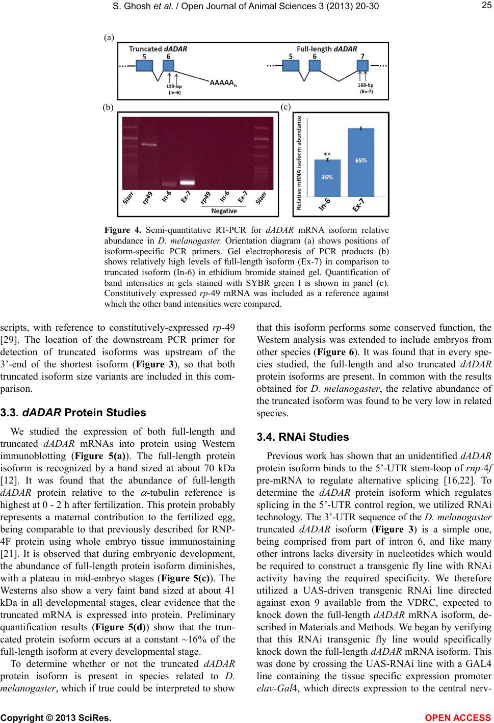
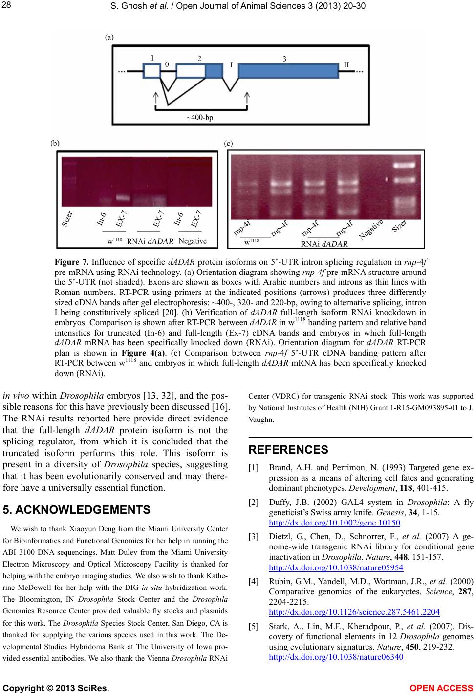
S. Ghosh et al. / Open Journal of Animal Sciences 3 (2013) 20-30 29
[6] Cooper, T.A., Wan, L. and Dreyfuss, G. (2009) RNA and
disease. Cell, 136, 777-793.
http://dx.doi.org/10.1016/j.cell.2009.02.011
[7] Bass, B.L. (2002) RNA editing by adenosine deaminases
that act on RNA. Annual Review of Biochemistry, 71,
817-846.
http://dx.doi.org/10.1146/annurev.biochem.71.110601.13
5501
[8] Nishikura, K. (2009) Functions and regulation of RNA
editing by ADAR deaminases. Annual Review of Bio-
chemistry, 79, 321-349.
http://dx.doi.org/10.1146/annurev-biochem-060208-1052
51
[9] Jepson, J.E.C. and Reenan, R.A. (2008) RNA editing in
regulating gene expression in the brain. Biochimica et
Biophysica Acta, 1779, 459-470.
http://dx.doi.org/10.1016/j.bbagrm.2007.11.009
[10] Petschek, J.P., Mermer, M.J., Scheckelhoff, M.R., Simone,
A.A. and Vaughn, J.C. (1996) RNA editing in Drosophila
4f-rnp gene nuclear transcripts by multiple A-to-G con-
versions. Journal of Molecular Biology, 259, 885-890.
http://dx.doi.org/10.1006/jmbi.1996.0365
[11] Petschek, J.P., Scheckelhoff, M.R., Mermer, M.J. and
Vaughn, J.C. (1997) RNA editing and alternative splicing
generate mRNA transcript diversity from the Drosophila
4f-rnp locus. Gene, 204, 267-276.
http://dx.doi.org/10.1016/S0378-1119(97)00465-4
[12] Palladino, M.J., Keegan, L.P., O’Connell, M.A., and Re-
enan, R.A. (2000) dADAR, a Drosophila double-stranded
RNA-specific adenosine deaminase is highly develop-
mentally regulated and is itself a target for RNA editing.
RNA, 6, 1004-1018.
http://dx.doi.org/10.1017/S1355838200000248
[13] Ma, E., Tucker, M.C., Chen, Q. and Haddad, G.G. (2002)
Developmental expression and enzymatic activity of
pre-mRNA deaminase in Drosophila melanogaster. Mo-
lecular Brain Research, 102, 100-104.
http://dx.doi.org/10.1016/S0169-328X(02)00186-9
[14] Hogg, M., Paro, S., Keegan, L.P. and O’Connell, M.A.
(2011) RNA editing by mammalian ADARs. Advances in
Genetics, 73, 87-119.
http://dx.doi.org/10.1016/B978-0-12-380860-8.00003-3
[15] Paro, S., Li, X., O’Connell, M.A. and Keegan, L.P. (2011)
Regulation and functions of ADAR in Drosophila. Cur-
rent Topics in Microbiology and Immunology, 353, 221-
236. http://dx.doi.org/10.1007/82_2011_152
[16] Chen, J., Lakshmi, G.G., Hays, D.L., McDowell, K.M.,
Ma, E. and Vaughn, J.C. (2009) Spatial and temporal ex-
pression of dADAR mRNA and protein isoforms during
embryogenesis in Drosophila melanogaster. Differentia-
tion, 78, 312-320.
http://dx.doi.org/10.1016/j.diff.2009.08.003
[17] Hess, K.A., Simone, A.A. and Petschek, J.P. (1996) Spa-
tial and temporal expression of 4f-rnp gene in Drosophila
melanogaster. Differentiation, 61, 103-111.
http://dx.doi.org/10.1046/j.1432-0436.1996.6120103.x
[18] Bell, M., Schreiner, S., Damianov, A., Reddy, R. and
Bindereif, A. (2002) p110, a novel human U6 snRNP pro-
tein and U4/U6 snRNP recycling factor. The EMBO
Journal, 21, 2724-2735.
http://dx.doi.org/10.1093/emboj/21.11.2724
[19] Bae, E., Reiter, N.J., Bingman, C.A., Kwan, S.S., Lee, D.,
Phillips, G.N., Butcher, S.E., and Brow, D.A. (2007)
Structure and interactions of the first three RNA recogni-
tion motifs of splicing factor Prp24. Journal of Molecular
Biology, 367, 1447-1458.
http://dx.doi.org/10.1016/j.jmb.2007.01.078
[20] Fetherson, R.A., Strock, S.B., White, K.N. and Vaughn,
J.C. (2006) Alternative pre-mRNA splicing in Drosophila
spliceosomal assembly factor RNP-4F during develop-
ment. Gene, 371, 234-245.
http://dx.doi.org/10.1016/j.gene.2005.12.025
[21] Chen, J., Concel, V.J., Bhatla, S., Rajeshwaran, R., Smith,
D.L.H., Varadarajan, M., Backscheider, K.L., Bockrath,
R.A., Petschek, J.P. and Vaughn, J.C. (2007) Alternative
splicing of an rnp-4f mRNA isoform retaining an evolu-
tionarily-conserved 5’-UTR intronic element is develop-
mentally regulated and shown via RNAi to be essential
for normal central nervous system development in Dro-
sophila melanogaster. Gene, 399, 91-104.
http://dx.doi.org/10.1016/j.gene.2007.04.038
[22] Lakshmi, G.G., Ghosh, S., Jones, G.P., Parikh, R., Rawlins,
B.A. and Vaughn, J.C. (2012) An RNA electrophoretic
mobility shift and mutational analysis of rnp-4f 5’-UTR
intron splicing regulatory proteins in Drosophila reveals a
novel new role for a dADAR protein isoform. Gene, 511,
161-168. http://dx.doi.org/10.1016/j.gene.2012.09.088
[23] Peters, N.T., Rohrbach, J.A., Zalewski, B.A., Byrkett,
C.M. and Vaughn, J.C. (2003) RNA editing and regula-
tion of Drosophila 4f-rnp expression by sas-10 antisense
readthrough mRNA transcripts. RNA, 9, 698-710.
http://dx.doi.org/10.1261/rna.2120703
[24] Campos-Ortega, J.A. and Hartenstein, V. (1997) The em-
bryonic development of Drosophila. Springer-Verlag,
Berlin.
[25] Wolff, T. (2000) Histological techniques for the Droso-
phila eye. Part I: Larva and pupa. In: Sullivan, W.,
Ashburner, M. and Hawley, R.S., Eds., Drosophila Pro-
tocols, Cold Spring Harbor Laboratory Press, Cold Spring
Harbor, New York, 201-227.
[26] Ashburner, M., Golic, K.G. and Hawley, R.S. (2005)
Drosophila: A laboratory handbook, second edition. Cold
Spring Harbor Laboratory Press, Cold Spring Harbor,
New York.
[27] Tadros, W., Goldman, A.L., Babak, T., Menzies, F., Vardy,
L., Orr-Weaver, T., Hughes, T.R., Westwood, J.T.,
Smibert, C.A. and Lipshitz, H.D. (2007) SMAUG is a
major regulator of maternal mRNA destabilization in
Drosophila and its translation is activated by the PAN GU
kinase. Developmental Cell, 12, 143-155.
http://dx.doi.org/10.1016/j.devcel.2006.10.005
[28] Graber, J.H., Cantor, C.R., Mohr, S.C. and Smith, T.F.
(1999) In silico detection of control signals: mRNA
3’-end processing sequences in diverse species. Proceed-
ings of the National Academy of Sciences, 96, 14055-
14060. http://dx.doi.org/10.1073/pnas.96.24.14055
[29] O’Connell, P. and Rosbash, M. (1984) Sequence, struc-
ture, and codon preference of the Drosophila ribosomal
Copyright © 2013 SciRes. OPEN ACCESS
S. Ghosh et al. / Open Journal of Animal Sciences 3 (2013) 20-30
Copyright © 2013 SciRes. OPEN ACCESS
30
protein 49 gene. Nuclei c Aci ds Research, 12, 5495-5513.
http://dx.doi.org/10.1093/nar/12.13.5495
[30] Mignone, F., Gissi, C., Liuni, S. and Pesole, G. (2002)
Untranslated regions of mRNAs. Genome Biology, 3,
1-10.
[31] Semotok, J.L., Cooperstock, R.L., Pinder, B.D., Vari, H.K.
and Lipshitz, H.D. (2005) Smaug recruits the CCR4/
POP2/NOT deadenylase complex to trigger maternal
transcript localization in the early Drosophila embryo.
Current Biology, 15, 284-294.
http://dx.doi.org/10.1016/j.cub.2005.01.048
[32] Hanrahan, C.J., Palladino, M.J., Ganetzky, B. and Reenan,
R.A. (2000) RNA editing of the Drosophila para Na+
channel transcript: Evolutionary conservation and devel-
opmental regulation. Genetics, 155, 1149-1160.