 Advances in Bioscience and Biotechnology, 2013, 4, 24-29 ABB http://dx.doi.org/10.4236/abb.2013.411A2004 Published Online November 2013 (http://www.scirp.org/journal/abb/) Chronic exposure to low doses of ozone produces a state of oxidative stress and blood-brain barrier damage in the hippocampus of rat Selva Rivas-Arancibia1*, Luis Fernando Hernández-Zimbrón1, Erika Rodríguez-Martínez1, Gabino Borgonio-Pérez1, Varsha Velumani1, Josefina Durán-Bedolla2 1Departamento de Fisiología, Facultad de Medicina, Universidad Nacional Autónoma de México, México D. F., México 2Centro de Investigación Sobre Enfermedades Infecciosas, Instituto Nacional de Salud Pública, Cuernavaca, México Email: *srivas@unam.mx Received 26 September 2013; revised 25 October 2013; accepted 3 November 2013 Copyright © 2013 Selva Rivas-Arancibia et al. This is an open access article distributed under the Creative Commons Attribution License, which permits unrestricted use, distribution, and reproduction in any medium, provided the original work is properly cited. ABSTRACT Chronic exposure to low doses of ozone similar to a day of high pollution in Mexico City causes a state of oxidative stress. This produces a progressive neu- rodegeneration in hippocampus of rats exposed to the gas. The aim of this study was to analyze the effect of chronic exposure on the changes in the blood-brain barrier in rats exposed to low doses of ozone. Method: each group received one of the following treatments, control group received air without ozone, and groups 2, 3, 4, 5, and 6 received ozone doses of 0.25 ppm for 4 h daily during 7, 15, 30, 60 and 90 days respectively. Each group was processed to inmunohistochemical technique against of the following antibody: blood- brain barrier, guanylyl cyclase, Iba-1, GFAP, NFκ-B, TNF-α. The results show that there is a correlation between the time exposure of ozone and the progres- sive damage, on the blood-brain barrier rupture, fi- nally causing edema of endothelial cell, increase in guanylyl cyclase type 1, thickening of the processes and astrocytes foot, and an increase in the expression of factors NFκ-B and TNF-α at 30 and 60 days of ex- posure to this gas. All the above indicates that the chronic state of oxidative stress causes a neurodegen- eration process, accompanied by disruption of the blood-brain barrier likely to occur in the Alzheimer’s disease. Keywords: Ozone; Oxidative Stress; Blood-Brain Barrier; Neurodegenerative Diseases 1. INTRODUCTION The environmental contamination is a major health prob- lem in the densely populated and industrialized cities [1,2]. The ozone, which is a product of the photochemi- cal air pollution, is the principal contaminant [3]. High levels of air contamination have been associated with an increase and deteriorating of chronic degenerative respi- ratory illness and also cardiopulmonary and neurodegen- erative disease [4-6]. It is well established that an acute as well as chronic inhalation of this gas produces a state of oxidative stress. When a redox imbalance occurs by an acute exposition to high doses of ozone (0.8 - 1.0 ppm) for 4 h, it causes cell damage, accompanied with swelling, inflammation and changes in mitochondrial as in the endoplasmic reticu- lum occurs [6,7]. All these changes are reversible, since the increase in reactive oxygen species (ROS) in the or- ganisms induces the stimulation of antioxidants systems which increases their activities [7]. In addition these sys- tems also have the reparation function that permits to revert the pathological changes that have been found. The changes in the redox balance are also produced dur- ing the respiratory burst by the activation of immune system as a response of an acute defense of the organism against the pathogens [8]. Once the infection has been handed over the balance of the oxide reduction will be recovered. However, the chronic oxidative stress state is present in degenerative diseases; this state is character- ized by a chronic increase of reactive oxygen species (ROS), reactive nitrogen species (RNS), and reactive species of transition metals [9]. In this state, the antioxi- dant systems are unable to revert the oxidative damage and as they are progressing it also alters the intracellular signaling. As well as other physiological regulatory sys- tems such as the loss of regulation of the inflammatory response, this contributes to increase even more the oxi- dative stress state. Also there is a progressive mitochon- *Corresponding author. OPEN ACCESS 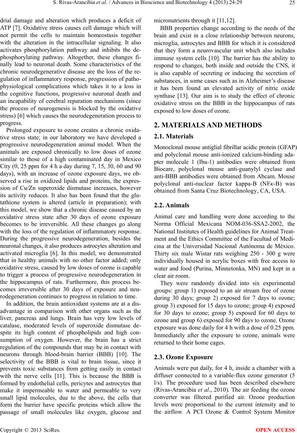 S. Rivas-Arancibia et al. / Advances in Bioscience and Biotechnology 4 (2013) 24-29 25 drial damage and alteration which produces a deficit of ATP [7]. Oxidative stress causes cell damage which will not permit the cells to maintain homeostasis together with the alteration in the intracellular signaling. It also activates phosphorylation pathway and inhibits the de- phosphorylating pathway. Altogether, these changes fi- nally lead to neuronal death. Some characteristics of the chronic neurodegenerative disease are the loss of the re- gulation of inflammatory response, progression of patho- physiological complications which takes it to a loss in the cognitive functions, progressive neuronal death and an incapability of cerebral reparation mechanisms (since the process of neurogenesis is blocked by the oxidative stress) [6] which causes the neurodegeneration process to progress. Prolonged exposure to ozone creates a chronic oxida- tive stress state; in our laboratory we have developed a progressive neurodegeneration animal model. When the animals are exposed chronically to low doses of ozone similar to those of a high contaminated day in Mexico City (0, 25 ppm for 4 h a day during 7, 15, 30, 60 and 90 days), with an increase of ozone exposure days, we ob- served a rise in oxidized lipids and proteins, the expres- sion of Cu/Zn superoxide dismutase increases, however its activity reduces. It also has been found that the glu- tathione system is altered (article in preparation); with this model, we show that a chronic disease caused by an oxidative stress state after 30 days of ozone exposure becomes to be irreversible. All these changes go along with the loss of the regulation of inflammatory response. During the progressive neurodegeneration, besides the neuronal changes, it also produces astrocytes alteration and activated microglia [6]. In this model, we demonstrated that in healthy animals with no other factor added; only oxidative stress, caused by low doses of ozone is capable to trigger a process of progressive neurodegeneration in the hippocampus of rats. Furthermore, this process be- comes irreversible after 30 days of exposure and neu- rodegeneration continues to progress in relation to time. In addition, the brain antioxidant systems are at a dis- advantage in comparison with other organs such as the liver, pancreas and lungs. Brain has very low levels of catalase, moderated levels of superoxide dismutase de- spite its high content of phospholipids and high con- sumption of oxygen. However, the brain has a strict regulation of the compounds that may be in contact with neurons through blood-brain barrier (BBB) [10]. The selectivity of the BBB is vital to brain tissue, since it prevents toxic substances from getting easily in contact with the nerve cells [11]. This is because the BBB is formed by endothelial cells, pericytes and astrocytes that make it impermeable to water and permeable to very small lipid molecules, due to the above, the cells that form the barrier have specific proteins which allow the passage of small molecules like oxygen, glucose and micronutrients through it [11,12]. BBB properties change according to the needs of the brain and exist in a close relationship between neurons, microglia, astrocytes and BBB for which it is considered that they form a neurovascular unit which also includes immune system cells [10]. The barrier has the ability to respond to changes, both inside and outside the CNS, it is also capable of secreting or inducing the secretion of substances, in some cases such as in Alzheimer’s disease it has been found an elevated activity of nitric oxide synthase [13]. Our aim is to study the effect of chronic oxidative stress on the BBB in the hippocampus of rats exposed to low doses of ozone. 2. MATERIALS AND METHODS 2.1. Materials Monoclonal mouse antiglial fibrillar acidic protein (GFAP) and polyclonal mouse anti-ionized calcium-binding ada- pter molecule 1 (Iba-1) antibodies were obtained from Biocare, polyclonal mouse anti-guanylyl cyclase and anti-BBB antibodies were obtained from Abcam. Mouse polyclonal anti-nuclear factor kappa-B (NFκ-B) was obtained from Santa Cruz Biotechnology, CA, USA. 2.2. Animals Animal care and handling were done according to the Norma Official Mexicana NOM-036-SSA2-2002, the National Institutes of Health guidelines for Animal Treat- ment and the Ethics Committee of the Facultad of Medi- cina at the Universidad Nacional Autónoma de México. Thirty six male Wistar rats weighing 250 - 300 g were individually housed in acrylic boxes with free access to water and food (Purina, Minnetonka, MN) and kept in a clear air room. They were randomly divided into six experimental groups: group 1) exposed to an air stream free of ozone during 30 days; group 2) exposed for 7 days to ozone; group 3) exposed for 15 days to ozone; group 4) exposed for 30 days to ozone; group 5) exposed for 60 days to ozone and group 6) exposed for 90 days to ozone. Ozone exposure was done daily for 4 h with a dose of 0.25 ppm. Immediately after the exposure to ozone, animals were returned to their home cages. 2.3. Ozone Exposure Animals were put daily, for 4 h, inside a chamber with a diffuser connected to a variable-flux ozone generator (5 l/s). The procedure used has been described elsewhere (Rivas-Arancibia et al., 2010). The air feeding the ozone converter was filtered purified air. Ozone production levels were proportional to the current intensity and to the airflow. A PCI Ozone & Control System Monitor Copyright © 2013 SciRes. OPEN ACCESS  S. Rivas-Arancibia et al. / Advances in Bioscience and Biotechnology 4 (2013) 24-29 26 (PCI Ozone & Control Systems, West Caldwell, NJ) was used to measure the ozone concentration inside the chamber during the experiment and to keep the ozone concentration constant. Air exposure: The same chamber was used for treating the control group where a flow of ozone-free purified air was used. 2.4. Immunohistochemistry Techniques After the completions of the treatments, animals from each group were anesthetized with sodium pentobarbital (50 mg/kg ip) and intracardially perfused with 4% para- formaldehyde (Sigma-Aldrich, St Louis, MO). In 0.1M phosphate buffer (PB) pH 7.4 (Tecsiquim, México), the brains were postfixed with 10% formaldehyde for 24 h and embedded in paraffin (Merck, Darmstadt, Germany). Sagittal brain slices containing the hippocampus of these animals were cut at 5 μm on a microtome, mounted on slides, and processed for immunohistochemistry. The immunohistochemistry for GFAP, NFκ-B, Iba-1, Guany- lyl cyclase, tumor necrosis factor α (TNF-α) and BBB was done as follows: Slices of each brain containing the hippocampus had the paraffin removed, treated with heat-retrieval solution (Biocare Medical, Concord, CA), and put into an electric pressure cooker (Decloaking Chamber; Biocare Medical) for 5 min. After being washed with distilled water and treated with hydrogen peroxide (J.T. Baker) (diluted 1:5) for 5 min, the slices were rinsed again and treated with a blocking reagent (Background Sniper; Biocare Medical) for 10 min. They were washed with PBS (Merck), pH 7.4, and incubated overnight at 4 C with anti-GFAP (purified monoclonal mouse antibody, diluted 1:200; Biocare), anti-NFκ-B (purified polyclonal mouse antibody, diluted 1:200), anti-Iba-1 (purified poly- clonal goat antibody, diluted 1:300), anti-guanylyl cy- clase (purified polyclonal mouse antibody, diluted 1:200), anti-TNF-α or anti-BBB (purified monoclonal mouse antibody, diluted 1:200). Sections were rinsed with PBS and treated with a sec- ondary antibody using Trekkie Universal Link (Starr Trek Universal HRP Detection; Biocare Medical) for 1 h. The sections were later washed with PBS and then treated with Trekavidin-HRP Label (Starr Trek Universal HRP Detection; Biocare Medical) for 30 min or the bound antibody was visualized using 3, 3-diaminoben- zidine (DAB Substrate Kit; ScyTek, Logan, UT) as the chromogen. The slices were washed with distilled water and contrasted with hematoxylin buffer solution (ScyTek, Concord, CA.). For double immunofluorescence, fol- lowing primary antibody incubation the sections were washed with TBS and incubated with the appropriate Alexa, 488, or 594-conjugated secondary antibody (Invi- trogen). Representative brain slices from each group were processed in parallel. After cover slipping with Entellan (F/550 ml; Merck), the slices were examined with an Olympus BX41 Microscope (Olympus, Japan) using a 100× magnification and photographed (Evolution VF-F- CLR-12, Media Cybernetics camera; Bethesda, MD). 3. RESULTS 3.1. Double Fluorescent Immunohistochemistry against Blood-Brain Barrier and Guanylyl Cyclase The results show that the blood-brain barrier damage increases with the time of exposure to ozone as shown in Figure 1. We can also observe alterations in the endothe- lial cells that are expressing guanylyl cyclase. There is an increase in the expression of this enzyme after sixty days of exposure as well as an increase in endothelial cells volume and at 90 days of exposure to ozone, BBB and endothelial cells are completely destroyed. 3.2. Double Fluorescent Immunohistochemistry against Astrocytes and Microglia We found an increase in the immunoreactivity of Iba-1 and GFAP protein. The results indicate the activation of both microglia and astrocytes with respect to exposure time. 3.3. Immunohistochemistry against astrocytes, NFκ-B and TNF-α The results show an increase in the thickness of the Figure 1. Effects of ozone treatment on Blood Brain Barrier (green) and Guanylyl cyclase (red) double immunofluorescence in the dentate gyrus of rats. Photomicrographs show not mor- phological alterations of endothelial cells and blood brain bar- rier (arrows) in control animals (A) and show s morphological alterations of endothelial cells and blood brain barrier (arrows) getting different times of ozone administration in the dentate gyrus of rats treated with air only (A), 7 days of ozone expo- sure (B),15 days of ozone exposure (C), 30 days of ozone ex- posure (D), 60 days of ozone exposure (E), and 90 days of ozone exposure (F). Magnification 100×, n = 6 per group. Copyright © 2013 SciRes. OPEN ACCESS 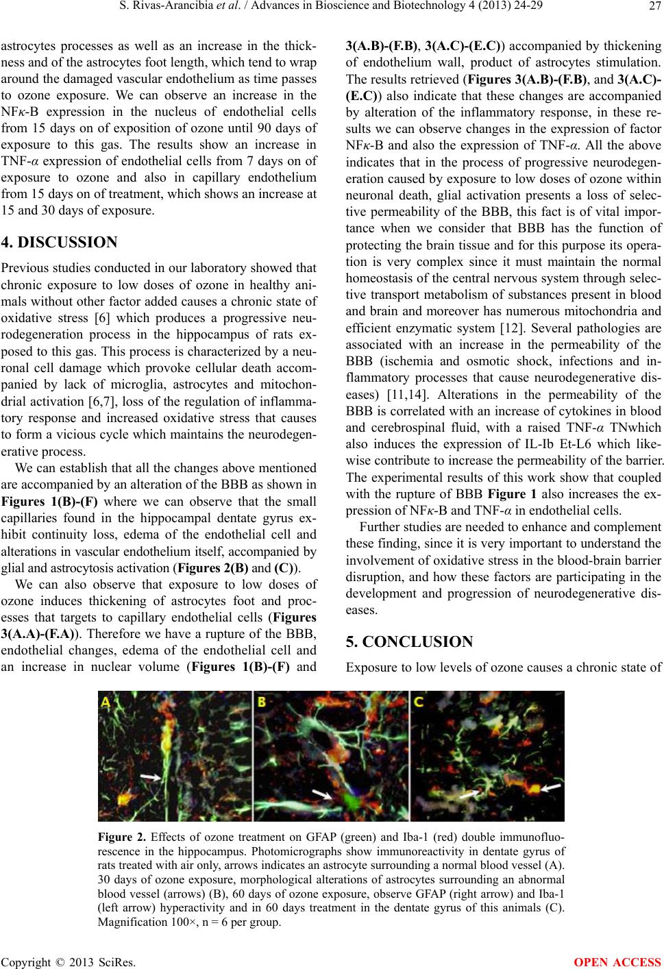 S. Rivas-Arancibia et al. / Advances in Bioscience and Biotechnology 4 (2013) 24-29 Copyright © 2013 SciRes. 27 3(A.B)-(F.B), 3(A.C)-(E.C)) accompanied by thickening of endothelium wall, product of astrocytes stimulation. The results retrieved (Figures 3(A.B)-(F.B), and 3(A.C)- (E.C)) also indicate that these changes are accompanied by alteration of the inflammatory response, in these re- sults we can observe changes in the expression of factor NFκ-B and also the expression of TNF-α. All the above indicates that in the process of progressive neurodegen- eration caused by exposure to low doses of ozone within neuronal death, glial activation presents a loss of selec- tive permeability of the BBB, this fact is of vital impor- tance when we consider that BBB has the function of protecting the brain tissue and for this purpose its opera- tion is very complex since it must maintain the normal homeostasis of the central nervous system through selec- tive transport metabolism of substances present in blood and brain and moreover has numerous mitochondria and efficient enzymatic system [12]. Several pathologies are associated with an increase in the permeability of the BBB (ischemia and osmotic shock, infections and in- flammatory processes that cause neurodegenerative dis- eases) [11,14]. Alterations in the permeability of the BBB is correlated with an increase of cytokines in blood and cerebrospinal fluid, with a raised TNF-α TNwhich also induces the expression of IL-Ib Et-L6 which like- wise contribute to increase the permeability of the barrier. The experimental results of this work show that coupled with the rupture of BBB Figure 1 also increases the ex- pression of NFκ-B and TNF-α in endothelial cells. astrocytes processes as well as an increase in the thick- ness and of the astrocytes foot length, which tend to wrap around the damaged vascular endothelium as time passes to ozone exposure. We can observe an increase in the NFκ-B expression in the nucleus of endothelial cells from 15 days on of exposition of ozone until 90 days of exposure to this gas. The results show an increase in TNF-α expression of endothelial cells from 7 days on of exposure to ozone and also in capillary endothelium from 15 days on of treatment, which shows an increase at 15 and 30 days of exposure. 4. DISCUSSION Previous studies conducted in our laboratory showed that chronic exposure to low doses of ozone in healthy ani- mals without other factor added causes a chronic state of oxidative stress [6] which produces a progressive neu- rodegeneration process in the hippocampus of rats ex- posed to this gas. This process is characterized by a neu- ronal cell damage which provoke cellular death accom- panied by lack of microglia, astrocytes and mitochon- drial activation [6,7], loss of the regulation of inflamma- tory response and increased oxidative stress that causes to form a vicious cycle which maintains the neurodegen- erative process. We can establish that all the changes above mentioned are accompanied by an alteration of the BBB as shown in Figures 1(B)-(F) where we can observe that the small capillaries found in the hippocampal dentate gyrus ex- hibit continuity loss, edema of the endothelial cell and alterations in vascular endothelium itself, accompanied by glial and astrocytosis activation (Figures 2(B) and (C)). Further studies are needed to enhance and complement these finding, since it is very important to understand the involvement of oxidative stress in the blood-brain barrier disruption, and how these factors are participating in the development and progression of neurodegenerative dis- eases. We can also observe that exposure to low doses of ozone induces thickening of astrocytes foot and proc- esses that targets to capillary endothelial cells (Figures 3(A.A)-(F.A)). Therefore we have a rupture of the BBB, endothelial changes, edema of the endothelial cell and an increase in nuclear volume (Figures 1(B)-(F) and 5. CONCLUSION Exposure to low levels of ozone causes a chronic state of Figure 2. Effects of ozone treatment on GFAP (green) and Iba-1 (red) double immunofluo- rescence in the hippocampus. Photomicrographs show immunoreactivity in dentate gyrus of rats treated with air only, arrows indicates an astrocyte surrounding a normal blood vessel (A). 30 days of ozone exposure, morphological alterations of astrocytes surrounding an abnormal blood vessel (arrows) (B), 60 days of ozone exposure, observe GFAP (right arrow) and Iba-1 (left arrow) hyperactivity and in 60 days treatment in the dentate gyrus of this animals (C). Magnification 100×, n = 6 per group. OPEN ACCESS 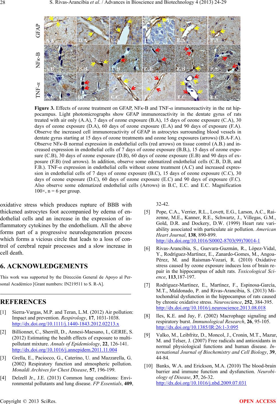 S. Rivas-Arancibia et al. / Advances in Bioscience and Biotechnology 4 (2013) 24-29 28 TNF-α NF -B GFAP Figure 3. Effects of ozone treatment on GFAP, NFκ-B and TNF-α immunoreactivity in the rat hip- pocampus. Light photomicrographs show GFAP immunoreactivity in the dentate gyrus of rats treated with air only (A.A), 7 days of ozone exposure (B.A), 15 days of ozone exposure (C.A), 30 days of ozone exposure (D.A), 60 days of ozone exposure (E.A) and 90 days of exposure (F.A). Observe the increased cell immunoreactivity of GFAP in astrocytes surrounding blood vessels in dentate gyrus starting at 15 days of ozone treatments and ozone long exposures (arrows) (B.A-F.A). Observe NFκ-B normal expression in endothelial cells (red arrows) on tissue control (A.B.) and in- creased expression in endothelial cells of 7 days of ozone exposure (B.B,), 15 days of ozone expo- sure (C.B), 30 days of ozone exposure (D.B), 60 days of ozone exposure (E.B) and 90 days of ex- posure (F.B) (red arrows). In addition, observe some edematized endothelial cells (C.B, D.B, and F.B.). TNF-α expression in endothelial cells without ozone treatment (A.C) and increased expres- sion in endothelial cells of 7 days of ozone exposure (B.C), 15 days of ozone exposure (C.C), 30 days of ozone exposure (D.C), 60 days of ozone exposure (E.C) and 90 days of exposure (F.C). Also observe some edematized endothelial cells (Arrows) in B.C, E.C. and E.C. Magnification 100×, n = 6 per group. oxidative stress which produces rupture of BBB with thickened astrocytes foot accompanied by edema of en- dothelial cells and an increase in the expression of in- flammatory cytokines by the endothelium. All the above forms part of a progressive neurodegeneration process which forms a vicious circle that leads to a loss of con- trol of cerebral repair processes and a slow increase in cell death. 6. ACKNOWLEDGEMENTS This work was supported by the Dirección General de Apoyo al Per- sonal Académico [Grant numbers: IN219511 to S. R-A]. REFERENCES [1] Sierra-Vargas, M.P. and Teran, L.M. (2012) Air pollution: Impact and prevention. Respirology, 17, 1031-1038. http://dx.doi.org/10.1111/j.1440-1843.2012.02213.x [2] Billionnet, C., Sherrill, D., Annesi-Maesano, I., GERIE, S. (2012) Estimating the health effects of exposure to multi- pollutant mixture. Annals of Epidemiology, 22, 126-141. http://dx.doi.org/10.1016/j.annepidem.2011.11.004 [3] Grella, E., Paciocco, G., Caterino, U. and Mazzarella, G. (2002) Respiratory function and atmospheric pollution. Monaldi Archives for Chest Disease, 57, 196-199. [4] Delzell Jr., J.E. (2013) Common lung conditions: Envi- ronmental pollutants and lung disease. FP Essentials, 409, 32-42. [5] Pope, C.A., Verrier, R.L., Lovett, E.G., Larson, A.C., Rai- zenne, M.E., Kanner, R.E., Schwartz, J., Villegas, G.M., Gold, D.R. and Dockery, D.W. (1999) Heart rate vari- ability associated with particulate air pollution. American Heart Journal, 138, 890-899. http://dx.doi.org/10.1016/S0002-8703(99)70014-1 [6] Rivas-Arancibia, S., Guevara-Guzmán, R., López-Vidal, Y., Rodríguez-Martínez, E., Zanardo-Gomes, M., Angoa- Pérez, M. and Raisman-Vozari, R. (2010) Oxidative stress caused by ozone exposure induces loss of brain re- pair in the hippocampus of adult rats. Toxicological Sci- ence, 113,187-197. [7] Rodríguez-Martínez, E., Martínez, F., Espinosa-García, M.T., Maldonado, P. and Rivas-Arancibia, S. (2013) Mi- tochondrial dysfunction in the hippocampus of rats caused by chronic oxidative stress. Neuroscience, 252, 384-395. http://dx.doi.org/10.1016/j.neuroscience.2013.08.018 [8] Iles, K.E. and Jay, F. (2002) Macrophage signaling and respiratory burst. Immunological Research, 26, 95-105. http://dx.doi.org/10.1385/IR:26:1-3:095 [9] Valko, M., Leibfritz, D., Moncol, J., Cronin, M.T., Mazur, M. and Telser, J. (2007) Free radicals and antioxidants in normal physiological functions and human disease. In- ternational Journal of Biochemistry and Cell Biology, 39, 44-84. [10] Banks, W.A. and Erickson, M.A. (2010) The blood-brain barrier and immune function and dysfunction. Neurobi- ology of Disease, 37, 26-32. http://dx.doi.org/10.1016/j.nbd.2009.07.031 Copyright © 2013 SciRes. OPEN ACCESS  S. Rivas-Arancibia et al. / Advances in Bioscience and Biotechnology 4 (2013) 24-29 29 [11] Stolp, H.B., Liddelow, S.A., Sá-Pereira, I., Dziegielewska, K.M. and Saunders, N.R. (2013) Immune responses at brain barriers and implications for brain development and neurological function in later life. Frontiers in Integrative Neuroscience, 7, 61. http://dx.doi.org/10.3389/fnint.2013.00061 [12] Enciu, A.M., Gherghiceanu, M. and Popescu, B.O. (2013) Triggers and effectors of oxidative stress at blood-brain barrier level: Relevance for brain ageing and neurode- generation. Oxidative Medicine and Cellular Longevity, 2013, 297512. http://dx.doi.org/10.1155/2013/297512 [13] Dorheim, M.A., Tracey. W.R., Pollock, J.S. and Gram- mas, P. (1994) Nitric oxide synthase activity is elevated in brain microvessels in Alzheimer’s disease. Biochemi- cal and Biophysical Research Communications, 30, 659- 665. http://dx.doi.org/10.1006/bbrc.1994.2716 [14] Erickson, M.A. and Banks, W.A. (2013) Blood-brain bar- rier dysfunction as a cause and consequence of Alz- heimer’s disease. Journal of Cerebral Blood Flow & Me- tabolism, 33, 1500-1513. http://dx.doi.org/10.1038/jcbfm.2013.135 Copyright © 2013 SciRes. OPEN ACCESS
|