Journal of Cancer Therapy, 2013, 4, 1426-1428
Published Online November 2013 (http://www.scirp.org/journal/jct)
http://dx.doi.org/10.4236/jct.2013.49169
Open Access JCT
Do All Prostate Cancers Behave the Same?
Dissanayake Thusitha1*, Arze Elizabeth2, Rogers Mailien3
1Department of Internal Medicine, James H. Quillen College of Medicine, East Tennessee State University, Johnson City, USA;
2Department of Pathology, James H. Quillen College of Medicine, East Tennessee State University, Johnson City, USA; 3Division of
Hematology-Oncology, James H. Quillen College of Medicine, East Tennessee State University, Johnson City, USA.
Email: *thusithard007@gmail.com
Received August 27th, 2013; revised September 25th, 2013; accepted October 3rd, 2013
Copyright © 2013 Dissanayake Thusitha et al. This is an open access article distributed under the Creative Commons Attribution
License, which permits unrestricted use, distribution, and reproduction in any medium, provided the original work is properly cited.
ABSTRACT
Two different immunohistochemical types suggestive of Large Cell Neuroendocrine (NE) carcinoma and Adenocarci-
noma in a patient with known diffusely metastatic, ho rmone refractory prostate carcinoma are rarities. Interestingly, our
patient had documented history of exposure to Agent Orange during his time of service. The use of routinely used im-
munohistochemical stains for pathological diagnosis was a challenge in this case, though throughout his disease course,
the diagnosis was confirmed as Adenocarcinoma of prostate with biopsies from all various sites of metastases. Systemic
chemotherapy has been historically suboptimal in management of aggressively behaved prostate carcinomas. Finding
any association of Agent Orange as a causative etiology and improving diagnosis and management of such aggressive
hormone refractory prostate carcinoma need further investigations.
Keywords: Large Cell Neuroendocrine Carcinoma; Adenocarcinoma of Prostate; Hormone Refractory Prostate
Carcinoma; Agent Orange
1. Introduction
A 62-year-old white male was diagnosed with metastatic
hormone refractory adenocarcinoma of prostate and was
treated with luteinizing hormone releasing hormone ago-
nist. The patient gave a history of melanoma and remote
history of tobacco abuse. Several months after treatment
was initiated, the patient was presented to the emergency
room for stroke like symptoms and was found to have
brain metastases secondary to his prostate.
Further workup revealed a mediastinal lymph node
wi th inconclusive biopsy result for a primary though adeno-
carcinoma was primarily suspected.
We present an interesting yet complicated case raising
the question of prostate cancer with exposure to Agent
Orange, to which our patient was exposed during the
Vietnam War.
2. Case Report and Discussion
A Vietnam veteran was originally diagnosed with Stage
IV Prostate Adenocarcinoma July 2011 when he pre-
sented with new onset urinary retention.
He underwent cystoscopy July 26th, 2011 and 4 out of
4 core biopsies were reported as adenocarcinoma of
prostate with Gleason score of 8.
His bone scan showed extensive bony metastases.
At the time of diagnosis, his PSA was only mildly
elevated at 5.66. He was started on an drogen depriv ation
therapy (ADT) with Zoladex in jections.
In November 2011, patient presented to VA ER with
vomiting and left sided weakness. On suspicion of cere-
bral vascular accident, CT scanning revealed a 4-cm right
frontal white matter centrally necrotic mass with sur-
rounding vasogenic edema. He was transferred to local
level 4 tertiary care center and brain biopsy performed.
Due to rarity of adenocarcinoma of prostate metasta-
sizing to the brain, search for more common sources
were sought. Further investigation proved his history of
melanoma was actually squamous cell but due to history
of smoking, lung primary was also a possibility.
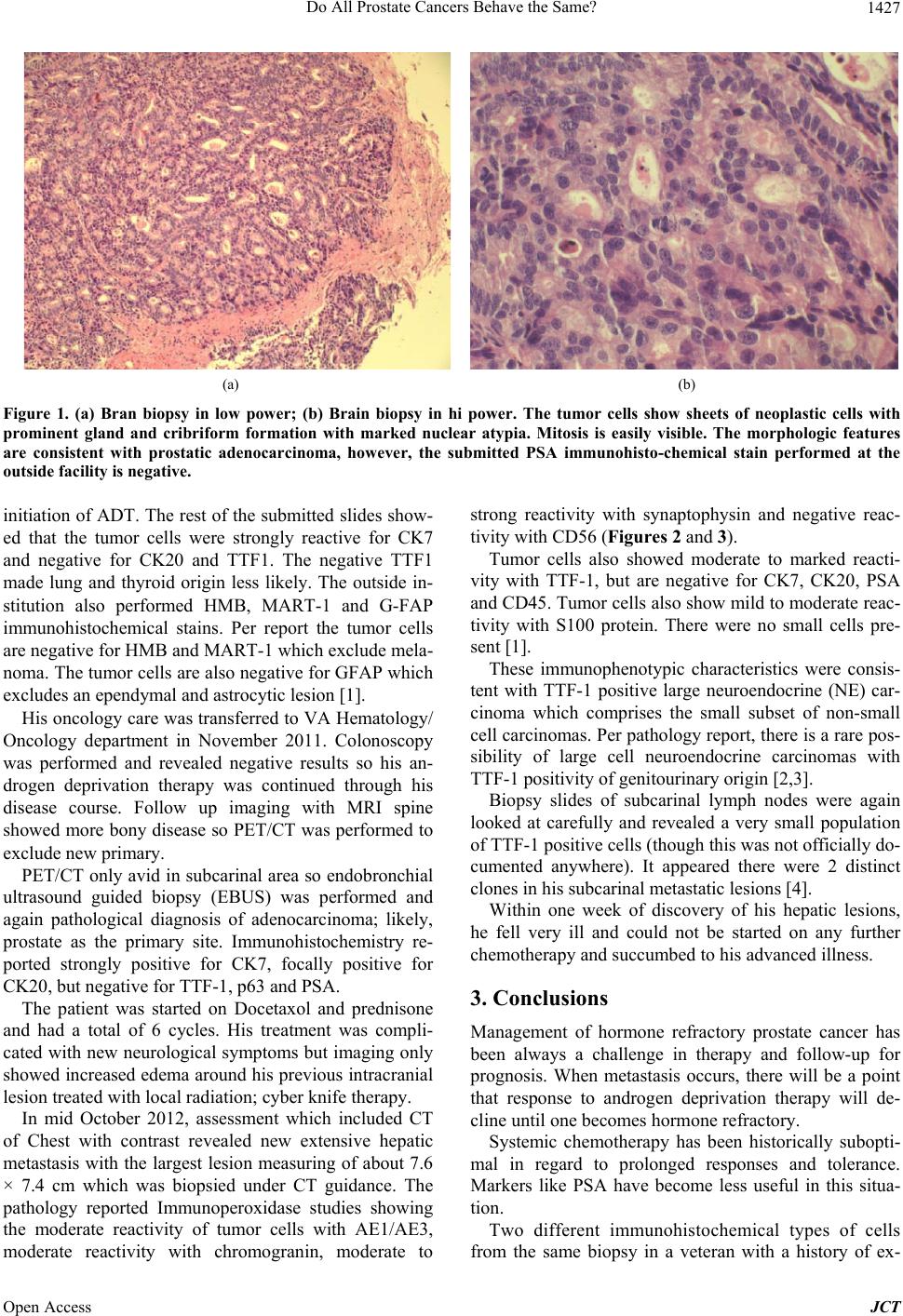
Pathology of brain biopsies reported as tumor cells
showing sheets of neoplastic cells with prominent gland
and cruciform formation with marked nuclear atypia with
easily visible mitosis. The morphological features were
consistent with prostate adenocarcinoma per pathology
report (Figures 1(a) and (b)).
The submitted PSA immunohistochemical stains at the
outside facility were negative which is common after
*Corresponding aut hor.
Do All Prostate Cancers Behave the Same? 1427
(a) (b)
Figure 1. (a) Bran biopsy in low power; (b) Brain biopsy in hi power. The tumor cells show sheets of neoplastic cells with
prominent gland and cribriform formation with marked nuclear atypia. Mitosis is easily visible. The morphologic features
are consistent with prostatic adenocarcinoma, however, the submitted PSA immunohisto-chemical stain performed at the
outside facility is negative.
initiation of ADT. The rest of the submitted slides show-
ed that the tumor cells were strongly reactive for CK7
and negative for CK20 and TTF1. The negative TTF1
made lung and thyroid origin less likely. The outside in-
stitution also performed HMB, MART-1 and G-FAP
immunohistochemical stains. Per report the tumor cells
are negative for HMB and MART-1 which exclude mela-
noma. The tumor cells are also negative for GFAP which
excludes an ependymal and astrocytic lesion [1].
His oncology care was transferred to VA Hematology/
Oncology department in November 2011. Colonoscopy
was performed and revealed negative results so his an-
drogen deprivation therapy was continued through his
disease course. Follow up imaging with MRI spine
showed more bony disease so PET/CT was performed to
exclude new primary.
PET/CT only avid in subcarinal area so endobronchial
ultrasound guided biopsy (EBUS) was performed and
again pathological diagnosis of adenocarcinoma; likely,
prostate as the primary site. Immunohistochemistry re-
ported strongly positive for CK7, focally positive for
CK20, but negative for TTF-1, p63 and PSA.
The patient was started on Docetaxol and prednisone
and had a total of 6 cycles. His treatment was compli-
cated with new neurological symptoms but imaging only
showed increased edema around his previous intracranial
lesion treated with local radiation; cyber knife therapy.
In mid October 2012, assessment which included CT
of Chest with contrast revealed new extensive hepatic
metastasis with the largest lesion measuring of about 7.6
× 7.4 cm which was biopsied under CT guidance. The
pathology reported Immunoperoxidase studies showing
the moderate reactivity of tumor cells with AE1/AE3,
moderate reactivity with chromogranin, moderate to
strong reactivity with synaptophysin and negative reac-
tivity with CD56 (Figures 2 and 3).
Tumor cells also showed moderate to marked reacti-
vity with TTF-1, but are negative for CK7, CK20, PSA
and CD45. Tumor cells also show mild to moderate reac-
tivity with S100 protein. There were no small cells pre-
sent [1].
These immunophenotypic characteristics were consis-
tent with TTF-1 positive large neuroendocrine (NE) car-
cinoma which comprises the small subset of non-small
cell carcinomas. Per pathology report, there is a rare pos-
sibility of large cell neuroendocrine carcinomas with
TTF-1 positivity of genitour inary origin [2,3].
Biopsy slides of subcarinal lymph nodes were again
looked at carefully and revealed a very small population
of TTF-1 positive cells (though this was no t officially do-
cumented anywhere). It appeared there were 2 distinct
clones in his subcarinal metastatic lesions [4].
Within one week of discovery of his hepatic lesions,
he fell very ill and could not be started on any further
chemotherapy and succumbed to his advanced illness.
3. Conclusions
Management of hormone refractory prostate cancer has
been always a challenge in therapy and follow-up for
prognosis. When metastasis occurs, there will be a point
that response to androgen deprivation therapy will de-
cline until one becomes hormone refractory.
Systemic chemotherapy has been historically subopti-
mal in regard to prolonged responses and tolerance.
Markers like PSA have become less useful in this situa-
tion.
Two different immunohistochemical types of cells
from the same biopsy in a veteran with a history of ex-
Open Access JCT
Do All Prostate Cancers Behave the Same?
1428
(a) (b)
Figure 2. (a) Liver biopsy in low power; (b) Liver biopsy in high power. Sections show fragments of fibrovasculer tissue in-
filtrated by a poorly differentiated carcinoma with zonal coagulation necrosis and moderate desmoplastic reaction. Tumor
cells have variable large nucleoli with coarse chromatin, occasional large nucleoli and scant cytoplasm. Mitotic activity is high,
including atypical mitosis and scattered tumor giant cells.
Fi gur e 3. Prostate biopsy . Four out of four core biopsy speci-
mens contain prostatic adenocarcinoma with a predominant
Gleason Grade of 4 + 4 = 8. There is also presented a focal
area with Gleason Grade 5 adenocarcinoma.
posure to Agent Orange are an unusual presentation.
As with our patient, the aggressiveness of the disease
and use of commonly available immunohistochemical
stains for pathological diagnosis can be a challenge.
Finding an association of Agent Orange as a causative
etiology, and improving diagnosis, and management of
such aggressive hormone refractory prostate carcinoma
need further research.
REFERENCES
[1] O. Kaufmann and M. Dietel, “Expression of Thy roid Tran-
scription Factor-1 in Pulmonary and Extrapulmonary
Small Cell Carcinomas and Other Neuroendocrine Car-
cinomas of Various Primary Sites,” Histopathology, Vol.
36, No. 5, 2000, pp. 415-420.
http://dx.doi.org/10.1046/j.1365-2559.2000.00890.x
[2] R. Mazzucchelli, D. Morichetti, A. Lopez-Beltran, L.
Cheng, M. Scarpelli, Z. Kirkali and R. Montironi, “Neu-
roendocrine Tumours of the Urinary System and Male
Gen ital Organs: Clinical Significance,” BJU International,
Vol. 103, No. 11, 2009, pp. 1465-1470.
http://dx.doi.org/10.1111/j.1464-410X.2009.08451.x
[3] K. Hiroshima, A. Iyoda, T. Shida, K. Shibuya, T. Iizasa,
H. Kishi, T. Tanizawa, T. Fujisawa and Y. Nakatani,
“Distinction of Pulmonary Large Cell Neuroendocrine
Carcinoma from Small Cell Lung Carcinoma: A Mor-
phological, Immunohistochemical, and Molecular Analy-
sis,” Modern Pathology, Vol. 19, No. 10, 2006, pp. 1358-
1368. http://dx.doi.org/10.1038/modpathol.3800659
[4] A. J. Evans, P. A. Humphrey, J. Belani, T. H. van der
Kwast and J. R. Srigley, “Large Cell Neuroendocrine
Carcinoma of Prostate: A Clinicopathologic Summary of
7 Cases of a Rare Manifestation of Advanced Prostate
Cancer,” American Journal of Surgical Pathology, Vol.
30, No. 6, 2006, pp. 684-693.
http://dx.doi.org/10.1097/00000478-200606000-00003
Open Access JCT