 J. Biomedical Science and Engineering, 2013, 6, 1040-1049 JBiSE http://dx.doi.org/10.4236/jbise.2013.611130 Published Online November 2013 (http://www.scirp.org/journal/jbise/) Some pharmacological studies on the methanolic extract of Inula graveolense L. Adnan J. M. Al-Fartosy Department of Chemistry, College of Science, University of Basra, Basra, Iraq Email: dr.adnanfrtosy@yahoo.com Received 25 September 2013; revised 21 October 2013; accepted 31 October 2013 Copyright © 2013 Adnan J. M. Al-Fartosy. This is an open access article distributed under the Creative Commons Attribution Li- cense, which permits unrestricted use, distribution, and reproduction in any medium, provided the original work is properly cited. ABSTRACT Inula graveolens L. is widely used in Iraq for the treatment of rheumatic fever, infant convulsions, toothache, blood sugar, and also to dissolve internal blood clots, and to aid digestion. However, the effi- cacy and mechanisms of action of the plant remain unclear. Therefore, the objective of the present study was to investigate the pharmacological effects of the methanolic extract (MEIG) of this plant belonging to compositae family. Anti-diarrheal and antipyretic ac- tivities of the extract were examined in rats. Anti-in- flammatory and antinociceptive were studied in mice. At the doses of 200 (P < 0.05) and 400 mg/kg body weight (P < 0.01), the extract displayed remarkable an- ti-diarrheal activity, evidence by a reduction in the rate of defecation as well as by retardation of intesti- nal transit of charcoal meal compared to normal sa- line control group, dose dependently similar to lope- ramide (5 mg/kg). The methanolic extract (400 mg/kg) showed a significant (P < 0.01) dose dependent anti- pyretic effect in yeast induced elevation of body tem- perature in experimental rats. The methanolic extract showed significant anti-inflammatory and antinoci- ceptive activity at the dose of 400 mg/kg (P < 0.01) as compared to standard drug diclofenac sodium (50 mg/kg). The extract inhibited paw and ear edema in a dose-related manner. A dose-dependent analgesic ac- tion was obtained against chemical (writhing test) and thermal (hot-plate test) stimuli indicated that antino- ciceptive activity may involve inhibition of pain by pe- ripheral and central mechanisms. Again, the metha- nolic extract (MEIG) was subjected for in vitro pro- tein anti-denaturation using Bovine serum albumin and anti-platelet aggregation of human blood activity. It was observed that the extract showed greater per- centage of inhibition of BSA (P < 0.01) at the highest concentration (400 µg/ml). The extract also showed potential platelet aggregation inhibitory activity in adose-dependent manner. The maximum inhibition was observed at the dose 400 µg/ml (P < 0.01) compar- ed to standard drug commercial heparin (20 µg/ml). Keywords: Pharmacological Activities; Antidiarrhae; Antipyretic; Anti-Inflammatory; Antinociceptive; Anti-Denaturation; Platelet Aggregation; Inula graveolens L. 1. INTRODUCTION Diarrhea is a gastrointestinal disorder, characterized by an increase in stool frequency and change consistency [1]. From a long time ago, plant kingdom played an im- portant role for discovering new drug source. A number of therapeutic drugs were isolated from plant species. For the treatment of diarrhea, medicinal plants are a potential source of antidiarrheal drugs [2]. Due to poor hygiene practices and malnutrition, children in developing coun- tries frequently suffer from various forms of infections which present as fevers [3]. These fevers are often ac- companied by aches and pain which all lead to morbidity and mortality. Herbal medicines are often used as reme- dies in these conditions since as a result of poverty or- thodox medicines may be unaffordable [4]. It is known that a large number of plant species contain various bio- active compounds that may have health-beneficial prop- erties, anti-inflammatory, anti-oxidant and antimicrobial effects, and their preventive and therapeutic use is in- creasing [5]. Pain is a sensorial modality and primarily protective in nature, but often causes discomfort. It is the most important symptom that brings the patient to physi- cian. Analgesics relieve pain as a symptom, without af- fecting its cause, currently available analgesic drugs such as opiates and NSAIDS are not useful in all cases due to their adverse effects [6]. Pain and inflammation are asso- ciated with many pathophysiologies of various clinical OPEN ACCESS  A. J. M. Al-Fartosy / J. Biomedical Science and Engineering 6 (2013) 1040-1049 1041 conditions like arthritis, cancer and vascular diseases. A number of natural products are used in various traditional medical systems to treat relief of symptoms from pain and inflammation [7]. The main cellular components sus- ceptible to damage by free radical and oxidative stress are lipids (peroxidation of unsaturated fatty acids in cell membrane), proteins (denaturation), carbohydrates and nucleic acids [8]. Both inflammation and free radical da- mage are inter-related aspects that influence each other. As said above, proteins are susceptible to undergo dena- turation by formation of free radical and the mechanism of inflammation injury is attributed in part to release of reactive oxygen species from activated neutrophil and macrophages [9]. Plant extracts are an alternative to the currently used anti-platelet agents, because they consti- tute a rich source of bioactive chemicals [10]. Inula gra- veolens L. is widely distributed in Mediterranean region and Middle East to West Pakistan. In Iraq (Basrah and lower Iraq), this plant is well known in Arabic and Eng- lish system as “Shuwaser, Suawaid” and “Strong-smel- lind Inula”, respectively [11]. Information gathered from some herbalists (in Basra governorate, Iraq) that the plant is useful to reduce rheumatic fever, infant convulsions, toothache, blood sugar, and also to dissolve internal blood clots, and to aid digestion. As there is no scientific report on the biological activity of the plant, the present investigation was done to evaluate the possible anti-dia- rrheal, antipyretic, anti-inflammatory, antinociceptive, an- ti-denaturation of protein and anti-platelet aggregation activities of the methanolic extract of Inula graveolens (MEIG). The findings from this work may add to the overall value of the medicinal potential of the plant. 2. MATERIALS AND METHODS 2.1. Chemicals All chemicals were purchased from Sigma-Aldrich Co. (St. Louis, MO), and solvents were from E. Merck (Darm- stadt, Germany). All of the reagents were prepared in deionized distilled water to eliminate the contamination of metal ions. 2.2. Plant Material and Extraction Procedure Inula graveolens L. plant, used in this study, was collected on october 2012 from Abu-Al-Khaseeb region (Southern of Basra), Iraq. The plant was botanically authenticated and voucher specimens 3897 were deposited in the Her- barium of Basra (Iraq, Basra, College of Science, Uni- versity of Basra). A quantity (100 g) of powdered plant was extracted in a Soxhelet apparatus with 80% metha- nol, for 24 h. The methanol extract was filtered and eva- porated to dryness under reduced pressure in a rotary evaporator to afford 9.47 g of dry extract. 2.3. Animals Albino rat (150 - 200 g) of both sex were used to study the anti-diarrhea and antipyretic activity. Healthy albino mice of either sex (20 - 30 g) were used for anti-in- flammatory and antinociceptive activity. The animals were housed in polypropylene cages (five in cage) under a 12 h light/12h dark cycle in a controlled temperature room (25 ± 2˚C). All the animals were acclimatized to the laboratory conditions for a week before use. They had free access to food and water. All studies were carried out by using five groups of six animals (3 males and 3 females). 2.4. Antidiarrheal Study 2.4.1. Caster Oil-Induc e d Diar rhea Test This test was carried out by using the method of [12]. The animals were fasted for 18 h prior to the test. Test, standard samples and control were administered orally 60 min before received of castor oil at a dose of 1 ml/animal (p.o). Then animals were placed in cages lined with adsorbent papers and observed for 4 h for the pres- ence of diarrhea defined as watery (wet), unformed stool. The control group result was considered as 100%. The anti-diarrhea effect was expressed as the average percent inhibition of defecation, which is calculated by the fol- lowing equation: % Inhibition1VtVc100 Where Vt and Vc represent mean number of defeca- tion caused by castor oil in control and standard or test, respectively. Control received normal saline (1 ml/kg, p.o) and loperamide (5 mg/kg, p.o) was used as standard drug. 2.4.2. Gastrointestinal M otility Test This test was performed according to the method previ- ously described using charcoal as a diet marker [13]. The animals were fasted for 18 h prior to the test. Test, stan- dard samples and control were administered orally 60 min before received of castor oil at a dose of 1 ml/animal (p.o). After 60 min of drug administration, all animals were received 1 ml of charcoal meal (10% charcoal in 5% gum acacia) orally. Sixty minute later, all animals were sacrificed, and the intestine was removed without stretching and placed lengthwise on moist filter paper. The gastrointestinal motility was expressed as the ave- rage percent retardation of intestinal transit, which is cal- culated by the following equation: % RetardationBA100 Where B and A represent the average distances (cm) travelled by the charcoal meal from the pylorus to caecum Copyright © 2013 SciRes. OPEN ACCESS  A. J. M. Al-Fartosy / J. Biomedical Science and Engineering 6 (2013) 1040-1049 1042 and total length (cm) of the small intestine, respectively. Control received normal saline (1 ml/kg, p.o) and lop- eramide (5 mg/kg, p.o) was used as standard drug. 2.5. Antipyretic Study The procedure described by [14] was adopted for this study. Fever was induced in the rats by injecting 20% (w/v) suspention of Brewer’s yeast (Saccharomyces cerevisiae) at a dosage of 1 ml/kg body weight subcu- taneously. The rectal temperature of each rat was re- corded by clinical thermometer before and after 18 h of yeast adminstration. Rats that did not show a minimum increase of 0.5˚C in temperature 18 h after yeast injection were discarded. Rectal temperature of all the rats was then recorded at the time of 1, 2 and 3 h after oral ad- ministration of both test and standard drugs. The control group result was considered as 100%. The antipyretic effect was expressed as the average percent reduction in rectal temperature, which is calculated by the following equation: n % ReductionBCBA100 Where B and A represent mean number of rectal tem- perature after and before 18 h of yeast adminstration re- spectively, while Cn represent rectal temperature after 1, 2 and 3 h oral administration of drug. Control received normal saline (1 ml/kg, p.o) and paracetamol (33 mg/kg, p.o) was used as standard drug. 2.6. Anti-Inflammtory Study 2.6.1. Carra geenan-Induced P aw Edema Test The test was determined according to the technique of [15]. After 0.5 h, 0.1 ml of 1% (w/v) carrageenan sus- pension was injected subcutaneously to the plantar sur- face of the left hind paw. The paw volume was measured using a plethysmometer (model 7140, Ugo Basile, Italy), immediately and 0.5, 1, 1.5 and 2 h after drug treatment. The anti-inflammatory effect is expressed as the average percent inhibition of edema, which is calculated by the following equation: % Inhibition1VtVc100 Where Vt and Vc represent the increase in paw vol- umes of mice treated with drug and control, respectively. Control received normal saline (1 ml/kg, p.o) and di- clofenac sodium (10 mg/kg, p.o) was used as standard drug. 2.6.2. Xylene-Induced Ear Edema Test A published method by [16] was adopted. After 0.5 h, 0.03 ml xylene was applied to the anterior and posterior surfaces of the right ear. The left ear was considered as control. Two hours after xylene application, mice were killed and both ears were removed. Circular sections were taken, using a cork borer with a diameter of 7 mm, and weighed. The increase in weight caused by the irritant was measured subtracting the weight of the untreated left ear section from that of the treated right ear sections. The anti-inflammatory effect is expressed as the average per- cent inhibition of writhes, which is calculated by the fol- lowing equation: % Inhibition1VtVc100 Where Vt and Vc represent the average writhes in the drug and control groups, respectively. Control received normal saline (1 ml/kg, p.o) and diclofenac sodium (10 mg/kg, p.o) was used as standard drug. 2.7. Antinociceptive Study 2.7.1. Hot- Pl a te Test The hot plate test was assessed according to the method described by [16], with minor modification. The tem- perature of a metal surface was maintained at 55˚C ± 0.5˚C. Latency to a discomfort reaction (jumping, with- drawal or licking of the paws) was determined before and after drug administration. A cut-off time was 15 sec, to avoid damage of the paw. Reaction time and the type of response were noted using a stopwatch. The latency was recorded before and 0.5, 1, 1.5 and 2 h after oral administration of both test and standard drugs. Average reaction times were then calculated and the percentage variation calculated using following relation: % Inhibition Before treatmentafter treatment1100 Control received normal saline (1 ml/kg, p.o) and di- clofenac sodium (10 mg/kg, p.o) was used as standard drug. 2.7.2. Writhin g Tes t The test was performed as described by [7]. Test, stan- dard samples and control were administered orally 30 min before intraperitoneal administration of 0.7% (v/v) acetic acid (volume of injection 0.1 ml/10g body weight). The mice were placed individually into glass beakers and 5 min were allowed to elapse. The number of stretching or writhing was recorded for the next 10 min. A per- centage reduction in the writhing number was considered evidence for analgesia, which is calculated by the fol- lowing equation: % Inhibition1VtVc100 Where Vt and Vc represent the average number of writhes in the drug and control groups, respectively. Control received normal saline (1 ml/kg, p.o) and di- Copyright © 2013 SciRes. OPEN ACCESS  A. J. M. Al-Fartosy / J. Biomedical Science and Engineering 6 (2013) 1040-1049 Copyright © 2013 SciRes. 1043 clofenac sodium (10 mg/kg, p.o) was used as standard drug. pernatant which contained platelets (i.e. platelet rich pla- sma (PRP)). The remaining blood was centrifuged at 1200 × g for 10 min to obtain another supernatant which did not contained platelets (i.e. platelet poor plasma (PPP)). The cuvettes were incubated at 37˚C for 5 min. The platelet aggregation was initiated by adding 20 µl of 10 µM adenosine di-phosphate (ADP) to 1 ml of PRP. The platelet aggregation was recorded for 5 min at 600 nm. The effect of different concentration (100, 200 and 400 µg/ml) of methanolic extract (MEIG) was studied by incubation with PRP at 37˚C for 5 min before the addition of ADP. The inhibition percentage of maximal aggrega- tion was calculated using the following equation: 2.8. Anti-Denaturation Study This test was carried out by using the method of [17] with slight modification. Briefly, 0.2% w/v bovine serum albumin (BSA) was prepared in Tris-buffer saline. pH was adjusted at 6.8 using glacial acetic acid. Stock solu- tion of 10,000 µg/ml of methanolic extract (MEIG) was prepared by using methanol as a solvent. From this stock solution 3 different concentrations of 100, 200 and 400 µg/ml were prepared by using methanol as a solvent. In Eppendorf tube, 50 µL of the extract and 5 mL of 0.2% (w/v) BSA solution and 50 µL of methanol were added. Test tubes containing the sample mixture were heated at 72˚C for 5 minutes. After cooling for 10 min, the ab- sorbance at 660 nm was measured. The experiment was performed in triplicate. The inhibition percentage of pre- cipitation (denaturation of the protein) was determined on % basis relative to the control using the following formula: % Inhibition1VtVc100 Where Vt and Vc represent the maximal aggregation of the drug and control groups, respectively. Control was the platelet poor plasma (PPP) and commercial heparin (20 µg/ml) was used as standard drug. 2.10. Statistical Analysis % Inhibition1VtVc100 The data were expressed as mean values ± SEM and test- ed with analysis of variance followed by Dunnett’s t-test. P-values < 0.05, 0.01 were considered to be statistically significant. Where Vt and Vc represent the average denaturation of the drug and control groups, respectively. Control was 50 µL of methanol and Ibuprofen (100 µg/ml) was used as standard drug. 3. RESULTS AND DISCUSSION 3.1. Antidiarrheal Study 2.9. Anti-Platelet Aggregation Study The results of this study are presented in Tabl es 1 and 2, respectively. The results revealed that the extract at the doses of 100, 200 and 400 mg/kg, produced a dose de- pendent decrease in the number of faecal matters and de- creased propulsion of charcoal meal of the gastrointesti- nal tract passed by the animals in castor oil-induced A published method by [18] was adopted. Blood was taken collected from healthy human volunteers who have not taken any medication two weeks prior to participa- tion in the study. The blood was mixed with 3.8% (w/v) sodium citrate solution in a ratio of 9:1 and centrifuged at 260 × g for 15 min at 20˚C in order to obtain the su- Table 1. Effect of the methanolic extract of Inula graveolens (MEIG) on castor oil-induced diarrhea in rats. Groups Dose mg/kg No. of faecal dr opping s in 4 h % Inhibition Group I Control 19.2 ± 1.21 - Group II Standard 5.7 ± 0.64** 70.31 Group III 100 13.6 ± 0.73 29.16 Group IV 200 9.4 ± 0.45* 51.04 Group V 400 6.1 ± 0.87** 68.22 N = 6, values are mean ± SEM, *P < 0.05, **P < 0.01, dunnet test as compared to control. Table 2. Effect of the methanolic extract of Inula graveolens (MEIG) on charcoal meal-stimulated gastrointestinal transit. Groups Dose mg/kg Intestinal length (cm) Distance traveled by charcoal (cm) % Retardation Group I Control 89.34 ± 1.31 76.61 ± 1.2 85.75 Group II Standard 84.25 ± 1.12 36.13 ± 1.08** 42.88 Group III 100 84.20 ± 1.26 52.31 ± 1.11 62.12 Group IV 200 86.18 ± 1.72 47.22 ± 1.49* 54.79 Group V 400 84.33 ± 1.34 39.15 ± 1.72** 46.42 N = 6, values are mean ± SEM, *P < 0.05, **P < 0.01, dunnet test as compared to control. OPEN ACCESS  A. J. M. Al-Fartosy / J. Biomedical Science and Engineering 6 (2013) 1040-1049 1044 diarrheal model, respectively. At doses (200 and 400 mg/kg) of the extract, a significant (P < 0.05 and P < 0.01) inhibitions (51.04% and 68.22%, respectively) of characteristic diarrheal feces were observed. The effect of the highest dose of the extract was similar to that of the standard drug, loperamide (5 mg/kg). Diarrhea results from an imbalance between the absorptive and secretory mechanisms in the intestinal tract accompanied by hurry resulting in an excess loss of fluid in the faeces. In some diarrhea the secretory component predominates while other diarrhea is characterized by hypermotility [19]. Cas- tor oil is a triglyceride characterized by a high content of the hydroxylated unsaturated fatty acid ricinoleic acid [20]. After oral ingestion of castor oil, ricinoleic acid is released by lipases in the lumen, and considerable amount of ricinoleic acid are absorbed in the intestine [21]. Pres- ence of ricinoleate in small intestine, the peristaltic activ- ity of small intestine increases as a result of permeability of Na+ and Cl− changed in the intestinal mucosa. Secre- tion of endogenous prostaglandin is also stimulated by ricinoleate [22]. Prostaglandins of the E series are con- sidered to be good diarrheogenic agents in experimental animals as well as in human beings. The inhibitors of prostaglandins biosynthesis are therefore considered to delay castor oil-induced diarrhea. Prostaglandins are as- sociated with changes in the bowel that stimulate diar- rhea. Recent study shows that the laxative effect of rici- noleic acid present in castor oil is due to the induction of contraction of intestinal smooth muscle which is medi- ated by activation of EP3 receptors on intestinal smooth muscle [23]. Many anti-diarrheal agents act by reducing the gastrointestinal motility and/or the secretion. Inhibi- tors of prostaglandin biosynthesis delay castor oil induc- ed diarrhea [12]. Methanolic extract of Inula graveolens (MEIG) exhibit significant anti-diarrheal activity. Earlier reports suggest that anti-diarrheal properties of medicinal plants might be ascribed to tannins, alkaloids, flavonoids, saponin and steroids [24]. The methanolic extract of Inula graveolens (MEIG) contain higher amount of both phe- nolic and flavonoid compounds which may be responsi- ble for its effect [25]. Flavonoids, present in plant extract, are reported to inhibit release of autacoids and prostag- landins, thereby may inhibit motility and secretion in- duced by castor oil [26]. However, the components res- ponsible for the anti-diarrheal activity of Inula graveolens L. are currently unclear. Therefore, further work must be carried out to isolate and identify these components. 3.2. Antipyretic Study The effect of methanolic extract of Inula graveolens L. is illustrated in Tab le 3 . It provoked dose dependent reduc- tion in body temperature when studied at 100, 200 and 400 mg/kg, p.o. during various assessment times (1 - 3 h). Maximum protection (82.14%) was observed after 3 h of drug administration at 400 mg/kg (P < 0.01). Standard drug, paracetamol produced 88.23% protection after 3 h drug treatment 33 mg/kg (p.o). Fever has been recog- nized as a major sign of diseased condition right from the very beginning of human civilization. The febrile re- sponse is coordinated by the central nervous system through endocrine, neurological, immunological and be- havioral mechanisms [27]. The initiation, manifestations and regulation of the febrile response are dependent on the pyrogenic and anti-pyrogenic properties of various exogenous and endogenous substances. There is a gen- eral consensus that fever is caused by a regular rise in body temperature above normal daily, fluctuations origi- nating in conjucation with an elevated thermoregulatory set point [28,29]. The neurons of thermoregulatory sys- tem center are sensitive not only to changes in blood temperature but to cold and warm receptors located in the skin and muscle and thus maintain an appropriate ba- lance between the heat production and loss [30]. In rou- tine practice, non-steroidal anti-inflammatory drugs (NSAID) are commonly prescribed for the treatment of different conditions. It has been suggested that pros- taglandins inhibition are involved in the antipyretic mecha- nisms of NSAID through cyclooxygenase pathway [31]. As a result, it could be speculated that the pharmacolo- gically active components from this extract (MEIG) might contain active principle(s) that demonstrated inhibitory action on cyclooxygenase and thus, they produced anti- pyretic activity by preventing the formation of prostag- landins or by increasing the concentration of the body’s Table 3. Effect of the methanolic extract of Inula graveolens (MEIG) on yeast-induced pyrexia in rats. Rectal temperature (˚C) Rectal temperature after administration of drug ( ˚C) % Reduction Groups Dose mg/kg Normal (A) 18 h after yeast administration (B)1 h (C1) 2 h (C2) 3 h (C3) 1 h (C1) 2 h (C2)3 h (C3) Group I Control 37.87 ± 0.14 38.60 ± 0.12 38.57 ± 0.1538.58 ± 0.1138.55 ± 0.12 - - - Group II Standard 37.62 ± 0.16 38.30 ± 0.17 37.87 ± 0.14** 37.80 ± 0.12** 37.70 ± 0.16** 63.23 73.52 88.23 Group III 100 37.67 ± 0.19 38.22 ± 0.18 38.12 ± 0.1838.02 ± 0.1738.00 ± 0.13 18.18 36.36 40.00 Group IV 200 37.37 ± 0.13 38.35 ± 0.15 38.01 ± 0.1237.85 ± 0.16*37.70 ± 0.20* 34.69 51.02 66.32 Group V 400 37.17 ± 0.20 38.01 ± 0.18 37.62 ± 0.15** 37.47 ± 0.16** 37.32 ± 0.14** 46.42 64.28 82.14 N = 6, values are mean ± SEM, *P < 0.05, **P < 0.01, dunnet test as compared to control. Copyright © 2013 SciRes. OPEN ACCESS 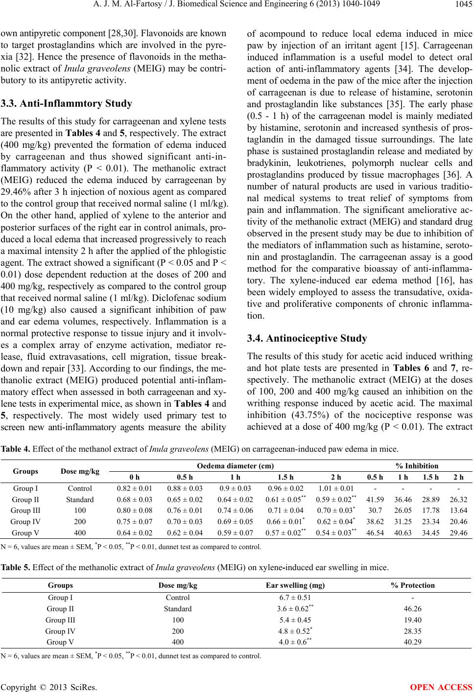 A. J. M. Al-Fartosy / J. Biomedical Science and Engineering 6 (2013) 1040-1049 1045 own antipyretic component [28,30]. Flavonoids are known to target prostaglandins which are involved in the pyre- xia [32]. Hence the presence of flavonoids in the metha- nolic extract of Inula graveolens (MEIG) may be contri- butory to its antipyretic activity. 3.3. Anti-Inflammtory Study The results of this study for carrageenan and xylene tests are presented in Tables 4 and 5, respectively. The extract (400 mg/kg) prevented the formation of edema induced by carrageenan and thus showed significant anti-in- flammatory activity (P < 0.01). The methanolic extract (MEIG) reduced the edema induced by carrageenan by 29.46% after 3 h injection of noxious agent as compared to the control group that received normal saline (1 ml/kg). On the other hand, applied of xylene to the anterior and posterior surfaces of the right ear in control animals, pro- duced a local edema that increased progressively to reach a maximal intensity 2 h after the applied of the phlogistic agent. The extract showed a significant (P < 0.05 and P < 0.01) dose dependent reduction at the doses of 200 and 400 mg/kg, respectively as compared to the control group that received normal saline (1 ml/kg). Diclofenac sodium (10 mg/kg) also caused a significant inhibition of paw and ear edema volumes, respectively. Inflammation is a normal protective response to tissue injury and it involv- es a complex array of enzyme activation, mediator re- lease, fluid extravasations, cell migration, tissue break- down and repair [33]. According to our findings, the me- thanolic extract (MEIG) produced potential anti-inflam- matory effect when assessed in both carrageenan and xy- lene tests in experimental mice, as shown in Tables 4 and 5, respectively. The most widely used primary test to screen new anti-inflammatory agents measure the ability of acompound to reduce local edema induced in mice paw by injection of an irritant agent [15]. Carrageenan induced inflammation is a useful model to detect oral action of anti-inflammatory agents [34]. The develop- ment of oedema in the paw of the mice after the injection of carrageenan is due to release of histamine, serotonin and prostaglandin like substances [35]. The early phase (0.5 - 1 h) of the carrageenan model is mainly mediated by histamine, serotonin and increased synthesis of pros- taglandin in the damaged tissue surroundings. The late phase is sustained prostaglandin release and mediated by bradykinin, leukotrienes, polymorph nuclear cells and prostaglandins produced by tissue macrophages [36]. A number of natural products are used in various traditio- nal medical systems to treat relief of symptoms from pain and inflammation. The significant ameliorative ac- tivity of the methanolic extract (MEIG) and standard drug observed in the present study may be due to inhibition of the mediators of inflammation such as histamine, seroto- nin and prostaglandin. The carrageenan assay is a good method for the comparative bioassay of anti-inflamma- tory. The xylene-induced ear edema method [16], has been widely employed to assess the transudative, oxida- tive and proliferative components of chronic inflamma- tion. 3.4. Antinociceptive Study The results of this study for acetic acid induced writhing and hot plate tests are presented in Tables 6 and 7, re- spectively. The methanolic extract (MEIG) at the doses of 100, 200 and 400 mg/kg caused an inhibition on the writhing response induced by acetic acid. The maximal inhibition (43.75%) of the nociceptive response was achieved at a dose of 400 mg/kg (P < 0.01). The extract Table 4. Effect of the methanol extract of Inula graveolens (MEIG) on carrageenan-induced paw edema in mice. Oedema diameter (cm) % Inhibition Groups Dose mg/kg 0 h 0.5 h 1 h 1.5 h 2 h 0.5 h 1 h 1.5 h2 h Group I Control 0.82 ± 0.01 0.88 ± 0.030.9 ± 0.03 0.96 ± 0.021.01 ± 0.01- - - - Group II Standard 0.68 ± 0.03 0.65 ± 0.020.64 ± 0.020.61 ± 0.05** 0.59 ± 0.02** 41.59 36.46 28.8926.32 Group III 100 0.80 ± 0.08 0.76 ± 0.010.74 ± 0.060.71 ± 0.040.70 ± 0.03*30.7 26.05 17.7813.64 Group IV 200 0.75 ± 0.07 0.70 ± 0.030.69 ± 0.050.66 ± 0.01*0.62 ± 0.04*38.62 31.25 23.3420.46 Group V 400 0.64 ± 0.02 0.62 ± 0.040.59 ± 0.070.57 ± 0.02** 0.54 ± 0.03** 46.54 40.63 34.45 29.46 N = 6, values are mean ± SEM, *P < 0.05, **P < 0.01, dunnet test as compared to control. Table 5. Effect of the methanolic extract of Inula graveolens (MEIG) on xylene-induced ear swelling in mice. Groups Dose mg/kg Ear swelling (mg) % Protection Group I Control 6.7 ± 0.51 - Group II Standard 3.6 ± 0.62** 46.26 Group III 100 5.4 ± 0.45 19.40 Group IV 200 4.8 ± 0.52* 28.35 Group V 400 4.0 ± 0.6** 40.29 N = 6, values are mean ± SEM, *P < 0.05, **P < 0.01, dunnet test as compared to control. Copyright © 2013 SciRes. OPEN ACCESS 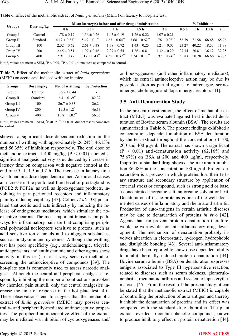 A. J. M. Al-Fartosy / J. Biomedical Science and Engineering 6 (2013) 1040-1049 1046 Table 6. Effect of the methanolic extract of Inula graveolens (MEIG) on latency to hot-plate test. Mean latency(s) before and after drug administration % Inhibition Groups Dose mg/kg 0 h 0.5 h 1 h 1.5 h 2 h 0.5 h 1 h 1.5 h2 h Group I Control 1.78 ± 0.17 1.56 ± 0.26 1.45 ± 0.19 1.26 ± 0.22 1.07 ± 0.21 - - - - Group II Standard 4.12 ± 0.33** 5.49 ± 0.1** 4.63 ± 0.64** 3.68 ± 0.62** 1.76 ± 0.08** 56.79 71.58 68.68 65.76 Group III 100 2.32 ± 0.62 2.61 ± 0.38 1.78 ± 0.72 1.43 ± 0.25 1.21 ± 0.07 23.27 40.22 18.53 11.88 Group IV 200 2.45 ± 0.51 1.97 ± 0.46 2.27 ± 0.54 1.86 ± 0.81 1.32 ± 0.20 27.34 20.81 36.1232.25 Group V 400 2.91 ± 0.47 3.17 ± 0.43** 4.35 ± 0.32** 2.24 ± 0.73** 1.97 ± 0.24** 38.83 50.78 66.66 43.75 N = 6, values are mean ± SEM, *P < 0.05, **P < 0.01, dunnet test as compared to control. Table 7. Effect of the methanolic extract of Inula graveolens (MEIG) on acetic acid-induced writhing in mice. Groups Dose mg/kg No. of writhing % Protection Group I Control 36.2 ± 0.44 - Group II Standard 6.4 ± 0.39** 82.32 Group III 100 26.7 ± 0.33* 26.24 Group IV 200 19.5 ± 1.2** 46.13 Group V 400 15.8 ± 1.02** 56.35 N = 6, values are mean ± SEM, *P<0.05, **P < 0.01, dunnet test as compared to control. showed a significant dose-dependent reduction in the number of writhing with approximately 26.24%, 46.13% and 56.35% of inhibition respectively. The oral dose of methanolic extract at 400 mg/kg (P < 0.01) elicited a significant analgesic activity as evidenced by increase in latency time on comparison with negative control at the end of 0.5, 1, 1.5 and 2 h. The increase in latency time was found in a dose dependent manner. Acetic acid causes an increase in the peritoneal fluid level of prostaglandins (PGE2 & PGF2a) as well as lipooxygenase products, in- volving in part peritoneal receptors and inflammatory pain by inducing capillary [37]. Collier et al. [38] postu- lated that acetic acid acts indirectly by inducing the re- lease of endogenous mediators, which stimulate the no- ciceptive neurons. The most important transmission path- ways for inflammatory pain are that comprising periph- eral polymodal nociceptors sensitive to protons, such as acid sensitive ion channels and to algogen substances, such as bradykinin and cytokines. Although the writhing test has poor specificity (e.g., anticholinergic, tricyclic antidepressants and antihistaminic and other agents show activity in this test), it is a very sensitive method of screening the antinociceptive of compounds [39]. The hot-plate test is commonly used to assess narcotic anal- gesia. Although the central and peripheral analgesics re- spond by inhibiting the number of contractions provoked by chemical pain stimuli, only the central analgesics in- crease the time of response in the hot plate test [40]. These observations tend to suggest that the methanolic extract of Inula graveolens (MEIG) may possess cen- trally- and peripherally-mediated antinociceptive proper- ties. The peripheral antinociceptive effect of the extract may be mediated via inhibition of cyclooxygenases and/ or lipooxygenases (and other inflammatory mediators), which its central antinociceptive action may be due its possible action as partial agonist of adrenergic, seroto- ninergic, cholinergic and dopaminergic recptors [41]. 3.5. Anti-Denaturation Study In the present investigation, the effect of methanolic ex- tract (MEIG) was evaluated against heat induced dena- turation of Bovine serum albumin (BSA). The results are summarized in Table 8. The present findings exhibited a concentration dependent inhibition of BSA denaturation by the test extract throughout the concentration of 100, 200 and 400 µg/ml. The extract has shown a significant (P < 0.01) anti-denaturation activity (62.16% and 75.67%) on BSA at 200 and 400 µg/ml, respectively. Ibuprofen a standard drug showed the maximum inhibi- tion 86.48% at the concentration 100 µg/ml. Protein de- naturation is a process in which proteins loss their terti- ary structure and secondary structure by application of external stress or compound, such as strong acid or base, a concentrated inorganic salt, an organic solvent or heat. Denaturation of tissue proteins is one of the well docu- mented causes of inflammatory and rheumatoid arthritis. Production of auto-antigens in certain arthritic diseases may be due to denaturation of proteins in vivo [42]. Agents that can prevent protein denaturation therefore, would be worthwhile for anti-inflammatory drug devel- opment. The mechanism of denaturation probably in- volves alteration in electrostatic, hydrogen, hydrophobic and disulphide bonding [43]. Several anti-inflammatory drugs have been reported to show dose dependent ability to inhibit thermally induced protein denaturation [44]. Bovine serum albumin (BSA) on denaturation expresses antigens associated to Type III hypersensitive reaction, related to diseases such as serum sickness, glomerulo- nephritis, rheumatoid arthritis and systemic lupus erythe- matosus [45]. From the result of the present study, it can be stated that the methanolic extract (MEIG) is capable of controlling the production of auto antigen and thereby it inhibit the denaturation of proteins and its effect was compared with the standard drug Ibuprofen. The plant extract revealed to contain phenolic compounds, known to produce inhibitory effect on protein denaturetion [44]. Copyright © 2013 SciRes. OPEN ACCESS 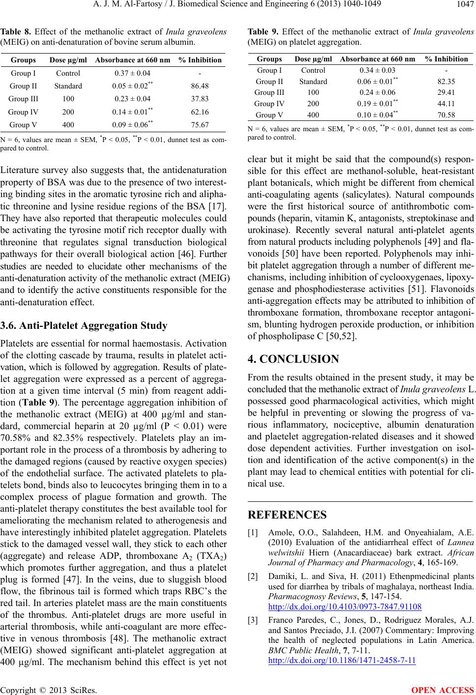 A. J. M. Al-Fartosy / J. Biomedical Science and Engineering 6 (2013) 1040-1049 1047 Table 8. Effect of the methanolic extract of Inula graveolens (MEIG) on anti-denaturation of bovine serum albumin. Groups Dose µg/ml Absorbance at 660 nm % Inhibition Group I Control 0.37 ± 0.04 - Group II Standard 0.05 ± 0.02** 86.48 Group III 100 0.23 ± 0.04 37.83 Group IV 200 0.14 ± 0.01** 62.16 Group V 400 0.09 ± 0.06** 75.67 N = 6, values are mean ± SEM, *P < 0.05, **P < 0.01, dunnet test as com- pared to control. Literature survey also suggests that, the antidenaturation property of BSA was due to the presence of two interest- ing binding sites in the aromatic tyrosine rich and alipha- tic threonine and lysine residue regions of the BSA [17]. They have also reported that therapeutic molecules could be activating the tyrosine motif rich receptor dually with threonine that regulates signal transduction biological pathways for their overall biological action [46]. Further studies are needed to elucidate other mechanisms of the anti-denaturation activity of the methanolic extract (MEIG) and to identify the active constituents responsible for the anti-denaturation effect. 3.6. Anti-Platelet Aggregation Study Platelets are essential for normal haemostasis. Activation of the clotting cascade by trauma, results in platelet acti- vation, which is followed by aggregation. Results of plate- let aggregation were expressed as a percent of aggrega- tion at a given time interval (5 min) from reagent addi- tion (Table 9). The percentage aggregation inhibition of the methanolic extract (MEIG) at 400 µg/ml and stan- dard, commercial heparin at 20 µg/ml (P < 0.01) were 70.58% and 82.35% respectively. Platelets play an im- portant role in the process of a thrombosis by adhering to the damaged regions (caused by reactive oxygen species) of the endothelial surface. The activated platelets to pla- telets bond, binds also to leucocytes bringing them in to a complex process of plague formation and growth. The anti-platelet therapy constitutes the best available tool for ameliorating the mechanism related to atherogenesis and have interestingly inhibited platelet aggregation. Platelets stick to the damaged vessel wall, they stick to each other (aggregate) and release ADP, thromboxane A2 (TXA2) which promotes further aggregation, and thus a platelet plug is formed [47]. In the veins, due to sluggish blood flow, the fibrinous tail is formed which traps RBC’s the red tail. In arteries platelet mass are the main constituents of the thrombus. Anti-platelet drugs are more useful in arterial thrombosis, while anti-coagulant are more effec- tive in venous thrombosis [48]. The methanolic extract (MEIG) showed significant anti-platelet aggregation at 400 µg/ml. The mechanism behind this effect is yet not Table 9. Effect of the methanolic extract of Inula graveolens (MEIG) on platelet aggregation. Groups Dose µg/mlAbsorba nce at 660 nm % Inhibition Group I Control 0.34 ± 0.03 - Group IIStandard 0.06 ± 0.01** 82.35 Group III100 0.24 ± 0.06 29.41 Group IV200 0.19 ± 0.01** 44.11 Group V400 0.10 ± 0.04** 70.58 N = 6, values are mean ± SEM, *P < 0.05, **P < 0.01, dunnet test as com- pared to control. clear but it might be said that the compound(s) respon- sible for this effect are methanol-soluble, heat-resistant plant botanicals, which might be different from chemical anti-coagulating agents (salicylates). Natural compounds were the first historical source of antithrombotic com- pounds (heparin, vitamin K, antagonists, streptokinase and urokinase). Recently several natural anti-platelet agents from natural products including polyphenols [49] and fla- vonoids [50] have been reported. Polyphenols may inhi- bit platelet aggregation through a number of different me- chanisms, including inhibition of cyclooxygenaes, lipoxy- genase and phosphodiesterase activities [51]. Flavonoids anti-aggregation effects may be attributed to inhibition of thromboxane formation, thromboxane receptor antagoni- sm, blunting hydrogen peroxide production, or inhibition of phospholipase C [50,52]. 4. CONCLUSION From the results obtained in the present study, it may be concluded that the methanolic extract of Inula graveolens L. possessed good pharmacological activities, which might be helpful in preventing or slowing the progress of va- rious inflammatory, nociceptive, albumin denaturation and plaetelet aggregation-related diseases and it showed dose dependent activities. Further investgation on isol- tion and identification of the active component(s) in the plant may lead to chemical entities with potential for cli- nical use. REFERENCES [1] Amole, O.O., Salahdeen, H.M. and Onyeahialam, A.E. (2010) Evaluation of the antidiarrheal effect of Lannea welwitshii Hiern (Anacardiaceae) bark extract. African Journal of Pharmacy and Pharmacology, 4, 165-169. [2] Damiki, L. and Siva, H. (2011) Ethenpmedicinal plants used for diarrhea by tribals of maghalaya, northeast India. Pharmacognosy Reviews, 5, 147-154. http://dx.doi.org/10.4103/0973-7847.91108 [3] Franco Paredes, C., Jones, D., Rodriguez Morales, A.J. and Santos Preciado, J.I. (2007) Commentary: Improving the health of neglected populations in Latin America. BMC Public Health, 7, 7-11. http://dx.doi.org/10.1186/1471-2458-7-11 Copyright © 2013 SciRes. OPEN ACCESS  A. J. M. Al-Fartosy / J. Biomedical Science and Engineering 6 (2013) 1040-1049 1048 [4] Soucat, A., Levy Bruhi, D., De Bethune, X., Gbedonou, P., Lamarque, J.P., Bangoura, O., Gandaho, T., Ortiz, C., Kaddar, M. and Knippenberg, R. (1997) Affordability, cost-effectiveness and efficiency of primary health care: The Bamako initiative experience in Benin and Guinea. The International Journal of Health Planning and Man- agement, 12, 81-108. http://dx.doi.org/10.1002/(SICI)1099-1751(199706)12:1+ <S81::AID-HPM467>3.3.CO;2-X [5] Juhas, S., Bujnakova, D., Rehak, P., Cikos, S., Czikkova, S. and Koppel, J. (2008) Anti-inflammatory effects of thyme essential oil in mice. Acta Vet Brno, 77, 327-334. http://dx.doi.org/10.2754/avb200877030327 [6] Mate, G.S., Naikwade, N.S., Chowki, C.S.A. and Patil, S.B. (2008) Evaluation of antinociceptive activity of Cis- sus quadrangularis on albino mice. International Journal of Green Pharmacy, 2, 118-121. http://dx.doi.org/10.4103/0973-8258.41186 [7] Ahmed, F., Selim, M.S.T., Das, A.K. and Choudhuri, M.S.K. (2004) Anti-inflammatory and antinociceptive ac- tivity of Lippa nodiflora L. Pharmazie, 59, 329-333. [8] Blokhina, O., Virolainen, E. and Fagerstedt, K.V. (2003) Antioxidants, oxidative damage and oxygen deprivation stress: A Review. Annals Botany, 91, 179-194. http://dx.doi.org/10.1093/aob/mcf118 [9] Verma, A., Kumar, A., Kavitha, D. and Anurag, K.B. (2001) Antidenaturation and antioxidant activities of an- nona cherimola in-vitro. International Journal of Pharma and Bio Sciences, 2, 1-6. [10] Cho, J.H., Son, D.J., Lee, C.H. and Park, Y.H. (2004) Anti- platelet activity of phenyl propanoids isolated from Eu- genia caryophyllata leaf oil. Food Science and Biotech- nology, 13, 315-317. [11] Chakravarty, H.L. (1976) Plant wealth of Iraq. Ministry of Agriculture & Agrarian Reform, Baghdad, 304. [12] Shoba, F.G. and Thomas, M. (2001) Study of antidiarrho- eal activity of four medicinal plants in castor-oil induced diarrhea. Journal of Ethnopharmacology, 76, 73-76. http://dx.doi.org/10.1016/S0378-8741(00)00379-2 [13] Mascolo, N., Izzo, A.A., Avotore, G., Barboto, F. and Ca- passo, F. (1994) Nitric oxide and castor oil included diar- rhea. Journal of Pharmacology and Experimental Thera- peutics, 268, 291-292. [14] Rao, R.R., Babu, R.M., Rao, M.R.V. and Babu, M.G.V (1997) Studies on antipyretic, analgesic and hypoglyce- mic activities of roots of Gynandropsis gynandra Linn. Indiana Drugs, 34, 690-693. [15] Winter, C.A., Risley, E.A. and Nuss, W. (1962) Carrage- enan induced edema in hind paw of rats as an assay for anti-inflammatory drugs. Proceedings of the Society for Experimental Biology and Medicine, 11, 544-547. [16] Hosseinzadeh, H., Ramezani, M. and Salmani, G.A. (2000) Antinociceptive, anti-inflammatory and toxicity effects of Zataria multiflora Boiss extract in mice and rats. Journal of Ethnopharmacology, 73, 379-380. http://dx.doi.org/10.1016/S0378-8741(00)00238-5 [17] Williams, L.A.D., Rosner, H., Conrad, J., Moller, W., Beifuss, U. and Chiba, K. (2002) Selected secondary me- tabolites from phytolaccaceae and their biological/phar- maceutical significance. Phytochemistry, 6, 13-68. [18] Subramaniam, A. and Satyanarayana, M.N. (1989) Influ- ence of certain dietary plant concentration on platelet ag- gregation. Journal of Food Safety, 9, 201-214. http://dx.doi.org/10.1111/j.1745-4565.1988.tb00521.x [19] Chitme, H.R., Chandra, R. and Kaushik, S. (2004) Studi- es on antidirrheal activity on calotropis gigantean R.BR. in experimental animals. Journal of Pharmaceutical Sci- ences, 7, 70-77. [20] Rahman, K., Barua, S., Islam, F., Islam, R., Abu Sayeed, M., Parvin, S. and Islam, E. (2013) Studies on the anti- diarrheal properties of leaf extract of Desmodium pu- chellum. Asian Pacific Journal of Tropical Medicine, 3, 639-643. http://dx.doi.org/10.1016/S2221-1691(13)60129-X [21] Watson, W.C. and Gordon, R.S. (1962) Studies on the di- gestion, absorption and metabolism of castor oil. Bioche- mical Pharmacology, 11, 229-236. http://dx.doi.org/10.1016/0006-2952(62)90078-3 [22] Palombo, E.A. (2006) Phytochemicals from traditional medicinal plants used in the treatment of diarrhea: Modes of action and effects on intestinal function. Phytotherapy Research, 20, 717-724. http://dx.doi.org/10.1002/ptr.1907 [23] Sorin, T., Till, F.A., Rolf, M.N., Martin, D. and Stefan, O. (2012) Castor oil induces laxation and uterus contraction via ricinoleic acid activating prostaglandin EP3 receptors. Proceedings of the National Academy of Sciences, 109, 9179-9184. http://dx.doi.org/10.1073/pnas.1201627109 [24] Balaji, G., Chalamaiah, M., Ramesh, B. and Amarnath, Y.R. (2012) Antidiarrheal activity of ethanol and aqueous extract of Carum copticum seeds in experimental rats. Asian Pacific Journal of Tropical Medicine, 2, 1151-1155. http://dx.doi.org/10.1016/S2221-1691(12)60376-1 [25] Al-Fartosy, A.J.M. (2011) Antioxidant properties of me- thanolic extract of Inula graveolens L. Turkish Journal of Agriculture and Forestry, 35, 591-596. [26] Veiga, V.F., Zunino, L., Calxto, J.B., Pititucci, M.L. and Pinato, A.C. (2001) Phtochemical and antiodematogenic studies of commercial copaiba oils available in Brazil. Phytotherapy Research, 15, 476-480. http://dx.doi.org/10.1002/ptr.976 [27] Reanmongkol, W., Noppapan, T. and Subhadhirasakul, S. (2009) Antinociceptive, antipyretic and anti-inflammato- ry activities of Putranjiva roxburghii Wall leaf extract in experimental animals. Journal of Natural Medicines, 63, 209-296. http://dx.doi.org/10.1007/s11418-009-0336-6 [28] Okokon, J.E. and Nwafor, P.A. (2010) Anti-inflamma- tory, analgesic and antipyretic activities of ethanolic root extract of Croton zambesicus. Pakistan Journal of Phar- maceutical Sciences, 23, 385-392. [29] Ogoina, D. (2011,) Fever, fever patterns and diseases called-fever-A review. The Journal of Infection and Pub- lic Health, 4, 108-124. http://dx.doi.org/10.1016/j.jiph.2011.05.002 [30] Zhu, Z., Ma, Z., Ran, K.J., Zhang, H., Zheng, C.J., Han, T., Zhang, Q.Y. and Qin, L.P. (2011) Analgesic, anti-in- flammatory and antipyretic activities of the petroleum Copyright © 2013 SciRes. OPEN ACCESS  A. J. M. Al-Fartosy / J. Biomedical Science and Engineering 6 (2013) 1040-1049 Copyright © 2013 SciRes. 1049 OPEN ACCESS ether fraction from the ethanol extract of Desmodium po- docarpum. Journal of Ethnopharmacology, 133, 1126- 1131. http://dx.doi.org/10.1016/j.jep.2010.11.042 [31] Taniguchi, Y., Yokoyama, K., Inui, K., Deguchi, Y., Fu- rukawa, K. and Noda, K. (1997) Inhinition of brain cyclo- oxygenaes-2 activity and the antipyretic action of nime- sulide. European Journal of Pharmacy, 330, 221-229. http://dx.doi.org/10.1016/S0014-2999(97)00183-0 [32] Rajnarayana, K., Reddy, M.S. and Chluvadi, M.R. (2006) Bioflavonoids classification, pharmacological, biochemi- cal effects and therapeutical potential. Indian Journal of Pharmace Science, 68, 380-384. [33] Vane, J.R. and Botting, R.M. (1995) New insights into the mode of action of anti-inflammtory drug. Inflam- matory Research, 44, 1-10. http://dx.doi.org/10.1007/BF01630479 [34] Di Rosa, M., Giround, J.P. and Willoughby, D.A. (1971) Studies of the acute inflammatory response induced in rats in different sites by carrageenan and turpentine. Jour- nal of Pathology, 104, 15-29. http://dx.doi.org/10.1002/path.1711040103 [35] Vinegar, R., Schreiber, W. and Hugo, R. (1969) Biphasic development of carrageenan in rats. Journal of Pharma- cology and Experimental Therapeutics, 66, 96-103. [36] Gupta, M., Mazumder, U.K., Gomathi Schreiber, P. and Thamilselvan, V. (2006) Anti-inflammatory evaluation of leaves of Plumeria acuminate. BMC Complementary and Alternative Medicine, 36, 1472-6882. [37] Choi, E.M. (2007) Antinociceptive and anti-inflammatory activities of pine (Pinus densflora) pollen extract. Phyto- therapy Research, 21, 471-475. http://dx.doi.org/10.1002/ptr.2103 [38] Collier, H.O.J., Dinneen, J.C., Johnson, C.A. and Schnei- der, C. (1968) The abdominal concentration response and its suppression by analgesic drug in the mouse. British Journal of Clinical Pharmacology, 32, 295-310. http://dx.doi.org/10.1111/j.1476-5381.1968.tb00973.x [39] Miranda, F.G., Vilar, J.C., Alves, J.A., Cavalcanti, S.C. and Antoniolli, A.R. (2001) Antinociceptive and antiede- matogenic properties and acute toxicity of Tabebuia avel- lanedae Lor. Ex Griseb. Inner bark aqueous extract. BMC Pharmacology, 1, 1-16. http://dx.doi.org/10.1186/1471-2210-1-6 [40] Garcia, M.D., Fernandez, M.A., Alvarez, A. and Saenz, M.T. (2004) Antinociceptive and anti-inflammatory effect of the aqueous extract from leaves of Pimenta racemosa var. ozua (Mirtaceae). Journal of Ethnopharmacology, 91, 69-73. http://dx.doi.org/10.1016/j.jep.2003.11.018 [41] Ojewole, J.A.O. (2006) Antinociceptive, anti-inflamma- tory and antidiabetic properties of Hypoxis hemerocalli- dea Fisch. & Mey CA. (Hypoxidaceae) corm [Africanpo- tato] aqueous extract in mice and rats. Journal of Ethno- pharmacology, 103, 126-134. http://dx.doi.org/10.1016/j.jep.2005.07.012 [42] Umapathy, E., Ndebia, EJ., Meeme, A., Adam, B., Men- ziwa, P., Nkeh Chungag, M.N. and Iputo, J.E. (2010) An experimental evaluation of Albuca seosa aqueous extract on membrane staibilzatio, protein denaturation and white blood cell migration during acute inflammation. Journal of Medicinal Plants Research, 4, 789-795. [43] Deshpande, V., Jadhav, V.M. and Kadam, V.J. (2009) In vitro anti-arthritic activity of Abutilon indicum (Linn.) Sweet. Indian Journal of Physiology and Pharmacology, 2, 644-645. [44] Duganath, N., Kumar, R.S., Kumanan, R. and Jayaveera, K.N. (2010) Evaluation of anti-denaturatin property and anti-oxidant activity of traditionally used medicinal plants. International Journal of Pharma and Biosciences, 1, 1-7. [45] Ahmad, F., Khan, A.R. and Rasheed, S. (1992) Study of analgesic and anti-inflammatory activity from plant ex- tracts of lactuca scariola and artemisia absinthium. Jour- nal of Islamic Academy of Sciences, 5, 111-114. [46] Rosner, H., Williams, L.A.D., Jung, A. and Kraus, W. (2001) Disassembly of micro-tubules and inhibition of neurite outgrowth, neuroblastoma cell proliferation, and MAP kinase tyrosine dephosphorylation by dibenzyl tri- sulphide. Biochimica et Biophysica Acta, 1540, 166-177. http://dx.doi.org/10.1016/S0167-4889(01)00129-X [47] Lavanya, R., Maheshwari, U.S., Harish, G., Raj, B.J., Ka- mali, S., Hemamalani, D., Varma, B.J., Reddy, U.C. (2010) Investigation of in vitro anti-inflammatory, anti-platelet and anti-arthritic activities in the leaves of Anisomeles malabarica Linn. Research Journal of Pharmaceutical, 1, 745-752. [48] Lin, C.C. and Huamg, P.C. (2002) Antioxidant and hepa- toprotective effects of Acanthopanay Senticosus. Phyto- therapy Research, 14, 489-494. http://dx.doi.org/10.1002/1099-1573(200011)14:7<489:: AID-PTR656>3.0.CO;2-G [49] Luceri, C., Giannini, L., Lodovici, M., Antonucci, E., Ab- bate, R., Masini, E. and Dolara P. (2007) Coumaric acid a common dietary phenol, inhibits platelet activity in vitro and in vivo. British Journal of Nutrition, 97, 458-463. http://dx.doi.org/10.1017/S0007114507657882 [50] Pingatelli, P., Lenti, L., Pulcinelli, F.M., Catasca, R., Sac- cani, G., Germano, G., Marcoccia, A., Silvestri, M.A., Ghi- selli, A. and Violi, F. (2002) Red and white wine differ- ently affect collagen-induced platelet aggregation. Patho- physiology of Haemostasis and Thrombosis, 32, 356-358. http://dx.doi.org/10.1159/000073599 [51] Hong, J., Smith, T.J., Ho, C.T., August, D.A. and Yang, C.S. (2001) Effect of purified green and black tea poly- phenolls on cyclooxygenase and lpioxygenase-dependent metabolism of arachidonic acid in human colon mucosa and colon tumor tissues. Biochemical Pharmacology, 62, 1175-1183. http://dx.doi.org/10.1016/S0006-2952(01)00767-5 [52] Buck, R., Pastore, J.J., Giraud, F., Sulpice, J.C. and Jan- mey, P.A. (2003) Flavonoid inhibition of platelet proco- agulant activity and phosphoinositide synthesis. Journal of Thrombosis and Haemostasis, 1, 1820-1828. http://dx.doi.org/10.1046/j.1538-7836.2003.00294.x
|