International Journal of Otolaryngology and Head & Neck Surgery, 2013, 2, 286-289
Published Online November 2013 (http://www.scirp.org/journal/ijohns)
http://dx.doi.org/10.4236/ijohns.2013.26059
Open Access IJOHNS
Use of Diode Laser in Excising Bilateral Inverted
Papilloma of Paranasal Sinuses
Saurabh Agarwal, Mohan Jagade, Avinash Borade, Anoop A, Rajesh Kar, Sunita Bage,
Shubhangi Kedar
Department of ENT, Grant Medical College & Sir J.J. Hospital, Mumbai, India
Email: dr.saurabhagarwal@yahoo.com
Received June 7, 2013; revised July 8, 2013; accepted August 1, 2013
Copyright © 2013 Saurabh Agarwal et al. This is an open access article distributed under the Creative Commons Attribution License,
which permits unrestricted use, distribution, and reproduction in any medium, provided the original work is properly cited.
ABSTRACT
The inverted papilloma is an u ncommon unilateral benign unilateral tumor of the nose & p aranasal sinuses. Although a
benign pathology, it is associated with aggressive local destruction and recurrence after removal and malignancy. The
clinical picture presents non-specific signs and symptoms, such as unilateral nasal obstruction, anosmia and headache.
Treatment is essentially surgical. This report has the objective of presenting an uncommon bilateral nasal inverted
papilloma and the use of diode laser in surg ical excision & making a literature rev iew. The uniqueness in our case was
the use of diode laser in excision of inverted papilloma.
Keywords: Inverted Papilloma; Laser; Fess; Paranasal Sinuses
1. Introduction
WHO defines inverted papilloma as a benign epithelial
tumor composed of well differentiated columnar or cili-
ated respiratory epithelium having variable squamous
differentiation. The inverted papilloma (IP) is a rare and
benign nasosinusal tumor, bearing an incidence of 0.75
to 1.5 cases per 100,000/year. It comprises 0.5% to 4%
of all primary nasal tumors [1-3]. 91% to 99% of the
inverted papillomas are unilateral [4]. Inverted papillo-
mas originate from the lateral wall of the nasal cavity,
and they secondarily affect the maxillary, ethmoidal,
frontal and sphenoid sinuses.
The common symptoms are nasal obstruction, epis-
taxis and headache in some patients complaining of nasal
discharge, facial pain, chronic sinusitis, visual symptoms
and epiphora.
Surgery is the treatment of choice of which lateral
rhinotomy, medial maxillectomy, endoscopic endonasal
excision or combined approach is usually used. We
hereby report a rare case of bilateral inverted papilloma
which was excised with the help of a diode laser. The
uniqueness in our case was the use of the diode laser in
the excision which helped us to attain a bloodless field
during excision with minimal intra operative bleeding.
2. Case Report
A 40 years old, male patient presented with chief com-
plaints of blocking sensation through both nostrils for 4
years. It was associated with recurrent headache, recur-
rent attacks of cold & sneezing. Patient was operated
twice elsewhere at peripheral hospital but symptoms re-
curred again. He gave history of addiction to cigarette
smoking for 20 years. On anterior rhinoscopy examina-
tion, a reddish, nonglistening, polypoidal mass was seen
filling both nostrils. Maxillary & frontal sinu s tenderness
was present bilaterally. On diagnostic nasal endoscopy,
reddish polypoidal mass was seen around middle meatus
in both nostrils.
On CT Paranasal sinuses lobulated enhancing soft tis-
sue mass was seen involving right frontal & adjacent part
of right ethmoid, left frontal, left anterior ethmoid & bi-
lateral superior nasal cavities &right maxillary ostium
(Figure 1). Endoscopic guided biopsy from both nostrils
was suggestive of inverted papilloma. Patient was
planned for excision of papilloma under general anaes-
thesia (Figure 2). Right sided Lateral rhinotomy incision
was taken. Right medial maxillectomy was done & nasal
mass was excised with diode laser in toto (Figure 3).
Left sided mass was excised endoscopically.
S. AGARWAL ET AL. 287
Figure 1. Axial CT PNS showing mass in both nostrils.
Figure 2. Pre-op photograph showing nasal mass.
His postoperative period was uneventful and histopa-
thological examination was confirmatory of inverted
papilloma. Patient was followed up for 1 year with no
signs of disease recurrence.
3. Discussion
Inverted papilloma of th e nose and parana sal sinuses is a
rare tumor of the head and neck region. Inverted papil-
loma remains still poorly understood because of the high
recurrence rate and the association with carcinoma. The
mean age of the patients was between 52 and 59 years
with significantly higher incidence in males (male to
female ratio about 4:1 to 3:1) [5-7] with greater preva-
lence in Caucasians. Although benign, the inverted papil-
Figure 3. Inta-op photograph showing excision of mass with
diode laser.
loma is characterized by an aggressive growth, great in-
vasion potential [4,8,9], being multicentric (12%), high
recurrence rates [1-3] and malignization (2% to 53%).
About 10% of the IP cases with cellular atypia are asso-
ciated with squamous cells carcinoma [3,10-13]. IP origi-
nate from the lateral wall of nasal cavity. The primary
involvement of the paranasal sinuses is extremely rare,
happening only to 5% of the cases [11-13].
Inverted papilloma has many synonyms such as
schneiderian pailloma, transitional cell papillloma, villi-
form papilloma, cylindrical cell papillloma. The first
description of this entity was by Ward who described the
macroscopic aspect of a papillomatous neoplasm [14]. It
was named schneiderian papilloma in honour of Sir. C.
Victor Schneider who in the 1600s identified nasal mu-
cosa origin from ectoderm [15]. Ringertz coined the term
inverted papilloma revealing the tendency to invert into
the underlying stroma [16].
The main theory on IP etiology proposes that Schnei-
der membrane, which forms the nasosinusal tract mucosa,
originates from the ectodermal invasion of the olfactory
placoid. This membrane would then suffer a number of
structural changes, causing a greater predisposition for
neoplastic differentiation [3,17].
The typical histopathological feature is inversion of
the multilayer epithelium into the underlyin g oedematous
stroma. Squamous cell epithelium is frequently found,
but there is also tran sitional cell epithelium, cylinder cell
epithelium, or combinations of these. The basement
membrane is typically intact. Stroma is well vascularized
and infiltrated with lymphocytes and plasma cells [18].
The commonest symptom of sinonasal inverted papil-
loma is progressive unilateral nasal obstruction. Other
Open Access IJOHNS
S. AGARWAL ET AL.
288
symptoms include blood mixed nasal discharge, head-
ache, facial pain, frequent clearing of throat, decreased or
loss of smell, epiphora or symptoms suggestive of sinusi-
tis. Inverted papilloma generally occurs unilateral, but
the bilateral involvement of the sinonasal tract has been
reported in less than 1% to 9% patients [19-21 ].
The diagnosis must start by a detailed examination,
investigating environmental exposure, noxious habits,
allergies and associated diseases, and by complete otorhi-
nolaryngological exam. Endoscopic evaluation and ra-
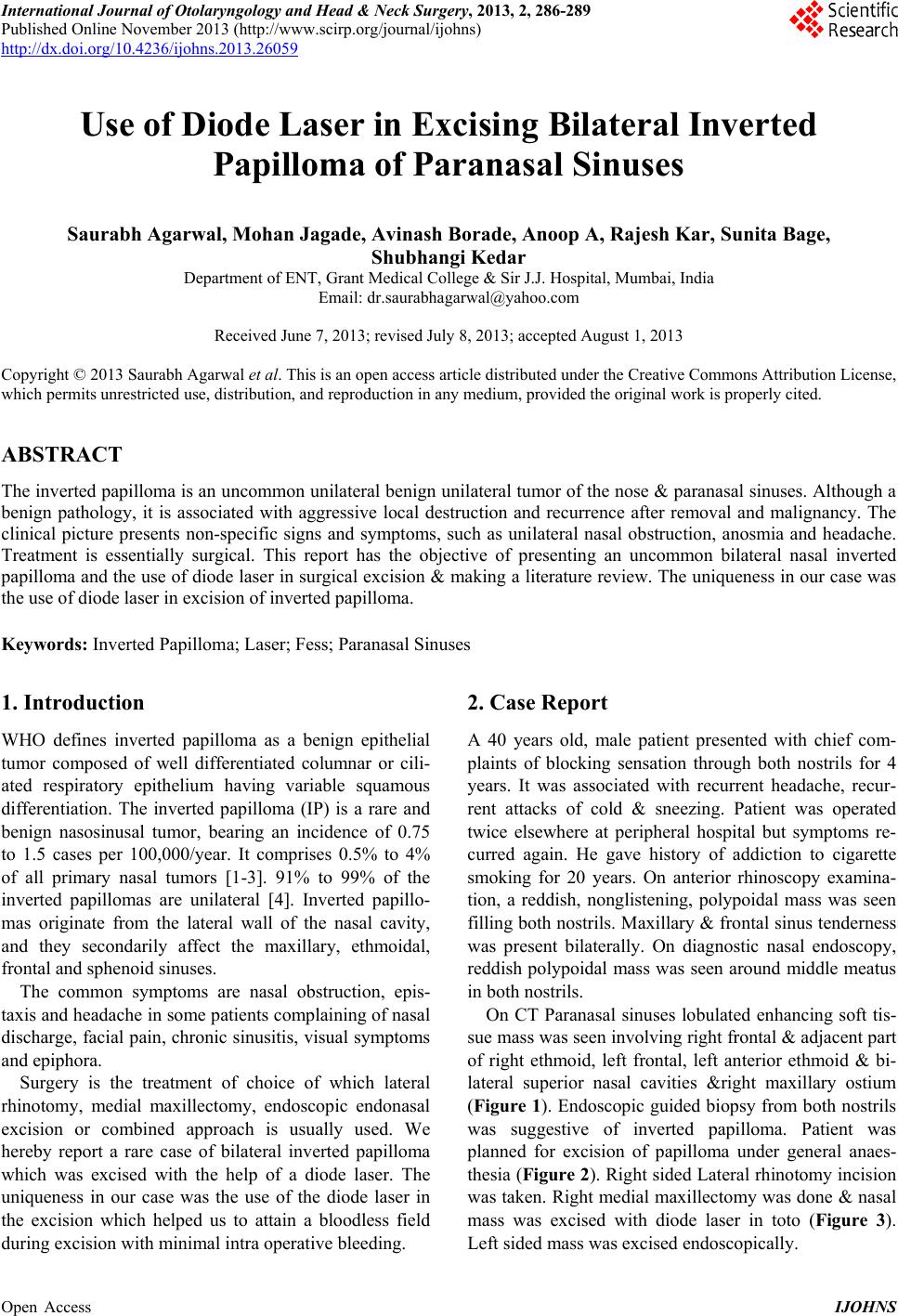
diological (CT and MRI) examinations are required for
tumor study and diagnosis. Biopsy together with histo-
pathology establishes the diagnosis. A unilateral mass
within the nasal cavity or paranasal sinuses with a sur-
face configuration that appears lobulated on CT is a new
sign that strongly suggests inverted papilloma as a pri-
mary diagnosis and also suggests inverted papilloma in
patients with tumor recurrence.
A columnar pattern is a reliable MRI indicator of IP
and reflects its histological architecture (positive predic-
tive value of 95.8%). The combination of this finding
with the absence of extended bone ero sion allows for the
confident discrimination of IPs from malignant tumours
[22].
Complete surgical removal is the first option for the
treatment of IP and is superior to radiation or chemo-
therapy [23,24]. Lateral rhinotomy has been regarded as
the traditional standard surgical approach to control IP
and to avoid recurrence [25]. It gives a good overview
and wide access to the surgical field and can also be per-
formed bilaterally [26]. Alternative techniques are the
midface degloving procedure & Denker’s approach. The
most recent developments in surgery are minimally inva-
sive transnasal endoscopic techniques [27,28]. In endo-
scopically accessible locations, recurrence rates for en-
doscopic vs. open surgery were similar [29]. However,
other studies reported higher recurrence rates of IP [30],
particularly in cases of peripheral extension, especially
into the maxillary sinus [31].
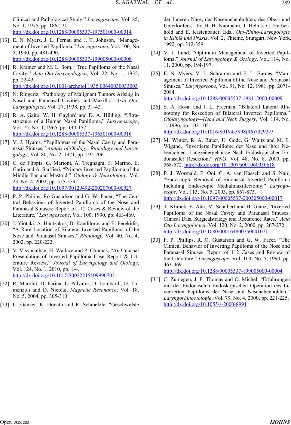
In our case lateral rhinotomy approach was used. The
diode laser was used for dissecting out the tumor from its
attachment along with the mucosa. Under direct vision,
the attachment was ablated with the laser in the cutting
and coagulation modes, and the tumor was then delivered.
The unique feature of our management was the use of the
diode laser. Inverted papillomas are notorious for exten-
sive bleeding, so a bloodless field was a great advantage.
The hemostatic property of the dode laser helped us to
achieve an almost bloodless field. This in turn helped us
visualize the exact site of tumor attachment. Laser was
essentially used in all three modes: contact, near-contact,
and noncontact to achieve cutting, vaporization and co-
agulation. The diode laser’s lesser depth of tissue pene-
tration and its capability to be used in contact, near con-
tact & non contact settings allows the laser to be safely
used near the lamina papyracea and skull base. So for
bilateral inverted papillomas limited to the nose and
paranasal sinuses, we believe our approach is a good
option in view of the low rates of intra operative blood
loss & minimal post operative morbidity.
4. Conclusion
Inverted papillomas are known for excessive bleeding
during tumour removal. The use of the diode laser in the
excision of inverted papillomas helps to achieve clear
intra operative field with no post operative blood transfu-
sion requirement.
REFERENCES
[1] D. P. Vrabec, “The Inverted Schneiderian Papilloma: 25-
Year Study,” Laryngoscope, Vol. 104, No. 5, 1994, pp.
582-608. http://dx.doi.org/10.1002/lary.5541040513
[2] T. T. Tsue, J. W. Bailet, D. W. Barlow and K. H.
Makielski, “Bilateral Sinusal Papilloma in Aplasic Maxi-
lar Sinuses,” American Journal of Otolaryngology, Vol.
l18, No. 4, 1997, pp. 263-268.
[3] M. C. Weissle r, W. W. Montgomery and S. K. Montgom-
ery, “Inverted Papiloma,” The Annals of Otolo gy, Rhinology,
and Laryngology, Vol. 95, 1986, pp. 215-221.
[4] K. Oikawa, Y. Furuta, N. Oridate, T. Nagahashi, A. Homma,
T. Ryu and S. Fukuda, “Preoperative Staging of Sinonasal
Inverted Papilloma by Magnetic Resonance Imaging,”
Laryngoscope, Vol. 133, No. 11, 2003, pp. 1983-1987.
[5] M. M. Lesperance and M. E. Ramon, “Squamous Cell
Carcinoma Arising in Inverted Papilloma,” Laryngoscope,
Vol. 105, No. 2, 1995, pp. 178-183.
http://dx.doi.org/10.1288/00005537-199502000-00013
[6] E. Raveh, R. Feinmesser, T. Shpitzer, E. Yaniv and K.
Segal, Israel Journal of Medical Sciences, Vol. 32, 1996,
p. 1162.
[7] D. P. Vrabec, “The Inverted Schneiderian Papilloma: A
25-Year Study,” Laryngoscope, Vol. 104, No. 5, 1994, pp.
502-605. http://dx.doi.org/10.1002/lary.5541040513
[8] L. Segal, E. Atar and C. Mor, “Inverted Papilloma of the
Nose and Paranasalsinuses,” Laryngoscope, Vol. 96, No.
4, 1996, pp. 394-398.
[9] J. A. Stankiewicz and S. J. Girs, “Endoscopic Surgical
Treatment of Nasal and Paranasal Sinus Inverted Papil-
loma,” Otolaryngology—Head and Neck Surgery, Vol.
109, No. 6, 1993, pp. 988-995.
[10] A. C. M. Alegre, A. H. C. Ramos, R. L. Voegels and F.
Romano, “Papiloma e Papilomainvertido,” In: C. A. Ca m-
pos and H. O. O. Costa, Eds., Tratado de Otorrinolarin-
gologia, Roca, São Paulo, 2003, pp. 126-132.
[11] J. H. Krouse, “Development of a Staging System for In-
verted Papilloma,” Laryngoscope, Vol. 110, No. 6, 2000,
pp. 965-968.
http://dx.doi.org/10.1097/00005537-200006000-00015
[12] D. P. Vrabec, “The Inverted Schneiderian Papilloma: A
Open Access IJOHNS
S. AGARWAL ET AL.
Open Access IJOHNS
289
Clinical and Pathological Study,” Laryngoscope, Vol. 85,
No. 1, 1975, pp. 186-221.
http://dx.doi.org/10.1288/00005537-197501000-00014
[13] E. N. Myers, J. L. Fernau and J. T. Johnson, “Manage-
ment of Inverted Papilloma,” Laryngoscope, Vol. 100, No.
5, 1990, pp. 481-490.
http://dx.doi.org/10.1288/00005537-199005000-00008
[14] R. Kramer and M. L. Som, “True Papilloma of the Nasal
Cavity,” Acta Oto-Laryngologica, Vol. 22, No. 1, 1935,
pp. 22-43.
http://dx.doi.org/10.1001/archotol.1935.00640030033003
[15] N. Ringertz, “Pathology of Malignant Tumors Arising in
Nasal and Paranasal Cavities and Maxilla,” Acta Oto-
Laryngologica, Vol. 27, 1938, pp. 31-42.
[16] R. A. Gaito, W. H. Gaylord and D. A. Hilding, “Ultra-
structure of a Human Nasal Papilloma,” Laryngoscope,
Vol. 75, No. 1, 1965, pp. 144-152.
http://dx.doi.org/10.1288/00005537-196501000-00016
[17] V. J. Hyams, “Papillomas of the Nasal Cavity and Para-
nasal Sinuses,” Annals of Otology, Rhinology and Laryn-
gology, Vol. 80, No. 2, 1971, pp. 192-206.
[18] C. de Flippis, G. Marioni, A. Tregnaghi, F. Marino, E.
Gario and A. Staffieri, “Primary Inverted Papilloma of the
Middle Ear and Mastoid,” Otology & Neurotology, Vol.
23, No. 4, 2002, pp. 555-559.
http://dx.doi.org/10.1097/00129492-200207000-00027
[19] P. P. Philips, Ro Gustafson and G. W. Facer, “The Cen-
tral Behaviour of Inverted Papilloma of the Nose and
Paranasal Sinuses: Report of 112 Cases & Review of the
Literature,” Laryngoscope, Vol. 100, 1990, pp. 463-469.
[20] J. Yiotaki, A. Hantzakos, D. Kandiloros and E. Ferekidis,
“A Rare Location of Bilateral Inverted Papilloma of the
Nose and Paranasal Sinuses,” Rhinology, Vol. 40, No. 4,
2002, pp. 220-222.
[21] V. Visvanathan, H. Wallace and P. Chumas, “An Unusual
Presentation of Inverted Papilloma Case Report & Lit-
erature Review,” Journal of Laryngology and Otology,
Vol. 124, No. 1, 2010, pp. 1-4.
http://dx.doi.org/10.1017/S0022215109990703
[22] R. Maroldi, D. Farina, L. Palvaini, D. Lombardi, D. To-
menzoli and D. Nicolai, Magnetic Resonance, Vol. 18,
No. 5, 2004, pp. 305-310.
[23] U. Ganzer, K. Donath and R. Schmelzle, “Geschwulste
der Inneren Nase, der Nasennebenhohlen, des Ober- und
Unterkiefers,” In: H. H. Naumann, J. Helms, C. Herber-
hold and E. Kastenbauer, Eds., Oto-Rhino-Laryngologie
in Klinik und Praxis, Vol. 2, Thieme, Stuttgart, New York,
1992, pp. 312-359.
[24] V. J. Lund, “Optimum Management of Inverted Papil-
loma,” Journal of Laryngology & Otology, Vol. 114, No.
11, 2000, pp. 194-197.
[25] E. N. Myers, V. L. Schramm and E. L. Barnes, “Man-
agement of Inverted Papilloma of the Nose and Paranasal
Sinuses,” Laryngoscope, Vol. 91, No. 12, 1981, pp. 2071-
2084.
http://dx.doi.org/10.1288/00005537-198112000-00009
[26] S. A. Hosal and J. L. Freeman, “Bilateral Lateral Rhi-
notomy for Resection of Bilateral Inverted Papilloma,”
Otolaryngology—Head and Neck Surgery, Vol. 114, No.
1, 1996, pp. 103-105.
http://dx.doi.org/10.1016/S0194-5998(96)70292-9
[27] M. Winter, R. A. Rauer, U. Gode, G. Waitz and M. E.
Wigand, “Invertierte Papillome der Nase und ihrer Ne-
benhohlen. Langzeitergebnisse Nach Endoskopischer En-
donasaler Resektion,” HNO, Vol. 48, No. 8, 2000, pp.
568-572. http://dx.doi.org/10.1007/s001060050618
[28] P. J. Wormald, E. Ooi, C. A. van Hasselt and S. Nair,
“Endoscopic Removal of Sinonasal Inverted Papilloma
Including Endoscopic Medialmaxillectomy,” Laryngo-
scope, Vol. 113, No. 5, 2003, pp. 867-873.
http://dx.doi.org/10.1097/00005537-200305000-00017
[29] T. Klimek, E. Atai, M. Schubert and H. Glanz, “Inverted
Papilloma of the Nasal Cavity and Paranasal Sinuses:
Clinical Data, Surgicalstrategy and Recurrence Rates,” Acta
Oto-Laryngologica, Vol. 120, No. 2, 2000, pp. 267-272.
http://dx.doi.org/10.1080/000164800750001071
[30] P. P. Phillips, R. O. Gustafson and G. W. Facer, “The
Clinical Behavior of Inverting Papilloma of the Nose and
Paranasal Sinuses: Report of 112 Cases and Review of
the Literature,” Laryngoscope, Vol. 100, No. 5, 1990, pp.
463-469.
http://dx.doi.org/10.1288/00005537-199005000-00004
[31] C. Zumegen, J. P. Thomas and O. Michel, “Erfahrungen
mit der Endonasalen Endoskopischen Operation des In-
vertierten Papilloms der Nase und Nasenebenhohlen,”
Laryngorhinootologie, Vol. 79, No. 4, 2000, pp. 221-225.
http://dx.doi.org/10.1055/s-2000-8991