Pharmacology & Pharmacy, 2013, 4, 578-583
Published Online November 2013 (http://www.scirp.org/journal/pp)
http://dx.doi.org/10.4236/pp.2013.48083
Open Access PP
One-Step Preparation of Poly-Lactic-Co-Glycolic-Acid
Microparticles to Prevent the Initial Burst Release of
Encapsulated Water-Soluble Proteins
Hiroyuki Takabe1,2, Moriyuki Ohkuma1, Yasunori Iwao2, Shuji Noguchi2, Shigeru Itai2*
1Drug Formulation Department, Central Research Laboratories, Kaken Pharmaceutical Co., Ltd. Shizuoka, Japan; 2Department of
Pharmaceutical Engineering, Graduate School of Pharmaceutical Sciences, University of Shizuoka, Shizuoka, Japan.
Email: *s-itai@u-shizuoka-ken.ac.jp
Received September 18th, 2013; revised October 12th, 2013; accepted October 22nd, 2013
Copyright © 2013 Hiroyuki Takabe et al. This is an open access article distributed under the Creative Commons Attribution License,
which permits unrestricted use, distribution, and reproduction in any medium, provided the original work is properly cited.
ABSTRACT
An initial burst is often observed during the release of active pharmaceutical ingredients (APIs) from poly-lactic-co-
glycolic-acid (PLGA) microparticles (MPs) which have been prepared by the emulsion-solvent evaporation method.
Herein, we describe the development of a simple one-step coating method that suppresses the initial burst release proc-
ess. This new method involves coating the PLGA-MPs with PLGA, with the coating process being performed through
the phase separation of PLGA on the surface of PLGA-MPs using the emulsion-solvent evaporation method. Bovine
serum albumin (BSA) was encapsulated in the PLGA-MPs as a model API. The coated MPs were spherical in shape
with no pores on their smooth surface, whereas the non-coated PLGA-MPs had porous surfaces. An in vitro release
study showed that the residual levels of BSA in the coated and non-coated PLGA-MPs after 1 h were about 99% and
16% of the original loads, respectively. The one-step coating method therefore represents a useful method for preparing
PLGA-MPs that do not give an initial burst release of proteinaceous APIs.
Keywords: Poly-Lactic-Co-Glycolic-Acid; Microparticle; Suppression of Initial Burst Release; Coating; Bovine Serum
Albumin
1. Introduction
Biocompatible and biodegradable synthetic polymers
such as poly-lactic-co-glycolic-acid (PLGA) have been
used as base materials in a number of different functional
materials, such as absorbable biomaterial implants [1,2],
absorbable surgical sutures [3,4] and microparticles (MPs)
for the sustained release of active pharmaceutical ingre-
dients (APIs) including bioactive proteins [5]. When
highly water-soluble proteins are encapsulated in MPs,
an initial burst release of the proteins has been often ob-
served [6,7]. The development of methods capable of
suppressing these initial burst releases is necessary to
enhance the therapeutic effects of APIs and reduce their
side effects. To date, several methods have been devel-
oped to suppress the initial burst release of APIs from
PLGA-MPs, including the replacement of PLGA with
different materials with slower degradation rates [8-10],
and the coating of the MPs with cationic compounds
[11-14] or water-soluble polymers [11-15]. Ahmed et al.
[16] recently reported a new method for coating PLGA-
MPs with PLGA. According to this method, however, the
PLGA-MPs had to be dispersed in peanut oil to enable
the formation of an oil layer on their surface to prevent
the dissolution and aggregation of PLGA-MPs during the
PLGA coating, indicating that this coating method still
required a series of time-consuming steps. In addition,
the application of this method could lead to significant
difficulties in terms of the associated cleaning processes,
because of oil in oil mixing, and concerns therefore re-
main that the final products could be contaminated with
oil. One of the major advantages of PLGA coatings is
that the encapsulated APIs can be released from the
PLGA-MPs at a constant rate because the degradation
rates of PLGA are similar in the MPs and the coating
layer. Taken together, there is an urgent need in the field
of pharmaceutical research for a simple and effective
*Corresponding author.
One-Step Preparation of Poly-Lactic-Co-Glycolic-Acid Microparticles to Prevent the Initial Burst Release of
Encapsulated Water-Soluble Proteins
579
method of coating PLGA-MPs with PLGA [17].
In the current study, we have developed a simple one-
step method for coating PLGA-MPs with PLGA during
the final step of the emulsion-solvent evaporation method.
With regards to the emulsion-solvent evaporation method,
PLGA phase separation occurred at the interface between
the PLGA and aqueous solutions during the preparation
of the PLGA-MPs [6,8-10]. Following the preparation of
the PLGA-MPs, the addition of a PLGA solution effec-
tively induced PLGA phase separation at the surface of
the PLGA-MPs, and the MPs were consequently coated
with PLGA. PLGA-MPs encapsulated with bovine serum
albumin (BSA) (BSA-MPs) were coated with PLGA
using the current method to evaluate the general utility of
this one-step coating method as a means of suppressing
the initial burst release of proteinaceous APIs (Figure 1),
and their physicochemical properties and protein-release
behaviors were also investigated.
2. Experimental
2.1. Materials
BSA (ELISA applications) and sodium dodecyl sulfate
(SDS) were purchased from Sigma-Aldrich (St. Louis,
MO, USA). PLGA-7510 and PLGA-7520 were pur-
chased from Wako Pure Chemical Industries Ltd. (Osaka,
Japan). Phosphate-buffered saline (PBS) tablets were
purchased from Takara Bio Inc. (Shiga, Japan). All of the
reagents used in the current study were purchased in the
highest grade commercially available.
2.2. Preparation of BSA-MPs
PLGA-7510 (1.0 g) was dissolved in dichloromethane
(DCM, 1.5 mL), and 0.25 mL of 16.0 mg/mL BSA
aqueous solution was added to the resulting solution and
homogenized at 16,000 × g for 30 seconds to obtain a
water-in-oil (W/O) emulsion. The W/O emulsion was
added to 150 mL of 0.1% PVA aqueous solution in a
drop-wise manner and homogenized at 7000 rpm for 60
seconds to prepare a water-in-oil-in-water (W/O/W)
emulsion. The DCM was then removed from the emul-
sion using the emulsion-solvent evaporation method un-
der stirring for 3 h at ambient temperature. The resulting
BSA-MPs were collected by centrifugation at 1600 × g
for 15 min, with the supernatant being decanted. Distilled
water was then added to the MPs to wash out any free
BSA and PVA, with the resulting material being sepa-
rated by centrifugation. This washing process was re-
peated five times, and the resulting washed BSA-MPs
were collected and lyophilized for 48 h.
2.3. Preparation of BSA-MPs 7510 Coating-1
and BSA-MPs 7520 Coating-1
The PLGA-7510 and PLGA-7520 coating solutions were
prepared by dissolving 100 mg of PLGA-7510 or PLGA-
7520 in 2.0 mL of acetone. The resulting PLGA-coating
solutions were added to the W/O/W emulsion prepared
as described above with stirring. These emulsions were
stirred for 3 h at ambient temperature to allow for the
removal of acetone by evaporation. The PLGA-coated
MPs (BSA-MPs 7510 coating-1 and BSA-MPs 7520
coating-1, respectively) were then washed and lyophi-
lized as in the BSA-MP preparation.
2.4. Preparation of BSA-MPs 7510 Coating-2
A portion of the lyophilized BSA-MPs as prepared in
Section 2.2 (1.0 g) was dispersed in 150 mL of a 0.1%
PVA aqueous solution, and the resulting dispersion was
added to 2.0 mL of a PLGA-7510 coating solution. The
mixture was then stirred for 3 h at 40˚C. The MPs coated
with PLGA-7510 (BSA-MPs 7510 coating-2) were
Figure 1. Schematic representation of th e PLGA coating process using the one-step emulsion-solvent evaporation method.
Open Access PP
One-Step Preparation of Poly-Lactic-Co-Glycolic-Acid Microparticles to Prevent the Initial Burst Release of
Encapsulated Water-Soluble Proteins
580
washed and lyophilized as in the BSA-MP preparation.
2.5. Particle Size Distribution
The particle size distributions of the different PLGA-
MPs were determined in a wet process using a laser scat-
tering particle size analyzer (LDSA-1500A, Tonichi
Computer Applications Co., Ltd., Tokyo, Japan).
2.6. Scanning Electron Microscopy (SEM)
The surface morphologies of the different PLGA-MPs
were assessed by SEM (JSM-6390LA, JEOL Ltd., Tokyo,
Japan). The samples were sputter-coated with platinum
under vacuum prior to imaging.
2.7. Determination of the BSA Content in MPs
Four milligram samples of the MPs were added to a 4:1
(v/v) mixture of acetone and water (1 mL), and the re-
sulting solutions were agitated for 1 h at ambient tem-
perature. These solutions were centrifuged for 15 min-
utes at 16,000 × g to remove supernatants. The pellets
thus obtained were washed with 1.6 mL of acetone and
dried at ambient temperature for 30 minutes. The dried
pellets were dissolved in 0.1 mL of a 0.5% SDS solution
by sonication (US-105, SND Co., Ltd., Nagano, Japan)
over a 15 minute period. The concentrations of BSA in
the solutions were determined using a protein quantifica-
tion kit CBQCA (Molecular Probes, Eugene, Oregon).
The BSA contents of the PLGA-MPs, as well as the ac-
tual drug loading and encapsulation efficiency values of
the materials were calculated according to the following
equations.


Actual drugloading%
BSA inPLGAMPsmg100
MPs mg

(1)



Encapsulation efficiency%
Actual drugloading%100
Theoretical drug loading%

(2)
2.8. Release Studies
Four milligram samples of the different MPs were dis-
persed in 0.4 mL of a PBS buffer in a micro-tube and
incubated at 37˚C. The micro-tubes were subsequently
collected at pre-determined time points and centrifuged
for 15 minutes at 1600 × g to remove the supernatants.
The MPs were then washed with 1 mL of distilled water,
and the levels of residual BSA in the MPs were quanti-
fied using CBQCA as described above. The ratio of re-
sidual BSA in the MPs was calculated according to the
following equation.
3. Results and Discussion
BSA-MPs, which were prepared using the emulsion-
solvent evaporation method, were coated in a one-step
operation to suppress the initial burst process. More spe-
cifically, phase separation occurred at the surface of the
MPs when PLGA solution was added to a dispersion of
MPs in a poor solvent, and the MPs were consequently
coated with PLGA. Using this one-step coating methods,
three kinds of PLGA-coated BSA-MPs were prepared: 1)
BSA-MPs coated with PLGA-7510 with a lactide: gly-
colide monomer composition of 75:25, and an average
molecular weight of 10,000 that were subsequently
named BSA-MPs 7510 coating-1; 2) BSA-MPs coated
with PLGA-7520 with an average molecular weight of
20,000 that were subsequently named BSA-MPs 7520
coating-1; and 3) BSA-MPs that were coated with PLGA
7510 following a lyophilization process that were subse-
quently named BSA-MPs 7510 coating-2. The yield of
the BSA-MPs was approximately 75%, whereas those of
the coated BSA-MPs were in the range of 80% - 85%
(Table 1). The higher yields observed for the coated MPs
were attributed to the amount of PLGA coating. The
yield of BSA-MPs 7510 coating-2 was lower than that of
BSA-MPs 7510 coating-1, likely because of the incre-
mental lyophilization process.
The median diameter (X50) values of the BSA-MPs and
the BSA-MPs 7510 coating-2 particles were 24.4 and
23.2 μm, respectively, whereas the X50 of the BSA-MPs
7510 coating-1 particles was higher at 31.8 μm (Figure 2,
Table 1). A mean diameter in the range of 20 to 30 μm is
generally required for the MPs to possess good syringe-
ability. Pleasingly, the X50 values of the MPs involved in
the current study fell within or were just outside of this
range. The X50 of the BSA-MPs 7520 coating-1 particles,
however, was 39.1 μm, and therefore out of the preferred
range. The higher viscosity of PLGA 7520 [18-20] may
have enhanced its ability to adhere to the MP surfaces
during the coating step, and resulted in its large X50.
Although many pores of about 2 μm in diameter were
observed on the surfaces of the BSA-MPs, no pores were
found on the smooth surfaces of the spherical BSA-MPs
coated with PLGA (Figure 3). These results indicated
that the pores on the surfaces of the MPs had been effi-
ciently covered with PLGA as a consequence of this one-
step coating procedure.
The encapsulation efficiencies of the PLGA-coated
MPs were all lower than that of the BSA-MPs (Table 1),
likely because of the leakage of BSA during the coating
process. The encapsulation efficiencies of the three
coated MPs prepared during the course of the current
study were particularly low,pecially for BSA-MPs es
Open Access PP
One-Step Preparation of Poly-Lactic-Co-Glycolic-Acid Microparticles to Prevent the Initial Burst Release of
Encapsulated Water-Soluble Proteins
581
Table 1. Yields and particle properties of the BSA-MPs and BSA-MPs coated with PLGA.
Particle size distribution (µm)
MPs Yield* (%) X10 X
50 X
90 BSA loading (%) Encapsulation efficiency (%)
BSA-MPs
BSA-MPs 7510 coating-1
BSA-MPs 7520 coating-1
BSA-MPs 7510 coating-2
76.2
83.6
81.1
81.8
9.6 ± 1.1
12.0 ± 0.6
16.6 ± 0.2
10.2 ± 0.4
24.4 ± 0.9
31.8 ± 0.2
39.1 ± 0.1
23.2 ± 0.1
50.5 ± 0.6
52.8 ± 0.1
63.9 ± 0.1
55.1 ± 1.1
0.25 ± 0.02
0.23 ± 0.02
0.18 ± 0.02
0.16 ± 0.01
65.5 ± 5.4
57.3 ± 4.4
44.2 ± 4.0
41.0 ± 1.9
*


Recovered amount ofPLGA-MPsg
Yield of the PLGA-MPs%100.
input PLGAg

Figure 2. Particle size distribution of the MPs. (a) BSA-MPs; (b) BSA-MPs 7510 coating-1; (c) BSA-MPs 7520 coating-1; and
(d) BSA-MPs 7510 coating-2. The bar and line charts show the frequency (%) and cumulative values (%), respectively.
Figure 3. SEM images of MPs. (a) BSA-MPs; (b) BSA-MPs 7510 coating-1; (c) BSA-MPs 7520 coating-1; and (d) BSA-MPs
7510 coating-2.
7510 coating-2 and BSA-MPs 7520 coating-1. For the
preparation of BSA-MPs 7510 coating-2, an incremental
process including a lyophilization step was needed. For
the preparation of BSA-MPs 7520 coating-1, the PLGA
7520 in the coating solution might not solidify rapidly on
the core surface because of its high viscosity [18-20], and
this resulted in more BSA leaking from the MPs and the
observed low encapsulation efficiency.
The BSA-MPs showed an initial burst release with
84% of the BSA being released from the MPs following
1 h of the release study (Figure 4(a)). The initial burst
release process could be induced by the rapid penetration
Open Access PP
One-Step Preparation of Poly-Lactic-Co-Glycolic-Acid Microparticles to Prevent the Initial Burst Release of
Encapsulated Water-Soluble Proteins
582
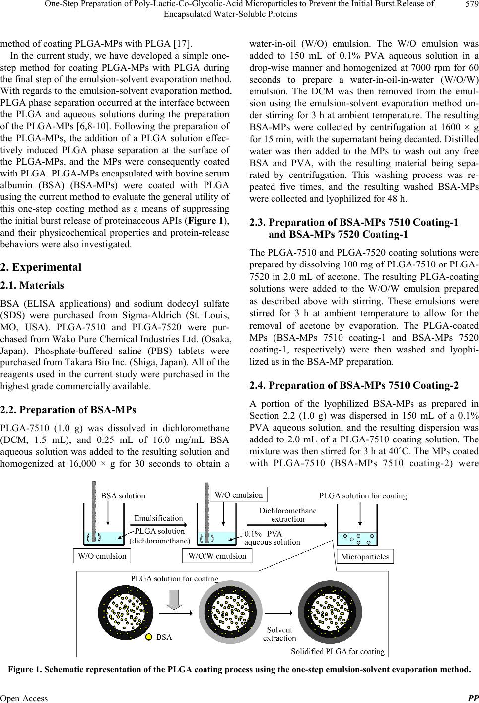
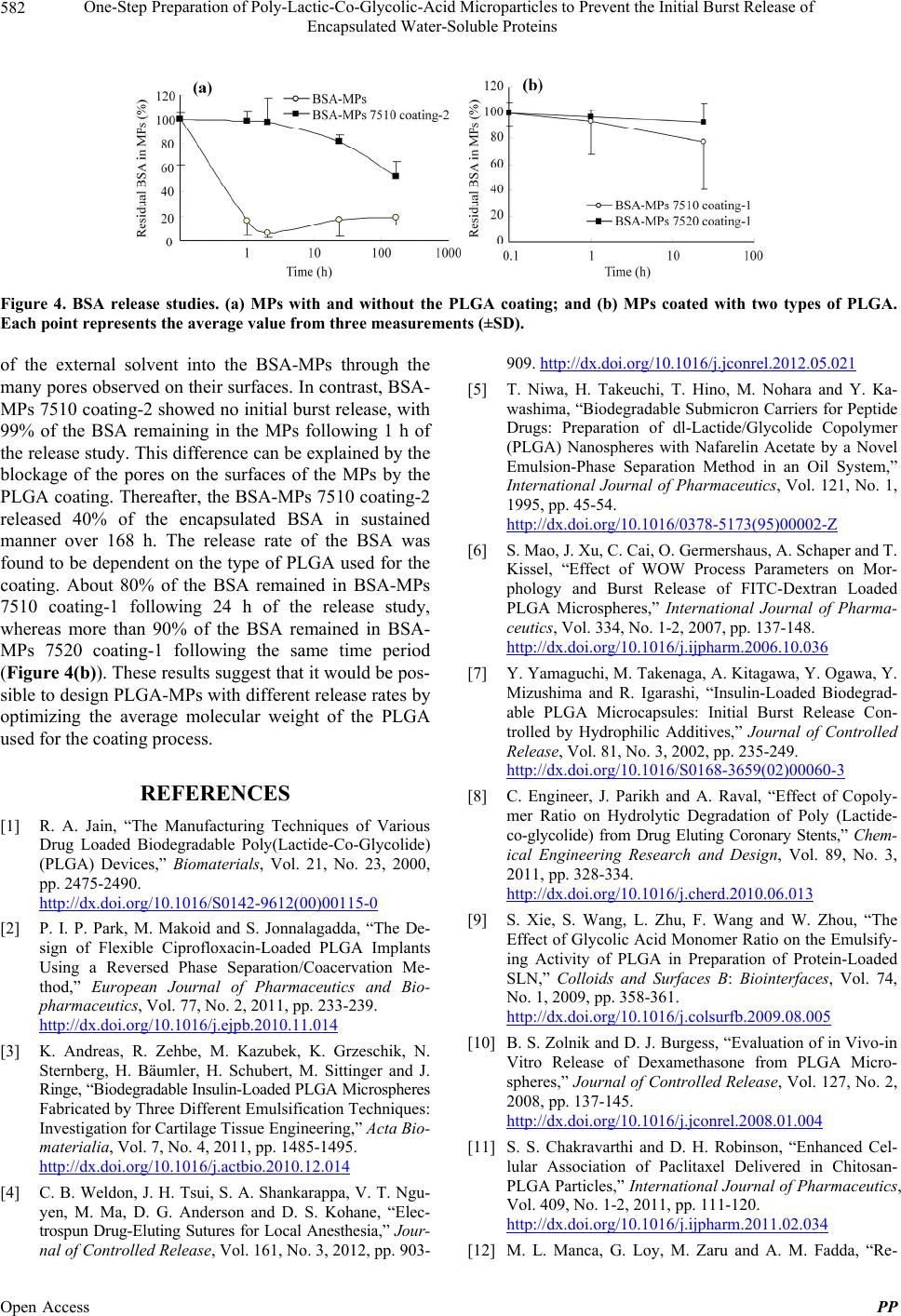
Figure 4. BSA release studies. (a) MPs with and without the PLGA coating; and (b) MPs coated with two types of PLGA.
Each point represents the average value from three measurements (±SD).
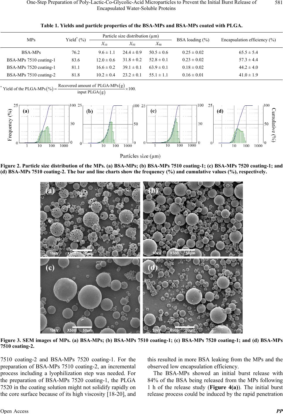
of the external solvent into the BSA-MPs through the
many pores observed on their surfaces. In contrast, BSA-
MPs 7510 coating-2 showed no initial burst release, with
99% of the BSA remaining in the MPs following 1 h of
the release study. This difference can be explained by the
blockage of the pores on the surfaces of the MPs by the
PLGA coating. Thereafter, the BSA-MPs 7510 coating-2
released 40% of the encapsulated BSA in sustained
manner over 168 h. The release rate of the BSA was
found to be dependent on the type of PLGA used for the
coating. About 80% of the BSA remained in BSA-MPs
7510 coating-1 following 24 h of the release study,
whereas more than 90% of the BSA remained in BSA-
MPs 7520 coating-1 following the same time period
(Figure 4(b)). These results suggest that it would be pos-
sible to design PLGA-MPs with different release rates by
optimizing the average molecular weight of the PLGA
used for the coating process.
REFERENCES
[1] R. A. Jain, “The Manufacturing Techniques of Various
Drug Loaded Biodegradable Poly(Lactide-Co-Glycolide)
(PLGA) Devices,” Biomaterials, Vol. 21, No. 23, 2000,
pp. 2475-2490.
http://dx.doi.org/10.1016/S0142-9612(00)00115-0
[2] P. I. P. Park, M. Makoid and S. Jonnalagadda, “The De-
sign of Flexible Ciprofloxacin-Loaded PLGA Implants
Using a Reversed Phase Separation/Coacervation Me-
thod,” European Journal of Pharmaceutics and Bio-
pharmaceutics, Vol. 77, No. 2, 2011, pp. 233-239.
http://dx.doi.org/10.1016/j.ejpb.2010.11.014
[3] K. Andreas, R. Zehbe, M. Kazubek, K. Grzeschik, N.
Sternberg, H. Bäumler, H. Schubert, M. Sittinger and J.
Ringe, “Biodegradable Insulin-Loaded PLGA Microspheres
Fabricated by Three Different Emulsification Techniques:
Investigation for Cartilage Tissue Engineering,” Acta Bio-
materialia, Vol. 7, No. 4, 2011, pp. 1485-1495.
http://dx.doi.org/10.1016/j.actbio.2010.12.014
[4] C. B. Weldon, J. H. Tsui, S. A. Shankarappa, V. T. Ngu-
yen, M. Ma, D. G. Anderson and D. S. Kohane, “Elec-
trospun Drug-Eluting Sutures for Local Anesthesia,” Jour-
nal of Controlled Release, Vol. 161, No. 3, 2012, pp. 903-
909. http://dx.doi.org/10.1016/j.jconrel.2012.05.021
[5] T. Niwa, H. Takeuchi, T. Hino, M. Nohara and Y. Ka-
washima, “Biodegradable Submicron Carriers for Peptide
Drugs: Preparation of dl-Lactide/Glycolide Copolymer
(PLGA) Nanospheres with Nafarelin Acetate by a Novel
Emulsion-Phase Separation Method in an Oil System,”
International Journal of Pharmaceutics, Vol. 121, No. 1,
1995, pp. 45-54.
http://dx.doi.org/10.1016/0378-5173(95)00002-Z
[6] S. Mao, J. Xu, C. Cai, O. Germershaus, A. Schaper and T.
Kissel, “Effect of WOW Process Parameters on Mor-
phology and Burst Release of FITC-Dextran Loaded
PLGA Microspheres,” International Journal of Pharma-
ceutics, Vol. 334, No. 1-2, 2007, pp. 137-148.
http://dx.doi.org/10.1016/j.ijpharm.2006.10.036
[7] Y. Yamaguchi, M. Takenaga, A. Kitagawa, Y. Ogawa, Y.
Mizushima and R. Igarashi, “Insulin-Loaded Biodegrad-
able PLGA Microcapsules: Initial Burst Release Con-
trolled by Hydrophilic Additives,” Journal of Controlled
Release, Vol. 81, No. 3, 2002, pp. 235-249.
http://dx.doi.org/10.1016/S0168-3659(02)00060-3
[8] C. Engineer, J. Parikh and A. Raval, “Effect of Copoly-
mer Ratio on Hydrolytic Degradation of Poly (Lactide-
co-glycolide) from Drug Eluting Coronary Stents,” Chem-
ical Engineering Research and Design, Vol. 89, No. 3,
2011, pp. 328-334.
http://dx.doi.org/10.1016/j.cherd.2010.06.013
[9] S. Xie, S. Wang, L. Zhu, F. Wang and W. Zhou, “The
Effect of Glycolic Acid Monomer Ratio on the Emulsify-
ing Activity of PLGA in Preparation of Protein-Loaded
SLN,” Colloids and Surfaces B: Biointerfaces, Vol. 74,
No. 1, 2009, pp. 358-361.
http://dx.doi.org/10.1016/j.colsurfb.2009.08.005
[10] B. S. Zolnik and D. J. Burgess, “Evaluation of in Vivo-in
Vitro Release of Dexamethasone from PLGA Micro-
spheres,” Journal of Controlled Release, Vol. 127, No. 2,
2008, pp. 137-145.
http://dx.doi.org/10.1016/j.jconrel.2008.01.004
[11] S. S. Chakravarthi and D. H. Robinson, “Enhanced Cel-
lular Association of Paclitaxel Delivered in Chitosan-
PLGA Particles,” International Journal of Pharmaceutics,
Vol. 409, No. 1-2, 2011, pp. 111-120.
http://dx.doi.org/10.1016/j.ijpharm.2011.02.034
[12] M. L. Manca, G. Loy, M. Zaru and A. M. Fadda, “Re-
Open Access PP
One-Step Preparation of Poly-Lactic-Co-Glycolic-Acid Microparticles to Prevent the Initial Burst Release of
Encapsulated Water-Soluble Proteins
583
lease of Rifampicin from Chitosan, PLGA and Chitosan-
Coated PLGA Microparticles,” Colloids and Surfaces B:
Biointerfaces, Vol. 67, No. 2, 2008, pp. 166-170.
http://dx.doi.org/10.1016/j.colsurfb.2008.08.010
[13] H. Nojehdehian, F. Moztarzadeh, H. Baharvand, H. Naza-
rian and M. Tahriri, “Preparation and Surface Characteri-
zation of Poly-l-Lysine-Coated PLGA Microsphere Scaf-
folds Containing Retinoic Acid for Nerve Tissue Engi-
neering: In Vitro Study,” Colloids and Surfaces B: Bio-
interfaces, Vol. 73, No. 1, 2009, pp. 23-29.
http://dx.doi.org/10.1016/j.colsurfb.2009.04.029
[14] J. Shen and D. J. Burgess, “Accelerated in Vitro Release
Testing of Implantable PLGA Microsphere/PVA Hy-
drogel Composite Coatings,” International Journal of
Pharmaceutics, Vol. 422, No. 1-2, 2012, pp. 341-348.
http://dx.doi.org/10.1016/j.ijpharm.2011.10.020
[15] A. Budhian, S. J. Siegel and K. I. Winey, “Controlling the
in Vitro Release Profiles for a System of Haloperidol-
Loaded PLGA Nanoparticles,” International Journal of
Pharmaceutics, Vol. 346, No. 1-2, 2008, pp. 151-159.
http://dx.doi.org/10.1016/j.ijpharm.2007.06.011
[16] A. R. Ahmed, K. Elkharraz, M. Irfan and R. Bodmeier,
“Reduction in Burst Release after Coating Poly(d,l-Lac-
tide-Co-Glycolide) (PLGA) Microparticles with a Drug-
Free PLGA Layer,” Pharmaceutical Development and
Technology, Vol. 17, No. 1, 2012, pp. 66-72.
http://dx.doi.org/10.3109/10837450.2010.513989
[17] A. R. Ahmed, A. Dashevsky and R. Bodmeier, “Reduc-
tion in Burst Release of PLGA Microparticles by Incor-
poration into Cubic Phase-Forming Systems,” European
Journal of Pharmaceutics and Biopharmaceutics, Vol. 70,
No. 3, 2008, pp. 765-769.
http://dx.doi.org/10.1016/j.ejpb.2008.07.008
[18] R. Jeyanthi, R. C. Mehta, B. C. Thanoo and P. P. Deluca,
“Effect of Processing Parameters on the Properties of
Peptide-Containing PLGA Microspheres,” Journal of Mi-
croencapsulation, Vol. 14, No. 2, 1997, pp. 163-174.
http://dx.doi.org/10.3109/02652049709015330
[19] H. K. Makadia and S. J. Siegel, “Poly Lactic-Co-Glycolic
Acid (PLGA) as Biodegradable Controlled Drug Delivery
Carrier,” Polymers, Vol. 3, No. 3, 2011, pp. 1377-1397.
http://dx.doi.org/10.3390/polym3031377
[20] G. Mittal, D. K. Sahana, V. Bhardwaj and M. N. V. Ravi
Kumar, “Estradiol Loaded PLGA Nanoparticles for Oral
Administration: Effect of Polymer Molecular Weight and
Copolymer Composition on Release Behavior in Vitro
and in Vivo,” Journal of Controlled Release, Vol. 119, No.
1, 2007, pp. 77-85.
http://dx.doi.org/10.1016/j.jconrel.2007.01.016
Open Access PP