Engineering, 2013, 5, 250-251
http://dx.doi.org/10.4236/eng.2013.510B051 Published Online October 2013 (http://www.scirp.org/journal/eng)
Copyright © 2013 SciRes. ENG
A New Method of Tracking of WM Crossing Fiber
Bundles Based on QBI*
Zhanxiong Wu
College of Electronic Information, Hangzhou Dianzi University, Hangzhou, China
Email: wzx@hdu.edu.cn
Received June 2013
ABSTRACT
Tracking of crossing WM fiber bundles can be resolved using diffusion MRI imaging. DTI can only resolve a single
fiber orientation within each voxel due to the constraints of the tensor model. DSI requires large pulse d field gradients
and time-intensive sampling. This paper puts forward with a new method based on QBI, which uses a spherical tomo-
graphic invers ion called Funk-Radon tran sform to get high angular resolution diffusion imaging signal. From the track-
ing results, we can get the conclusion that QBI-tracking can resolve crossing fiber time-savingly.
Keywords: Diffusio n Tensor Imaging; Diffusion Spectrum Imaging; q-Ball
1. Introduction
White matter is mainly composed of nerve fibers, which
are the connected channels of brain different function
areas. The tracking of nerve fibers is very important for
white matter disease, and is of great significance for cog-
nitive research. Diffusion MRI has been widely used to
assess the integrity of axonal fibers because of its unique
ability to map fiber orientations in vivo.
Diffusion tensor imaging (DTI) provides a powerful
tool for mapping neural histoarchitecture in vivo. How-
ever, DTI can only resolve a single fiber orientation
within each imaging voxel due to the constraints of the
tensor model. DTI cannot resolve fibers crossing, bend-
ing, or twisting within an individual voxel [1-4].
As to diffusion spectrum imaging (DSI), diffusion is
described with the probability density function (PDF)
which for each voxel specifies the 3D distribution of mi-
croscopic displacements of MR-visible spins that it con-
tains. Reconstruction of the diffusion PDF by Fourier
transform of the diffusion signal forms the basis of the
QSI method. But DSI requires large pulsed field gra-
dients and time-intensive sampling [5-7].
Q-ball imaging can resolve multiple orientations of
crossing fiber in one voxel and does not require any as-
sumptions on the di ffusion p r oc e s s o f water m olecules.
2. Method and Material
2.1. Reconstruction of the Diffusion Orientation
Distribution Function (ODF)
QBI is reconstructed based on the relationship of the in-
terested ODF vector and its orthogonal plane projected
on to the acquired q-space data. The ODF was directly
calculated from the attenuated echo signal on a shell in
the q-space with a fixed b-value based on the Funk-Ra-
don transform approach.
(1)
where u is the unit vector for the desired ODF direction,
and C is the normalization constant, and E is the atte-
nuated echo signal [8].
The diffusion MRI dataset was provided by Advanced
Biomedical MRI Lab of National Taiwan University
Hospital. The acquisition equipment was 3 Tesla Trio of
the company Siemens of Germany, 1.9 mm × 1.9 mm ×
1.9 mm, TR = 11,500 ms, TE = 31 ms, △/δ = 80/35 ms,
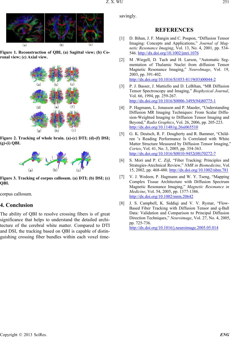
the maximal b value is 4000 s/mm2. Figure 1 shows the
reconstruction of QBI.
2.2. Tracking of Crossing Fiber Bundles
Firstly all of local maximums of ODF were got, then the
parameters of angle threshold and stepping size were set;
At last we found the smooth lines among the adjacent
voxels. Here the value of angle threshold was set to 60
degrees, and the value of stepping size was set to 1.4 mm.
The constraint of fiber bundle length was between 16
mm and 86 mm.
3. Results
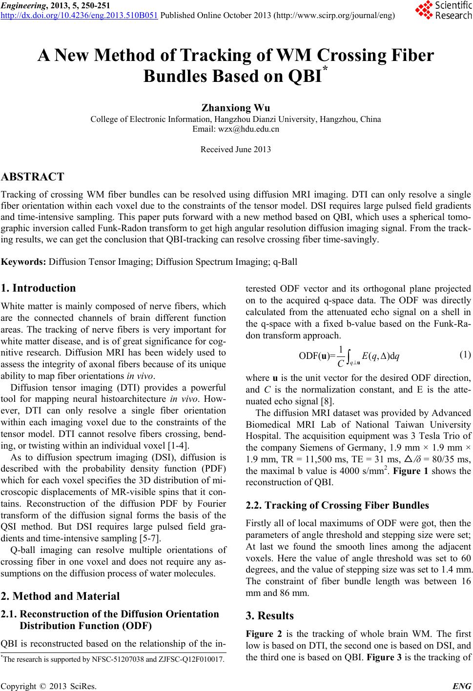
Figure 2 is the tracking of whole brain WM. The first
low is based on DTI, the second one is based on DSI, and
the third one is based on QBI. Figure 3 is the tracking of
*The research is supported by NFSC-51207038 and ZJFSC-Q12F010017.