Engineering, 2013, 5, 62-67
http://dx.doi.org/10.4236/eng.2013.510B013 Published Online October 2013 (http://www.scirp.org/journal/eng)
Copyright © 2013 SciRes. ENG
Relationships between a Thiol-Disulfide System and
Liposoluble Antioxidants with Cytogenetic Indices in
Humans Exposed to Low Doses Radiation
G. F. Ivanenko, E. B. Burlakova
Russian Academy of Sciences, Emanuel Institute of Biochemical Physics, Moscow, Russia
Email: galiv@sky.chph.ras.ru, seren@sky.chph.ras.ru
Received October 2012
ABSTRACT
This work presents the results of biochemical examination of people exposed to irradiation as a result of the Chernobyl
catastrophe. In remote period ( in 4, 5, 6 and 7 years ) after Chernobyl accident we stud ied the state of the redox syste m
of glutathione(GSH, GSSG) and the response of the system of essential lipid antioxidants (vitamin E, A) in b lood plas-
ma of people of various ages. An analysis of correlations between cytogenetic indices in lymphocytes and levels of re-
duced glutathione and liposoluble antioxidants in the plasma of peripheral blood in children born after the Chernobyl
accident and liquidators is presented. The cumulative doses for the examined group of children received by their moth-
ers from 0.8 to 70 cSv and liquidators received, on aver age, the highest irradiation doses from 0.1 to 150 cSv. A com-
plex relationship between lipo-and water-soluble antioxidants level in plasma in human population (children living in
radionuclide-contaminated regions and the Chernobyl liquidators) exposed to chronic low-level radiation after the
Chernobyl accident was demonstrated. The obtained experimental data indicate different responses of the human popu -
lation water-and fat-soluble antioxidants system to low (from 0.1 to 20 cSv) and high (from 20 to 150 cSv) doses of
ionizing radiation.
Keywords: Antioxidant; Radiation; Pla sma ; Human Population
1. Introduction
A large number of new facts concerning the effect of
low-level radiation on living organisms were provided
after the Chernobyl accident. The significance of low-
dose exposures for cell survival, tissue damage, and indi-
vidual health are poorly understood [1,2]. For a long pe-
riod of nuclear industry development, exposures to low
doses were considered safe. However, recent publica-
tions demonstrate a relatively high efficiency of low-
level radiation in the region of low radiation doses. Ac-
tive mutation process in various cells and tissues and
carcinogenesis promotion were observed after exposure
to low radiation doses [3]. Mutagenesis is accompanied
by chromosomal and genomic instability in cells of vari-
ous tissues. In the case of ionizing radiation, the primary
damage, namely double-strand breaks, induced both
structural chromosomal aberrations and microstructural
DNA damage detected as gene mutations [4].
Various biochemical and biophysical tests are being
developed in order to evaluate the effect of exposure to
low radiation doses; in particular, the antioxidant status
of the body is given considerable attention. Glutathione
is particularly important among water-soluble antioxi-
dants [5,6]. Resistance of the body to ionizing radiation
depends on both lipid antioxidant activity and the level of
sulfhydryl groups [7,8].
It is common knowledge th at thiols as well as vitamins
C, A, and E p rotect cells from oxidation and inhibit lipid
peroxidation. A relatively high contribution of water-
soluble antioxidants is commonly associated with rege-
neration. Glutathione was proposed to mediate the reduc-
tion of at least two vitamins, α-tocopherol and ascorbic
acid; it increases their effect and prevents lipid peroxida-
tion [9,10].
Below we present experimental data on the effect of
low-level radiation on the status of water-and lipo-so-
luble antioxidant in the liquidators and people living in
conditions of long-term exposure to low radiation doses
in the regions contaminated by radionuclides after the
Chernobyl accident (1 - 20 Ci/km2). We tried to establish
the relationship between levels of glutathione, v itamin E,
A and the level of radiation burden and to analyze the
dose-response relationships for certain types of genetic
damages such as the frequency chromatid breaks, in the
course of long-term monitoring. Below we analyze poss-
ible correlations between cytogenetic indices in peri-
G. F. IVANENKO, E. B. BUR L AKOVA
Copyright © 2013 SciRes. ENG
63
pheral blood lymphocytes, glutathione and lipid antioxi-
dant levels in blood plasma of people after the Chernobyl
accident.
2. Methods and Subjects
Children were examined from birth to the age of eight in
three regions with different levels of soil pollution with
radionuclides (largely 137Cs) after the Chernobyl accident.
Children (n = 125) from the Chechersk Region (15 - 20
Ci/km2) (Gomel Region of Belorussia) were examined 5
years after the accident; after six years children (n = 210)
from the Mtsensk Region (1 - 5 Ci/km2) and, after seven
years, children (n = 116) from the Bolkhov Region (5 -
10 Ci/km2) (both Orlov Region of Russia) were ex-
amined. Children at the age of 5 - 6 - 7 years (at the mo-
ment of examination) survived the accident during their
intrauterine development; other ones were born after the
accident (1990-1992).
Four years after the Chernobyl accident (1986), we
examined the liquidators (LI) that were working in the
first days after the accident and later moved to Slavutich
town outside the 30 km zone (n = 22). The radiation
doses of this group ranged from 2 to 150 cSv. Six years
later, we examined one more group of liquidators (L2, n
= 128) who worked fro m May 1986 to 1987 in the region
of Chernobyl accident and received radiation doses from
0.1 to 70 cSv. Were examined сhildren at the age 2 - 12
(n = 9) and of adults of Slavutich (n = 29) living in ra-
dionuclide-contaminated regions (1 - 5 Ci/km2). Resi-
dents of Moscow (n = 21) not exposed to radiation were
the comparison group.
The level of reduced and oxidized glutathione in plas-
ma was determined as described elsewhere [11,12] with
modifications. Glutathione level was determined in 20 -
100 µl plasma by spectrofluorometry at 350/420 nm. The
level of oxidized glutathione was calculated as the dif-
ference between the total numbers of SH groups before
and after the reduction using blank assays and standard
curves fo r GSH and G SSG . All results averaged for three
parallel experiments were expressed as µM. Tocopherol
and retinol were determined by spectrofluorometry at
295/325 and 330/470 nm, respectively, with appropriate
standards as desc ribed [13] .
The cytogenetic test relied on the traditional method
(analysis of unstable chromosomal aberrations). Whole
blood (0.5 ml) was cultivated as described by [14]. Both
chromosomal (dicentrics, centric rings, and paired frag-
ments) and chromatid (single fragments) aberrations
were evaluated. The fr equency of chr omatid b reaks (FC B)
calculated as a sum of single fragments (Fs), doubled
paired fragments (Fp), quadrupled dicentrics (Dc), and
rings (Cr) per 100 m e taphases .
Individual radiation doses of the Chernobyl liquidators
were determined as a total Chernobyl radiation loading.
Individual accumulated doses of radiation were deter-
mined from the age of children or as a total Chernobyl
radiation burden of their mothers before delivery. We
studied the effect of accumulated radiation dose of
mothers (Dm) after the Chernobyl accident on individual
cytogenetic and biochemical changes in peripheral blood
of their children.
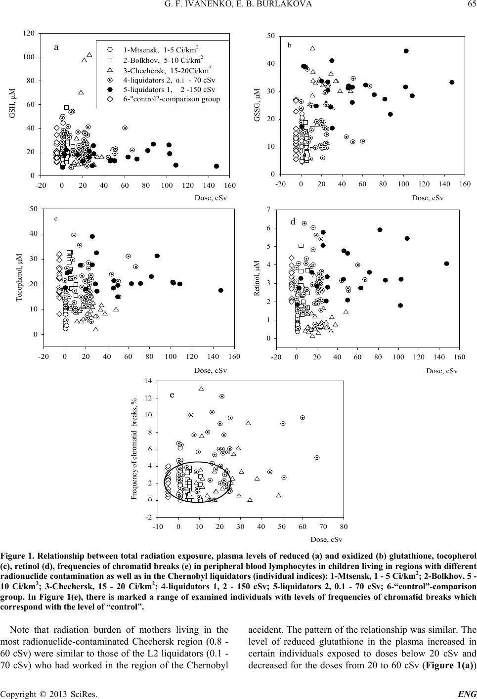
3. Results and Discussion
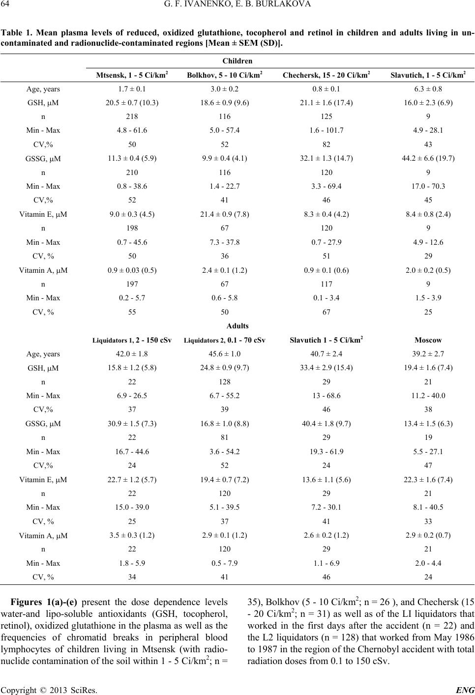
Table presents the mean level of water-and lipo-soluble
antioxidants (GSH, vitamins E and A), oxidized gluta-
thione in blood plasma of adults and children living in
uncontaminated and radionuclide-contaminated regions
after the Chernobyl accident (1 - 20 Ci/km2 by l37Cs) as
well as of the liquidators with th e radiation doses from 2
to 150 and from 0.1 to 70 cSv (group s LI and L2, respec-
tively). Table demonstrates the absence of significant
differences in the studied indices in children and adults
living in regions with different radionuclide contamina-
tion. Only the liquidators (LI) had a significantly lower
GSH and a significantly higher level of GSSG in the
blood plasma as compared to all studied residents (Table
1). However, the stability of the blood plasma indices in
children from Mtsensk (1 - 5 Ci/km2), Bolkhov (5 - 10
Ci/km2), and Chechersk (15 - 20 Ci/km2) with different
levels of soil contamination by radionuclides was consi-
derably lower as compared to people from uncontami-
nated regions. The χ2 test demonstrated that GSH level
follows the Poisson distribution. Poisson variables have
particularly high coefficients of variatio n (CV).
Table 1 shows that the CV exceeds 40% for GSH in
children living in these regions. The highest individual
variability was observed in children from the Chechersk
region with the highest radionuclide contamination (15 -
20 Ci/km2); the corresponding CV reflecting variability
of the indices was equal to 82% for GSH and 67% for
vitamin A. Levels of tocopherol and retinol in children
from Chechersk region was significantly lower than in
residents of uncontaminated regions. In addition to lipo-
peroxidative stress, children of Chechersk had a three
time s higher levels of GSSG in the blood plasma in
comparison to Mtsensk and of Bolkhov residents (Table
1).
Apparently, the high individual variability of the stu-
died indices in children depends on radiobiological prop-
erties of ionizing radiation in case of long-term exposure
of their mothers to low-level radiation. We studied the
effect of accumulated total radiation doses of mothers
(Dm) after the Chernobyl accident on long-term changes
of biochemical and cytogenetic indices in their children
in comparison with the dose effects in the Chernobyl
liquidators.
G. F. IVANENKO, E. B. BUR L AKOVA
Copyright © 2013 SciRes. ENG
64
Table 1. Mean plasma levels of reduced, oxidized glutathione, tocopherol and retinol in children and adults living in un-
contaminated and radionuclide-contaminated regions [Mean ± SEM (SD)].
Children
Mtsensk, 1 - 5 Ci/km2 Bolkhov, 5 - 10 Ci/km2 Chechersk, 15 - 20 Ci/km2 Slavutich, 1 - 5 Ci/km2
Age, years 1.7 ± 0.1 3.0 ± 0.2 0.8 ± 0.1 6.3 ± 0.8
GSH, µM 20.5 ± 0.7 (10.3) 18.6 ± 0.9 (9.6) 21.1 ± 1.6 (17.4) 16.0 ± 2.3 (6.9)
n 218 116 125 9
Min - Max 4.8 - 61.6 5.0 - 57.4 1.6 - 101.7 4.9 - 28.1
CV,% 50 52 82 43
GSSG, µM 11.3 ± 0.4 (5.9) 9.9 ± 0.4 (4.1) 32.1 ± 1.3 (14.7) 44.2 ± 6.6 (19.7)
n 210 116 120 9
Min - Max 0.8 - 38.6 1.4 - 22.7 3.3 - 69.4 17.0 - 70.3
CV,% 52 41 46 45
Vitamin E, µM 9.0 ± 0.3 (4.5) 21.4 ± 0.9 (7.8) 8.3 ± 0.4 (4.2) 8.4 ± 0.8 (2.4)
n 198 67 120 9
Min - Max 0.7 - 45.6 7.3 - 37.8 0.7 - 27.9 4.9 - 12.6
CV, % 50 36 51 29
Vitamin A, µM 0.9 ± 0.03 (0.5) 2.4 ± 0.1 (1.2) 0.9 ± 0.1 (0.6) 2.0 ± 0.2 (0.5)
n 197 67 117 9
Min - Max 0.2 - 5.7 0.6 - 5.8 0.1 - 3.4 1.5 - 3.9
CV, % 55 50 67 25
Adults
Liquidat or s 1, 2 - 150 cSv Liquida tor s 2, 0.1 - 70 cSv Slavutich 1 - 5 Ci/km2 Moscow
Age, years 42.0 ± 1.8 45.6 ± 1.0 40.7 ± 2.4 39.2 ± 2.7
GSH, µM 15.8 ± 1.2 (5.8) 24.8 ± 0.9 (9.7) 33.4 ± 2.9 (15.4) 19.4 ± 1.6 (7.4)
n 22 128 29 21
Min - Max 6.9 - 26.5 6.7 - 55.2 13 - 68.6 11.2 - 40.0
CV,% 37 39 46 38
GSSG, µM 30.9 ± 1.5 (7.3) 16.8 ± 1.0 (8.8) 40.4 ± 1.8 (9.7) 13.4 ± 1.5 (6.3)
n 22 81 29 19
Min - Max 16.7 - 44.6 3.6 - 54.2 19.3 - 61.9 5.5 - 27.1
CV,% 24 52 24 47
Vitamin E, µM 22.7 ± 1.2 (5.7) 19.4 ± 0.7 (7.2) 13.6 ± 1.1 (5.6) 22.3 ± 1.6 (7.4)
n 22 120 29 21
Min - Max 15.0 - 39.0 5.1 - 39.5 7.2 - 30.1 8.1 - 40.5
CV, % 25 37 41 33
Vitamin A, µM 3.5 ± 0.3 (1.2) 2.9 ± 0.1 (1.2) 2.6 ± 0.2 (1.2) 2.9 ± 0.2 (0.7)
n 22 120 29 21
Min - Max 1.8 - 5.9 0.5 - 7.9 1.1 - 6.9 2.0 - 4.4
CV, % 34 41 46 24
Figures 1(a)-(e) present the dose dependence levels
water-and lipo-soluble antioxidants (GSH, tocopherol,
retinol), oxidized glutathione in the plasma as well as the
frequencies of chromatid breaks in peripheral blood
lymphocytes of children living in Mtsensk (with radio-
nuclide contamination of the soil within 1 - 5 Ci/km2; n =
35), Bolkhov (5 - 10 Ci/km2; n = 26 ) , and Chechersk (15
- 20 Ci/km2; n = 31) as well as of the LI liquidators that
worked in the first days after the accident (n = 22) and
the L2 liquidators (n = 128) that worked from May 1986
to 1987 in the region of the Chernobyl accident with total
radiation doses from 0.1 to 150 cSv.
G. F. IVANENKO, E. B. BUR L AKOVA
Copyright © 2013 SciRes. ENG
65
Dose, cSv
-20 020406080100 120 140 160
GSSG, µM
0
10
20
30
40
50 b
Dose, cSv
-20 020406080100 120 140 160
Retinol, µM
0
1
2
3
4
5
6
7
d
Dose, cSv
-10 010 20 30 40 50 60 70 80
Frequency of chromatid breaks, %
-2
0
2
4
6
8
10
12
14
e
Figure 1. Relationship between total radiation exposure, plasma levels of reduced (a) and oxidized (b) glutathione, tocopherol
(c), retinol (d), frequencies of chromatid breaks (e) in peripheral blood lymphocytes in children living in regions with different
radionuclide contamination as well as in the Chernobyl liquidators (individual indices): 1-Mtsensk, 1 - 5 Ci/km2; 2-Bolkhov, 5 -
10 Ci/km2; 3-Chechersk, 15 - 20 Ci/km2; 4-liquidators 1, 2 - 150 cSv; 5-liquidators 2, 0.1 - 70 cSv; 6-“control”-comparison
group. In Figure 1(e), there is marked a range of examined individuals with levels of frequencies of chromatid breaks which
correspond with t h e l ev el o f “ control”.
Note that radiation burden of mothers living in the
most radionuclide-contaminated Chechersk region (0.8 -
60 cSv) were similar to those of the L2 liquidators (0.1 -
70 cSv) who had worked in the region of the Chernobyl
accident. The pattern of the relationship was similar. The
level of reduced glutathione in the plasma increased in
certain individuals exposed to doses below 20 cSv and
decreased for the doses from 20 to 60 cSv (Figure 1(a))
Dose, cSv
-20 020406080100 120 140 160
GSH, µM
0
20
40
60
80
100
120
1-Mtsensk, 1-5 Ci/km
2
2-Bolkhov, 5-10 Ci/km
2
3-Chechersk, 15-20Ci/km
2
4-liquidators 2, 0,1 - 70 cSv
5-liquidators 1, 2 -150 cSv
6-"control"-comparison group
a
0.1
Dose, cSv
-20 020406080100 120 140 160
Tocopherol, µM
0
10
20
30
40
50
ñ
c
G. F. IVANENKO, E. B. BUR L AKOVA
Copyright © 2013 SciRes. ENG
66
Further increase in the radiation burden to 20 - 150 cSv
increased GSSG level in the plasma (Figure l(b)) and
decreased GSH level in the plasma (Figure l(a)), which
decreased the glutathione potential. Decreased levels of
reduced glutathione under the influence of low-level
radiation (below 20 cSv) is accompanied by accelerated
consumption of vitamin E (Figure 1(c)) and increased
level of vitamin A in blood plasma (Figure 1(d)). The
deficiency of lipo-and water-soluble antioxidants reveal-
ed long after the accident (4 - 7 years) in the liquidators
and children of exposed mothers is an important health
marker for populations exposed to chronic low-level in
radiation. The system of water- and liposoluble antioxi-
dants differently responses to radiation exposure of low
intensi t y depending on the ex posure dose of ra diatio n.
Different responses to the studied systems in human
population to the effect of low-level radiation can be due
to an increased sensitivity of the systems to radiation
resulting from disturbed control mechanisms involving
free radicals. High stationery concentrations of active
oxygen forms at the low doses confirm proposed delay”
of the repairer systems [15]. Low efficiency of the re-
pairer systems increase the level of spontaneous chro-
mosomal disturbances, which induce mutations in genes
and increase the incidence of cancer [16].
This conclusion is confirmed by an increase in cyto-
genetic disturbances in lymphocytes of peripheral blood
determined upon an individual examination of this group
of people (children and liquidators). Figure 1(e) presents
the individual dose dependence of the frequencies of
chromatid breaks in peripheral blood lymphocytes of
children (Mtsensk, 1 - 5 Ci/km 2; Bolkhov, 5 - 10 Ci/km2;
and Chechersk, 15 - 20 Ci/km2) living in regions with
different levels of radionuclide contamination of the soil
after the Chernobyl accident as well as of liquidators 2,
(0.1 to 70 cSv.) These experiments demonstrate the com-
plex dependence of the frequencies of chromatid breaks
in lymphocytes in human population on radiation doses.
For instance, at low radiation doses (from 0.8 to 30 cSv),
some individuals have a considerably increased frequen-
cies of chromatid breaks (up to 12 per 100 cells; F igure
1(e)) in lymphocytes as compared to those in people not
exposed to ionizing radiation. A two-or three-fold in-
crease in the cytogenetic indices is observed in over 20%
people at low radiation doses. When the doses increased
from 30 to 60 c Sv, the frequencies of chromatid breaks in
peripheral blood lymphocytes also increased (up to 5 per
100 cells; Figure 1(e)). A 3 - 5-fold deviation of indi-
vidual cytogenetic and biochemical indices from the
control (Figure 1) indicates a high rate of mutations in
somatic cells of children and L2 even after a long period
after the accident (5 - 7 years). This fact can be consi-
dered as an epidemiological confirmation of the role of
chromosomal aberrations in carcinogenesis, since the
Figure 2. Relationship between frequencies of chromatid
breaks in peripheral blood lymphocytes and plasma levels of
reduced glutathione, in children living in regions with dif-
ferent radionuclide contamination as well as in the Cher-
nobyl liquidators 2 (individual indices). In Figure 2, there is
marked a range of examined individuals with levels of fre-
quencies of chromatid breaks and levels GSH of people
which correspon d with the level of “ co n t ro l” .
risk of cancer increases in individuals with high level of
spontaneous chromosomal aberrations [16].
It was substantiated the studies of antioxidant status
and role of cytogenetic examinations for predicting of the
delayed consequences of irradiation. The development of
various biochemical and the cytogenetic tests are of great
importance for evaluation and prediction of individual
radiosensitivity of the organism.
As an example, let us present data on the correlation
between cytogenetic indices in peripheral blood lym-
phocytes and plasma levels GSH of people exposed to
low doses of radiation (Figure 2). We observed unidirec-
tional and bidirectional changes frequencies of chromatid
breaks and biochemical indices of human blood (positive
and negative correlation).
Quantitatively different response of the body to low
(below 20 cSv) and high (from 20 to 150 cSv) doses of
ionizing radiation is an unbiased marker of radiation-
induced disorders and underlies development of far dis-
orders in human population. To the great probability, the
low-rate radiation possesses another mechanism com-
pared wit h hi gh doses of a c ute radia tion.
REFERENCES
[1] W. F. Morgan and J. L. Schwartz, “Environmental Muta-
gen Society Symposium on Risks of Low Dose, Low
Dose Rate Exposures of Ionizing Radiation to Humans,
International Journal of Radiation Biology, Vol. 83, 2007,
pp. 491-499.
http://dx.doi.org/10.1080/09553000701370886
0.1
G. F. IVANENKO, E. B. BUR L AKOVA
Copyright © 2013 SciRes. ENG
67
[2] A. V. Akleev “Tissue Reaction under Chronic Exposure
to Ionizing Radiation,Radiation Biology. Radioecology
(Russian), Vol. 49, No. 1, 2009, pp. 5-20.
[3] N. E. A. Crompton, “Programmed Cell Response to Io-
nizing Radiation Damage,Acta Oncologica, Vol. 37,
1998, pp. 129-142.
http://dx.doi.org/10.1080/028418698429685
[4] R. D. Govorun, “Cytogenetic Damage and Mutagenesis
in Mammalian and Human Cells Induced by Ionizing
Radiation with Varying LET,Radiation Biology. Radio-
ecology (Russian), Vol. 37, No. 4, 1997, pp. 539-548.
[5] J. Glavind, “Antioxidants in Animal Tissue,Acta Che-
mica Scandinavica, Vol. 17, 1963, pp. 1635-1640.
http://dx.doi.org/10.3891/acta.chem.scand.17-1635
[6] V. V. Sokolovskii, “A Thiol-Disul fide System in n Orga-
nism Reaction to Environmental Factors,” Nauka, Sankt-
Peterburg, 2008, p. 111.
[7] E. B. Burlakova A. V. Alesenko, E. M. Molochkina, N. P.
Palmina and N. G. Khrapova, “Biological Antioxidants in
Radiation Damage and Malignant Growth,” Nauka, Mos-
cow, 1975, p. 211.
[8] G. F. Ivanenko, “The Role of Antioxidant Activity of
Lipids and Endogenous Thiols in Radio Resistance of the
Organism,” Extended Abstract of Cand. Set (Biol.) Dis-
sertation, Moscow, 1985, p. 168.
[9] D. D. M. Wayner, G. W. Burton, K. U. Ingold, L. R. C.
Barclay and S. J. Locke “The Relative Contributions of
Vitamin E, Urate, Ascorbate and Proteins to the Peroxyl
Radical-Trapping Antioxidant Activity of Human Blood
Plasma,Biochemica et Biophysica Acta, Vol. 92 4, 1987,
pp. 408-419.
http://dx.doi.org/10.1016/0304-4165(87)90155-3
[10] F. B. Pruijn, G. R. M. M. Haenen and A. Bast “Integray
between Vitamin E, Glutathione and Dihydrolipoic Acid
in Protection against Lipid Peroxidation,Fat Science
Technology, Vol. 93, No. 6, 1991, pp. 216-221.
[11] G. L. Ellman and H. Lysko, “Disulfide and Sulfhydryl
Compounds in TCA Extracts of Human Blood and Plas-
ma,Journal of Laboratory and Clinical Medicine, Vol.
70, 1967, pp. 51 8-527.
[12] T. L. Mc Neil and L. Y. Beck, “Fluorometric Estimation
of GSH-OPT,” Analytical Biochemistry, Vol. 22, 1968,
pp. 431-441.
http://dx.doi.org/10.1016/0003-2697(68)90285-6
[13] L. G. Hansen and W. G. Warwick “A Fluorometric Mi-
cromethod for Serum Vitamins A and E,” Technical Bul-
letin of the Registry of Medical Technologists, Vol. 39,
No. 3, 1969, pp. 70-73.
[14] M. A. Bender, A. A. Awa, A. L. Brooks, P. G. Groer, L.
Littlefield, C. Pereira, R. J. Pre ston and B. W. Wachhol z,
“Current Status of Cytogenetic Procedures to Detect and
Quantify Previous Exposures to Radiation,Mutation Re-
search, Vol. 196, 1988, pp. 103-159.
http://dx.doi.org/10.1016/0165-1110(88)90017-6
[15] L. S. Vartyanyan, S. M. Gurevich, A. I. Kozachenko, L.
G. Nagler, E. L. Lozovskaya and E. B. Burlakova, “Changes
in Superoxide Production Rate and in Superoxide Dis-
mutase and Glutathione Peroxidase Activities in Subcel-
lular Organelles in Mouse Liver under Exposure to Low
Doses of Low-Intensity Radiation,Biokhimiya (Russian),
Vol. 65, No. 4, 2000, pp. 522-527.
[16] L. Hagmar, A. Brogger, I. L. Hansteen, S. Heim, B.
Hogstedt, K. Linnainma, F. Mitelman, I. Nordenson, et
al., “Cancer Risk in Humans Predicted by Increased Le-
vels of Chromosomal Aberration in Lymphocytes: Nordic
Study Group on the Health Risk of Chromosome Dam-
age,Cancer Res earch, Vol. 54, 1994, pp. 2919-2922.