L. Marques et al. / Case Reports in Clinical Medicine 2 (2013) 381-385
Copyright © 2013 SciRes. OPEN ACCESS
384
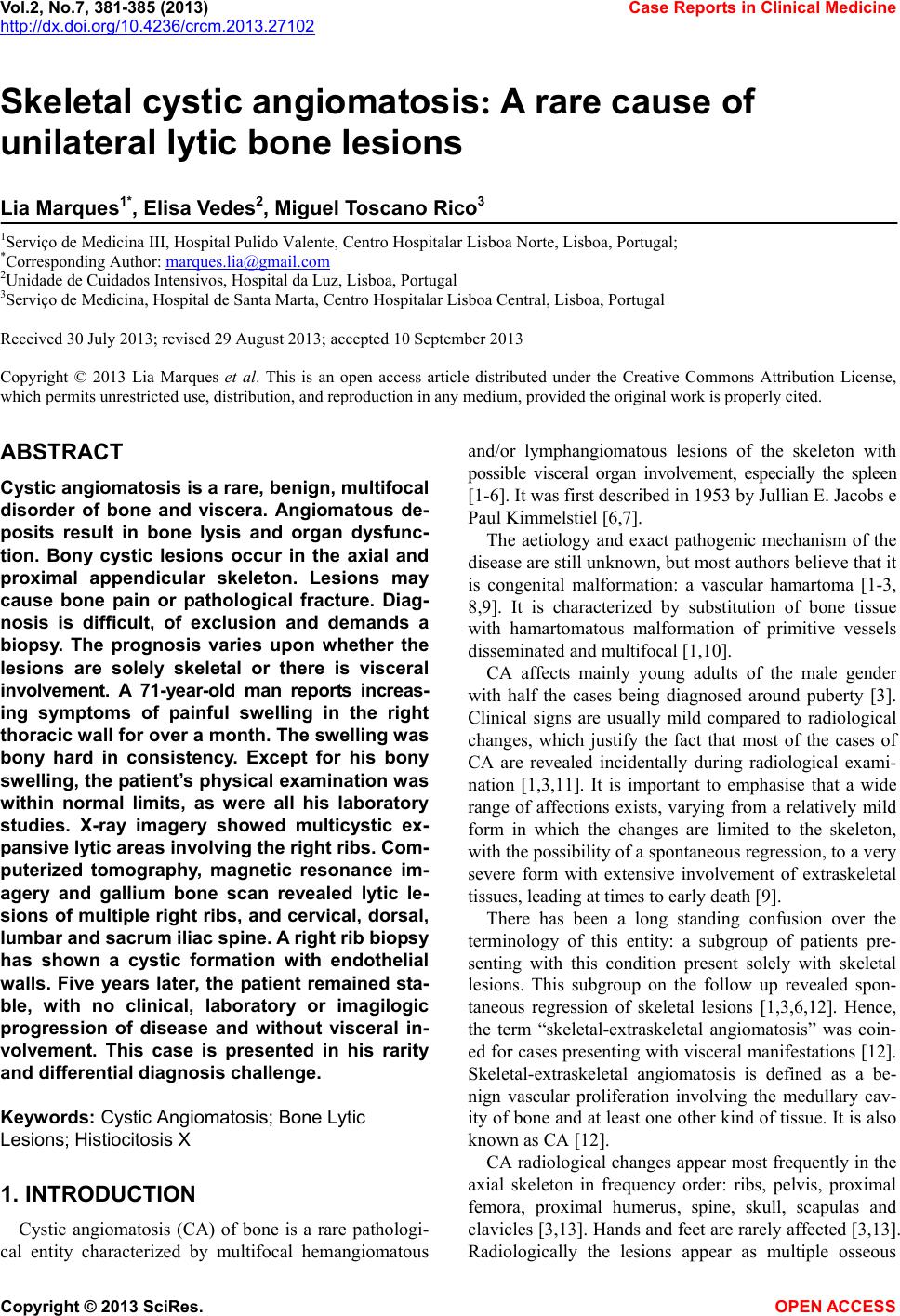
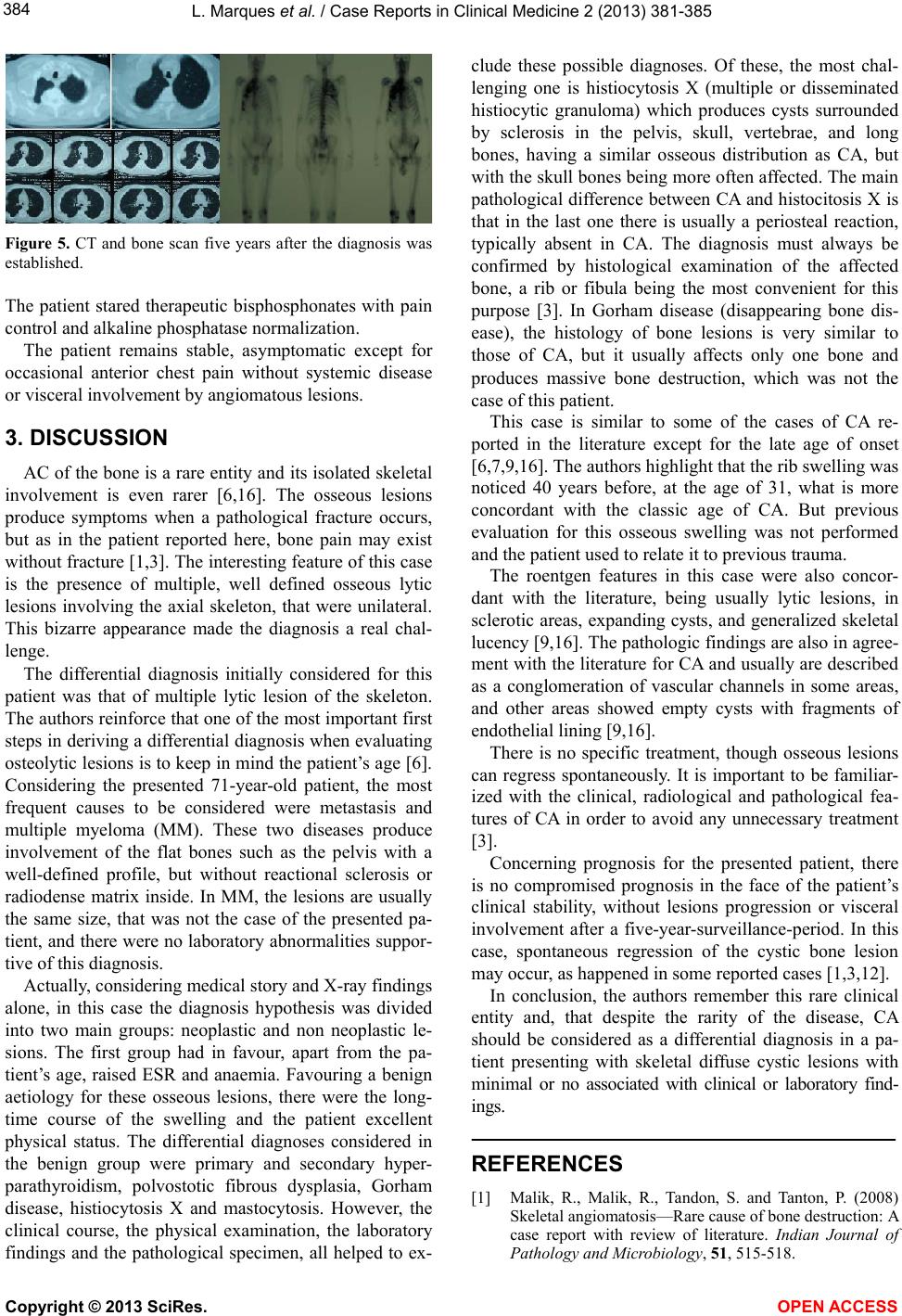
Figure 5. CT and bone scan five years after the diagnosis was
established.
The patient stared therapeutic bisphosphonates with pain
control and alkaline phosphatase normalization.
The patient remains stable, asymptomatic except for
occasional anterior chest pain without systemic disease
or visceral involvement by angiomatous lesions.
3. DISCUSSION
AC of the bone is a rare entity and its isolated sk eletal
involvement is even rarer [6,16]. The osseous lesions
produce symptoms when a pathological fracture occurs,
but as in the patient reported here, bone pain may exist
without fracture [1,3]. The interesting feature of this case
is the presence of multiple, well defined osseous lytic
lesions involving the axial skeleton, that were unilateral.
This bizarre appearance made the diagnosis a real chal-
lenge.
The differential diagnosis initially considered for this
patient was that of multiple lytic lesion of the skeleton.
The authors reinforce that one of the most important first
steps in deriving a differential diagnosis when evaluating
osteolytic lesions is to keep in mind the patient’s age [6 ].
Considering the presented 71-year-old patient, the most
frequent causes to be considered were metastasis and
multiple myeloma (MM). These two diseases produce
involvement of the flat bones such as the pelvis with a
well-defined profile, but without reactional sclerosis or
radiodense matrix inside. In MM, the lesions are usually
the same size, that was not the case of the presented pa-
tient, and there were no laborato ry abnormalities suppor-
tive of this diagnosis.
Actually, considering medical story and X-ray findings
alone, in this case the diagnosis hypothesis was divided
into two main groups: neoplastic and non neoplastic le-
sions. The first group had in favour, apart from the pa-
tient’s age, raised ESR and anaemia. Favouring a benign
aetiology for these osseous lesions, there were the long-
time course of the swelling and the patient excellent
physical status. The differential diagnoses considered in
the benign group were primary and secondary hyper-
parathyroidism, polvostotic fibrous dysplasia, Gorham
disease, histiocytosis X and mastocytosis. However, the
clinical course, the physical examination, the laboratory
findings and the pathological specimen, all helped to ex-
clude these possible diagnoses. Of these, the most chal-
lenging one is histiocytosis X (multiple or disseminated
histiocytic granuloma) which produces cysts surrounded
by sclerosis in the pelvis, skull, vertebrae, and long
bones, having a similar osseous distribution as CA, but
with the skull bone s being more often affected. The main
pathological difference between CA and histocitosis X is
that in the last one there is usually a periosteal reaction,
typically absent in CA. The diagnosis must always be
confirmed by histological examination of the affected
bone, a rib or fibula being the most convenient for this
purpose [3]. In Gorham disease (disappearing bone dis-
ease), the histology of bone lesions is very similar to
those of CA, but it usually affects only one bone and
produces massive bone destruction, which was not the
case of this patient.
This case is similar to some of the cases of CA re-
ported in the literature except for the late age of onset
[6,7,9,16]. The authors highligh t that the rib swelling was
noticed 40 years before, at the age of 31, what is more
concordant with the classic age of CA. But previous
evaluation for this osseous swelling was not performed
and the patient used to relate it to previous trauma.
The roentgen features in this case were also concor-
dant with the literature, being usually lytic lesions, in
sclerotic areas, expanding cysts, and generalized skeletal
lucency [9,16]. The pathologic findings are also in agree-
ment with the literature for CA and usua lly are described
as a conglomeration of vascular channels in some areas,
and other areas showed empty cysts with fragments of
endothelial lining [9,16].
There is no specific treatment, though osseous lesions
can regress spontaneously. It is important to be familiar-
ized with the clinical, radiological and pathological fea-
tures of CA in order to avoid any unnecessary treatment
[3].
Concerning prognosis for the presented patient, there
is no compromised prognosis in the face of the patient’s
clinical stability, without lesions progression or visceral
involvement after a five-year-surveillance-period. In this
case, spontaneous regression of the cystic bone lesion
may occur, as happened in some reported cases [1,3,12].
In conclusion, the authors remember this rare clinical
entity and, that despite the rarity of the disease, CA
should be considered as a differential diagnosis in a pa-
tient presenting with skeletal diffuse cystic lesions with
minimal or no associated with clinical or laboratory find-
ings.
REFERENCES
[1] Malik, R., Malik, R., Tandon, S. and Tanton, P. (2008)
Skeletal an gi o matosis—Rar e cause of bone destruction: A
case report with review of literature. Indian Journal of
Pathology and Microbiology, 51, 515-518.