M. M. Harutunian et al. / Open Journal of Stomatology 3 (2013) 335-337
336
to reproduce the greatest detail of all the impression ma-
terials, preparing electronic versions of their papers.
2. TECHNIQUE
A healthy 56-year-old male who had gone five years
without visiting a dentist presen ted to New York Univer-
sity College of Dentistry for dental treatment. A com-
prehensive examination was performed and he was
treatment planned to have porcelain veneers and crowns
to enhance his smile. It was noted from the start that he
presented with existing black triangles due to his mild
periodontal disease.
Following discussions of risks and benefits of the
treatment and obtaining informed consent, 4 teeth were
prepared for porcelain venee r s and 1 full ceramic crown.
Medium body polyvinyl siloxane material (Reprosil,
Denstply Caulk, Milford, DE) was used. The tray fit was
checked and loaded. This material was left to set in place
according to the manufacturer’s instructions, 3 minutes
working time and 3 minutes setting time. Once the sec-
ond phase of impression material set, the tray was re-
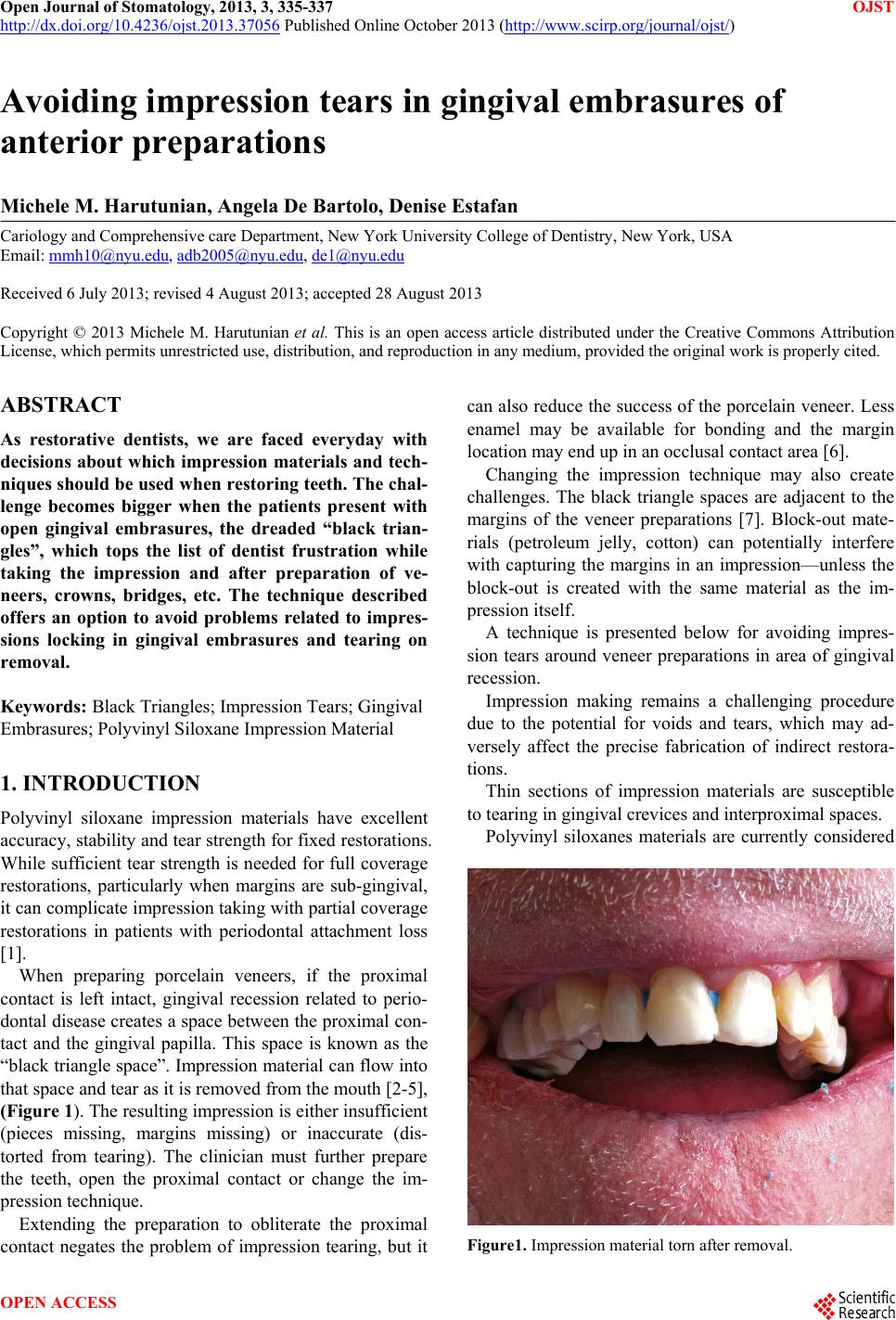
moved. The result was a torn impression in the cervical
areas of the teeth, (black triangles), shown in (Figure 1).
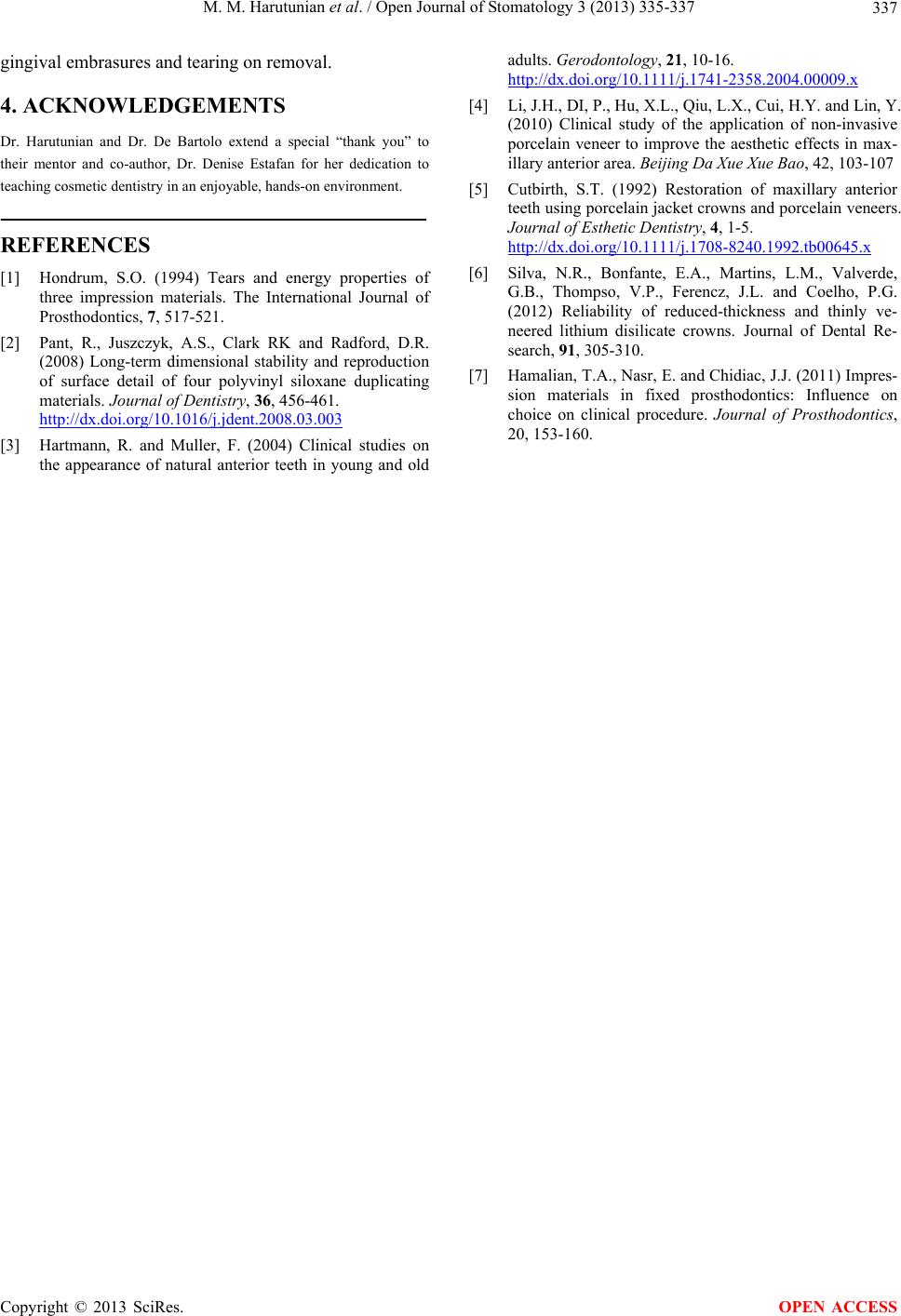
Medium body polyvinyl siloxane material (Reprosil,
Denstply Caulk, Milford, DE) was placed at the palatal
of the prepared teeth, short of the margins (Figure 2). It
was left to set in place according to the manufacturer’s
instructions. The tray was removed. The impression ma-
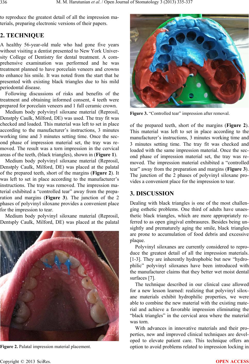
terial exhibited a “controlled tear” away from the prepa-
ration and margins (Figure 3). The junction of the 2
phases of polyvinyl siloxane provides a convenient place
for the impression to tear.
Medium body polyvinyl siloxane material (Reprosil,
Dentsply Caulk, Milford, DE) was placed at the palatal
Figure 2. Palatal impression material placement.
Figure 3. “Controlled tear” impression after removal.
of the prepared teeth, short of the margins (Figure 2).
This material was left to set in place according to the
manufacturer’s instructions, 3 minutes working time and
3 minutes setting time. The tray fit was checked and
loaded with the same impression material. Once the sec-
ond phase of impression material set, the tray was re-
moved. The impression material exhibited a “controlled
tear” away from the preparation and margins (Figure 3).
The junction of the 2 phases of polyvinyl siloxane pro-
vides a convenient place for the impression to tear.
3. DISCUSSION
Dealing with black triangles is one of the most challen-
ging esthetic problems. One third of adults have unaes-
thetic black triangles, which are more appropriately re-
ferred to as open gingival embrasures. Besides being un-
sightly and prematurely aging the smile, black triangles
are prone to accumulation of food debris and excessive
plaque.
Polyvinyl siloxanes are currently considered to repro-
duce the greatest detail of all the impression materials.
[1-3]. They are inherently hydrophobic but new “hydro-
philic” polyvinyl siloxanes have been introduced with
the manufacturer claims that they better wet moist dental
surfaces [7].
The technique described in our clinical case allowed
for a new lesson learned: realizing that polyvinyl silox-
ane materials exhibit hydrophilic properties, we were
able to combine the new material with the existing mate-
rial and achieve a favorable impression eliminating the
“black triangles” in the cervical area where the material
was torn.
With advances in innovative materials and their pro-
perties, new and improved clinical techniques are devel-
oped to elevate patient care. This technique offers an
option to avoid problems relate d to impression lo cking in
Copyright © 2013 SciRes. OPEN ACCESS