Advances in Bioscience and Biotechnology, 2013, 4, 945-948 ABB
http://dx.doi.org/10.4236/abb.2013.410125 Published Online October 2013 (http://www.scirp.org/journal/abb/)
Cortical columns (barrels) display normal size in the
brain’s primary somatosensory cortex of mice carrying
null mutations of the insulin receptor substrate 1 gene:
A preliminary report
Marta López-Santibáñez Guevara1, Eileen Uribe-Querol2, Alma Lilia Fuentes Farías3*,
Esperanza Meléndez-Herrera3, Agustine Joseph D’Ercole4, Gabriel Gutiérrez-Ospina1,5*
1Departamento de Biología Celular y Fisiología, Instituto de Investigaciones Biomédicas, Universidad Nacional Autónoma de
México, México D.F., México
2División de Estudios de Posgrado e Investigación, Facultad de Odontología, Universidad Nacional Autónoma de México, México
D.F., México
3Departamento de Zoología, Instituto de Investigaciones sobre los Recursos Naturales, Universidad Michoacana de San Nicolás de
Hidalgo, Morelia, México
4Department of Pediatrics, The University of North Carolina at Chapel Hill, Chapel Hill, USA
5Coordinación de Psicofisiología, Facultad de Psicología, Universidad Nacional Autónoma de México, México D.F., México
Email: *afuentes@umich.mx, *gabo@biomedicas.unam.mx
Received 10 August 2013; revised 10 September 2013; accepted 25 September 2013
Copyright © 2013 Marta López-Santibáñez Guevara et al. This is an open access article distributed under the Creative Commons At-
tribution License, which permits unrestricted use, distribution, and reproduction in any medium, provided the original work is prop-
erly cited.
ABSTRACT
Circuits in barrels of the rodent brain’s primary
somatosensory (S1) cortex build up following con-
structivist rules. Previous evidence in mice supports
that the precise addition of barrel neuropil is pro-
moted by insulin-like growth factor-1 (IGF-1). The
signaling cascades mediating this response remain
undetermined. To address whether the effects of IGF-
1 upon the growth of S1 circuits are mediated by in-
sulin receptor substrate-1 (IRS-1), we studied barrel
size in adult mice having the IRS-1 gene knocked out
(IRS-1 ko). Our results reveal that barrel size is simi-
lar between wild type and IRS-1 ko mice suggesting
that IRS-1 is not essential for barrel circuitry growth.
Hence, investigations aimed at exploring other sub-
strates activated by IGF-1, namely IRS-2 and IRS-4,
are needed to reveal signaling pathways that mediate
the precise addition of S1 neuronal circuitry.
Keywords: Barrel Cortex; Neu ronal Cir cuit As semblage;
Trophic Interactions; Constructivism
1. INTRODUCTION
Conventional wisdom claims that mature neuronal cir-
cuits arise from an initially redundant set of connections
which are later refined through a process of selective
pruning driven by neuronal activity and competition
[1-4]. Empirical evidence suggests, however, that this
principle is far from universal since circuits in the spinal
cord [5,6], the olfactory bulb [7,8] and in the primary
visual [9] and somatosensory (S1) [10-13] cortices of
different mammals (however, see [14]), are predomi-
nantly elaborated by adding up connections and neuronal
processes [15].
The ce reb ral co rtex is for med by colu mns th at r un ver-
tically through its entire thickness. In S1, these columns
are called barrels and they represent collections of me-
chanosensory receptors distributed throughout the body.
In previous reports, we confirmed that barrel neuropil
and synaptic contacts are added through postnatal de-
velopment. More importantly, the ad dition of barrel neu-
ropil/synapses depended upon the availability of insulin-
like growth factor-1 (IGF-1). Indeed, barrels in trans-
genic mice overexpressing IGF-1 were enlarged, whereas
those in transgenic mice having brain ectopic expression
of IGF-binding-protein-1 displayed reduced size [16].
Both enlarged and small sized barrels had similar syn-
apse density [17]. These results thus support the concept
that neuronal circuits are elaborated by the precise, dif-
ferential addition of new elements through non-competi-
tive, constructive processes that depend upon the avail-
ability of neurotrophic factors (see [11] and [18] for a
*Corresponding authors.
OPEN ACCESS
M. L.-S. Guevara et al. / Advances in Bioscience and Biotechnology 4 (2013) 945-948
946
similar conclusion).
Considerable research has been conducted to under-
stand the cellular and molecular mechanisms that under-
lie the competitive and selective pruning of neuronal
connections. However, much less is known about the
molecular underpinnings supporting the formation and
growth of neuronal connections through constructivist
processes. Although molecules governing axonal and
dendritic navigation and growth may readily explain pre-
cise targeting and directed growth [19], the molecular
underpinnings that promote the precise addition of neu-
ronal circuits remain unexplored. Given that IGF-1 over-
expression promotes this process in S1 barrels, it is pos-
sible that molecules along the IGF-1 signaling pathway
may well underlie constructivist events during S1 as-
semblage.
IGF-1 binds to the IGF receptor typ e 1 that, after acti-
vation, phosphorylates signaling proteins of the insulin
receptor substrate (IRS) family. IRSs, in turn, activate
effector proteins containing Src homology 2 domains, a
process that activates downstream MAP Kinase and the
phosphatidylinositol 3-kinase (PI3K) signaling cascades
[20-23]. A member of the IRS family is IRS-1. IRS-1
mRNA is expressed in the cerebral cortex [24] and its
null mutation retards brain growth and reduces brain
weight, presumably by impairing IGF-1 actions on brain
growth [25,26]. IRS-1 might then be involved in promot-
ing the precise and progressive addition of barrel neu-
ropil following IGF-1 binding to its receptor. Hence, by
combining succinic dehydrogenase (SDH) histochemis-
try and image analyses, we evaluated whether adult wild
type (wt) mice and those carrying null mutations of the
IRS-1 gene (IRS-1ko mice) displayed differences in bar-
rel size.
2. MATERIALS AND METHODS
2.1. Mice
Studies were performed in genotyped adult male wild
type (n = 3) and homozygous IRS-1 ko mice (n = 4) gen-
erously provided by Dr. C. Ronald Kahn (Joslin Diabetes
Center and Department of Medicine, Harvard Medical
School, Boston, MA). Upon arrival, mice were kept until
the day of sacrifice under 12:12 hours light/dark cycle at
22˚C having free access to food and water. Animal han-
dling and procedures followed the guidelines of the NIH
guide for the care and use of experimental animals and
were approved by the institutional review committee at
the University of North Carolina (UNC), Chapel Hill.
2.2. Histological Procedures
The anesthetized (ketamine/xylazine; 900/20 µg/gr body
weight) mice were perfused with physiological saline
followed by 10% glycerol. Brains were removed and
weighed and the cerebral cortex was dissected and frozen
flattened [13]. Serial tangential sections (30 µm) were
cut in a cryostat and stained for the activity o f the oxida-
tive enzyme, succinic dehydrogenase (SDH) by means of
SDH histochemistry [13]. Briefly, sections were dried on
the slides at 37˚C for 30 min; fixed in 10% formalin, pH
7.6, at 4˚C for 1 min and rinsed in distilled water. Then
they were stained by reacting them with 0.05 M sodium
succinate and 0.55 mM nitroblue tetrazolium in 0.05 M
phosphate buffer, pH 7.6. Slides were rinsed and cover-
slipped with DPX. Animal perfusion and sample staining
were carried out at UNC.
2.3. Estimation of Barrel Cross-Sectional and
Posteromedial Barrel Subfield Areas
As previously commented, S1 barrels in the mouse brain
represent collections of mechanosensory receptors lo-
cated along the body. The body has different segments
though. Barrels are then segregated into distinct body
representations. From these representations, the most
conspicuous is that known as the posteromedial barrel
subfield (PMBSF) in which each PMBSF barrel repre-
sents each facial vibrissae. The histological material ob-
tained from wt and IRS-1 ko mice was transferred to the
Universidad Nacional Autónoma de México where mor-
phometry of PMBSF barrels was conducted. Two dimen-
sional maps of PMBSF were drawn under bright field
microscope equipped with a camera lucida. Completed
maps were digitized and PMBSF and barrel cross-sec-
tional areas were estimated using Image-Pro (Media Cy-
bernetics, Silver Spring, MD), as described elsewhere
[13].
2.4. Data Analyses
Data are reported as means ± standard deviation for both
experimental groups. PMBSF barrel area and barrel
cross-sectional area were statistically compared between
wt and IRS-1 ko mice by using One-way ANOVA test
followed by a Tukey’s post hoc test (OriginPro v 7;
OriginLab Corporation) setting the significance value at
p < 0.05.
3. RESULTS
The number, shape and anatomical definition of the
PMBSF were fully comparable between wt and IR S-1 ko
mice (Figure 1). Although average barrel area (wt: 0.04761
± 0.00256 versus IRS-1 ko: 0.04481 ± 0.00076 both in
cm2), total barrel field area (wt: 1.5233 ± 0.08172 versus
IRS-1 ko: 1.4673 ± 0.03872 both in cm2) and PMBSF
area (wt: 1.8645 ± 0.16628 versus IRS-1 ko: 1.6626 ±
0.04809 both in cm2) tended to be reduced in IRS-1 ko
mice, such trends were not statistically significant.
Copyright © 2013 SciRes. OPEN ACCESS
M. L.-S. Guevara et al. / Advances in Bioscience and Biotechnology 4 (2013) 945-948 947
wt IRS-1 ko
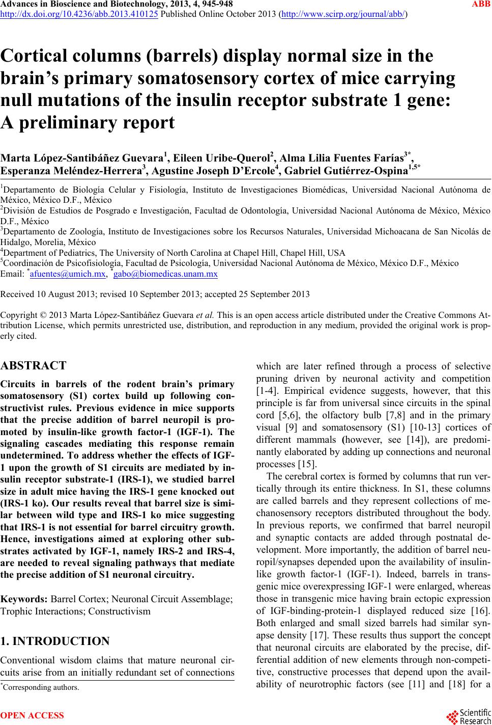
Figure 1. Representative camera lucida drawings of the barrel
field of wild type (wt) and IRS-1 knock out (IRS-1 ko) mice.
No qualitative differences were observed between mouse groups
(Scale = 1 mm).
4. DISCUSSION
Previous reports in rodents support that barrel circuitry in
S1 builds up following constructivist principles [10-13,
15]. We have shown that the precise and progressive ad-
dition of barrel neuropil is promoted by IGF-1 [15,16].
The signaling cascade involved in this event has not been
elucidated. We then explored the contribution of IRS-1 in
this process by comparing the adult size of SI barrels
between wt and IRS-1 ko mice. IRS-1 has been previ-
ously shown to be expressed in the cerebral cortex [24]
and its mutation retards brain growth and reduces brain
weight [25,26]. Unexpectedly, IRS-1 ko mice displayed
barrel and PMBSF areas fully comparable to those ob-
served in wt mice, thus supporting that IRS-1 is not es-
sential for promoting the precise addition of barrel neu-
ropil. In a previous report, Ye et al. [26] have shown that
IRS-2 and IRS-4 may compensate the lack of IRS-1 with
regard to myelination processes, and it is therefore con-
ceivable that either one or both could also “rescue” the
barrel’s phenotype in IRS-1 ko mice. In sum, IRS-1 does
not seem to mediate the trophic effects of IGF1 on the
barrel cortex. Future experiments must address whether
IRS-2 and/or IRS-4 participate in the construction of
barrel neuropil.
5. ACKNOWLEDGEMENTS
Authors thank Jesús Ramirez Santos, Ivonne Mora, Edel Pineda Lopez
and Raymundo Reyes for technical assistance. This work was sup-
ported in part by CONACyT (Grant No. 82879 to G. G. O.), PAPIIT-
UNAM (Grants Nos. IN203912-3 to G. G. O. and IA202013-2 to E. U.
Q.), CIC-UMSNH (Grant No. 8.37 to A. L. F. F.).
REFERENCES
[1] Bennet, M.R., Gibson, W.G. and Lemon, G. (2002) Neu-
ronal cell death, nerve growth factor and neurotrophic
models: 50 years on. Autonomic Neuroscience: Basic &
Clinical, 95, 1-23.
http://dx.doi.org/10.1016/S1566-0702(01)00358-7
[2] Blankenship, A.G. and Feller, M.B. (2010) Mechanisms
underlying spontaneous patterned activity in developing
neural circuits. Nature Reviews Neuroscience, 11, 18-29.
http://dx.doi.org/10.1038/nrn2759
[3] Buss, R.R., Sun, W. and Oppenheim, R.W. (2006) Adap-
tive roles of programmed cell death during nervous sys-
tem development. Annual Review of Neuroscience, 29,
1-35.
http://dx.doi.org/10.1146/annurev.neuro.29.051605.11280
0
[4] Zweifel, L.S., Kuruvilla, R. and Ginty, D.D. (2005) Func-
tions and mechanisms of retrograde neurotrophin signal-
ling. Nature Reviews Neuroscience, 6, 615-625.
http://dx.doi.org/10.1038/nrn1727
[5] Konstantinidou, A.D., Silos-Santiago, I., Flaris, N. and
Snider, W.D. (1995) Development of the primary afferent
projection in human spinal cord. Journal of Comparative
Neurology, 354, 11-12.
http://dx.doi.org/10.1002/cne.903540102
[6] Silos-Santiago, I., Jeng, B. and Snider, W.D. (1995) Sen-
sory afferents show appropriate somatotopy at the earliest
stage of projection to dorsal horn. Neuroreport, 6, 861-
865.
http://dx.doi.org/10.1097/00001756-199504190-00009
[7] Pomeroy, S.L., LaMantia, A.S. and Purves, D. (1990)
Postnatal construction of neural circuitry in the mouse
olfactory bulb. Journal of Neuroscience, 10, 1952-1966.
[8] Valle-Leija, P., Blanco-Hernández, E., Drucker-Colín, R.,
Gutiérrez-Ospina, G. and Vidaltamayo, R. (2012) Super-
numerary formation of olfactory glomeruli induced by
chronic odorant exposure: A constructivist expression of
neural plasticity. PloS ONE, 7, e35358.
http://dx.doi.org/10.1371/journal.pone.0035358
[9] Crowley, J.C. and Katz, L.C. (2002) Ocular dominance
development revisited. Current Opinion in Neurobiology,
12, 104-109.
http://dx.doi.org/10.1016/S0959-4388(02)00297-0
[10] Agmon, A., Yang, L.T., O’Dowd, D.K. and Jones, E.G.
(1993) Organized growth of thalamocortical axons from
the deep tier of terminations into layer IV of developing
mouse barrel cortex. Journal of Neuroscience, 13, 5365-
5382.
[11] Uribe-Querol, E., Martínez-Martínez, E., Hernández,
L.R., Padilla Cort és, P., Merchant-Larios, H. and, Gutiér-
rez-Ospina, G. (2013) Selective and constructive mecha-
nisms contribute to neural circuit formation in the barrel
cortex of the developing rat. Advances in Bioscience and
Biotechnology, 4, 785-797.
http://dx.doi.org/10.4236/abb.2013.47103
[12] Catalano, S.M., Robertson, R.T. and Killackey, H.P.
(1996) Individual axon morphology and thalamocortical
topography in developing rat somatosensory cortex. Jour-
nal of Comparative Neurology, 367, 36-53.
http://dx.doi.org/10.1002/(SICI)1096-9861(19960325)36
7:1<36::AID-CNE4>3.0.CO;2-K
[13] Riddle, D., Richards, A., Zsuppan, F. and Purves, D.
(1992) Growth of the rat somatic sensory cortex and its
Copyright © 2013 SciRes. OPEN ACCESS
M. L.-S. Guevara et al. / Advances in Bioscience and Biotechnology 4 (2013) 945-948
Copyright © 2013 SciRes.
948
OPEN ACCESS
constituent parts during postnatal development. Journal
of Neuroscience, 12, 3509-3524.
[14] Rebsam, A., Seif, I. and Gaspar, P. (2005) Dissociating
barrel development and lesion-induced plasticity in the
mouse somatosensory cortex. Journal of Neuroscience,
25, 706-710.
http://dx.doi.org/10.1523/JNEUROSCI.4191-04.2005
[15] Gutiérrez-Ospina, G., Calikoglu, A.S., Ye, P., D’Ercole
and A.J. (1996) In vivo effects of insulin-like growth fac-
tor-I on the development of sensory pathways: analysis of
the primary somatic sensory cortex (S1) of transgenic
mice. Endocrinology, 137, 5484-5492.
http://dx.doi.org/10.1210/en.137.12.5484
[16] Gutiérrez-Ospina, G., Uribe-Querol, E., Sánchez, N.,
Geovannini, H., Padilla, P. and Hernández-Echeagaray, E.
(2004) Similar synapse density in layer IV columns of the
primary somatosensory cortex of transgenic mice with
different brain size: Implications for mechanisms under-
lying the differential allocation of cortical space. Brain
Behavior and Evolution, 64, 61-69.
http://dx.doi.org/10.1159/000079116
[17] Quartz, S.R. and Sejnowski, T.J. (1997) The neural basis
of cognitive development: A constructivist manifesto.
The Behavioral and Brain Sciences, 20, 537-596.
http://dx.doi.org/10.1017/S0140525X97001581
[18] Hodge, R.D., D’Ercole, A.J. and O’Kusky, J.R. (2005)
Increased expression of insulin-like growth factor-I (IGF-
I) during embryonic development produces neocortical
overgrowth with differentially greater effects on specific
cytoarchitectonic areas and cortical layers. Developmen-
tal Brain Research, 154, 227-237.
http://dx.doi.org/10.1016/j.devbrainres.2004.10.016
[19] López-Bendito, G. and Molnár, Z. (2003) Thalamocorti-
cal development: How are we going to get there? Nature
Reviews Neuroscience, 4, 276-289.
http://dx.doi.org/10.1038/nrn1075
[20] White, M.F. (1997) The insulin signallling system and the
IRS proteins. Diabetologia, 40, S2-S17.
http://dx.doi.org/10.1007/s001250051387
[21] Mardilovich, K., Pankratz, S.L. and Shaw, L.M. (2009)
Expression and functions of the insulin receptor substrate
proteins in cancer. Cell Communication and Signaling, 7,
14. http://dx.doi.org/10.1186/1478-811X-7-14
[22] Withers, D.J. (2001) Insulin receptor substrate proteins
and neuroendocrine function. Biochemical Society Tran-
sactions, 29, 525-529.
http://dx.doi.org/10.1042/BST0290525
[23] Giovannone, B., Scaldaferre, M.L., Federici, M., Porzio,
O., Lauro, D., Fusco, A., Sbraccia, P., Borboni, P., Lauro,
R. and Sesti, G. (2000) Insulin receptor substrate (IRS)
transduction system: Distinct and overlapping signaling
potential. Diabetes/Metabolism: Research and Reviews,
16, 434-441.
http://dx.doi.org/10.1002/1520-7560(2000)9999:9999<::
AID-DMRR159>3.0.CO;2-8
[24] Folli, F., Bonfanti, L., Renard, E., Kahn, C.R. and Meri-
ghi, A. (1994) Insulin receptor substrate-1 (IRS-1) distri-
bution in the rat central nervous system. Journal of Neu-
roscience, 14, 6412-6422.
[25] Pete, G., Fuller, C.R., Oldham, J.M., Smith, D.R., D’Ercole,
A.J., et al. (1999) Postnatal growth responses to insu-
lin-like growth factor I in insulin receptor substrate-
1-deficient mice. Endocrinology, 140, 5478-5487.
http://dx.doi.org/10.1210/en.140.12.5478
[26] Ye, P., Li, L., Lund, P.K. and D’Ercole, A.J. (2002) De-
ficient expression of insulin receptor substrate-1 (IRS-1)
fails to block insulin-like growth factor-I (IGF-I) stimula-
tion of brain growth and myelination. Developmental
Brain Research, 136, 111-121.
http://dx.doi.org/10.1016/S0165-3806(02)00355-3