Journal of Signal and Information Processing, 2013, 4, 96-101
doi:10.4236/jsip.2013.43B017 Published Online August 2013 (http://www.scirp.org/journal/jsip)
Holographic Microwave Imaging Array for Brain Stroke
Detection
Lulu Wang1, A. M. Al-Jumaily1, Ray Simpkin2
1Institute of Biomedical Technologies, Auckland University of Technology, Auckland, New Zealand; 2Callaghan Innovation, Auck-
land, New Zealand
Email: luwang@aut.ac.nz, ahmed.al-jumaily@aut.ac.nzm, ray.simpkin@callaghaninnovation.govt.nz
Received May, 2013.
ABSTRACT
This paper proposes a new holographic microwave imaging array (HMIA) technique for brain stroke detection. This
approach is based on holographic microwave and aperture synthesis imaging techniques. The system is designed for
operation at a single frequency of 2.5 GHz. A realistic three dimensional (3D) head model that contains skin, fat, skull,
cerebrospinal fluid (CSF), grey matter, white matter and ischemic or hemorrhagic stroke area is developed using
MATLAB to demonstrate the proposed HMIA imaging algorithm. A matching solution medium is used between the
antennas and the head model. The study is conducted using HMIA computer simulations and 3D head model with-
stroke.The simulation results showed that small stroke area (5 mm in diameter) could be successfully detected with the
HMIA approach.
Keywords: Microwave Imaging; Holographic Microwave Imaging Array; Aperture Synthesis Imaging; Brain Imaging;
Brain Stroke
1. Introduction
A brain stroke is the third leading cause of death after the
heart disease and cancer [1]. There are two types of brain
stroke, ischemic and hemorrhagic. Ischemic stroke ac-
counts for approximately 87% of all strokes. Acute is
chemic strokes occur as a result of an obstruction within
a blood vessel supplying blood to the brain. Hemorrhagic
strokes result from bleeding within the brain or in the
space surrounding. Both medical conditions lead to death
in the intermediate future if left untreated. Moreover, the
symptoms can be similar between the two conditions;
however the medical treatment is significantly different.
An incorrect determination of the stroke can lead to the
death of the patient. The risk factors include old age, hy-
pertension, or transient ischemic attack, diabetes, high
cholesterol, cigarette smoking, and atrial fibrillation [2].
The current clinical imagingdiagnosis tools for stroke
detection include Computed Tomography (CT), Mag-
netic resonant Imaging scanning (MRI), Positron Emis-
sion Tomography (PET) and ultrasound [3].The main
clinical imaging tools for brain stroke detection are CT
and MRI. Unfortunately these tools are not suitable for
continuous monitoring of a stroke’s evolution due to cost,
time consuming imaging operations and the imaging
equipment is not portable. Moreover, CT imaging uses
ionizing radiation that is harmful to the patient. These
circumstances motivate the interest for new technologies
that can integrate with currently available imagining
technologies to improve the overall effectiveness of the
diagnosis [4].
Microwave based imaging techniques create a map of
electromagnetic wave scattering arising from the contrast
in the dielectric properties of different tissues and have
been investigated as one of the most promising medical
imaging tools for many years [5-6]. The advantages of
microwave imaging include a whole view of body tissue,
lower cost, more comfortable and no radiations. Recent
investigations [7-9] indicated that microwave imaging-
has the potential to determine perfusion related changes
in the human brain and microwave based imaging ap-
proaches could be developed as a useful new imaging
modality for stroke management.
Unlike these groups [7-9], we propose a new Holo-
graphic Microwave Imaging Array (HMIA) technique
for stroke detection, which is based on the aperture syn-
thesis far-field imaging technique. Recently, HMIA
technique for breast cancer detection has been proposed
[10-11]. HMIA of the brain presents a significant chal-
lenge, as the brain is an object of interest that is located
inside a high dielectric contrast shield, comprising the
skull (with low dielectric contrast ) and cere-
bral spinal fluid (with high dielectric contrast
ε10~15
ε55~60
)
Copyright © 2013 SciRes. JSIP
Holographic Microwave Imaging Array for Brain Stroke Detection 97
[7]. The aim of this study is to assess the feasibility and
potential performance characteristics of the HMIA sys-
tem for brain stroke detection.
2. Theory
Figure 1 shows the block diagram of the HMIA system.
The system contains an array of 16 small antennas, one is
the transmitter and others are receivers, which are located
around the head model in far-filed distance. The space
between the head model and antenna array is filled with
the matching solution medium. The system is designed
for operating at a single frequency of 2.5 GHz [7]. When
the transmitter transmits the electromagnetic field, the
other15 antennas receive the scattered electromagnetic
field. The HMIA measures the scattered radiation of the
head, which is composed of a set of correlation interfer-
ometer pairs. The electromagnetic signals received by
each pair are cross-correlated to get the visibility func-
tion. A head scattering intensity distribution is formed by
applying an inverse Fourier transform to the complex
visibility data. Then a 2D projection image on a 2D plane
of a 3D head is generated.
Figure 1. Block diagram of HMIA system.
Figure 2 shows the 3D geometry relevant to HMIA
system. If two points and are
assumed within the head, the visibility function of the
backscattered electric field
(, ,)Pxyz (, ,)Pxyz

s
cat
Ewithin the head for any
two antennasi
A
and located at
j
Ai
rand
j
ris defined:
*
(,Gr )( )
ijscati scatj
rr ()r EE (1)
Where * denotes the complex conjugate and <> stands
for the expected value (time average).
It is well-known that the scattered electric field can be
represented as an integral over the volume of the scat-
terer involving the induced polarisation currents that
arise from the complex permittivity contrast with the host
medium [12]. In the far-field of the antenna, the scattered
field can then be written as follows:
 

b
jk s r
2
0
tot
e
E(s) dV
4
scat b
V
k
Er s






s
r
(2)
Where in (2) ()
tot
Es=Total electric field (incident
plus scattered) at a point inside the head with position
vector,
s
,
1j
00
2/k
2/kbb
0
=Wavelength in free space
b
=Wavelength in host medium
()
s
=Complex relative permittivity distribution of
object
b
=Complex relative permittivity of host medium
r=Position vector from a point in the head to the re-
ceiving antenna
Figure 2. 3D geometry of HMIA measurement.
Substituting for the scattered fields in (1) using (2)
gives the following six-fold integral for the complex
visibility function:






2
2*
0
tot s
VV
jkR R
*
tot
G(r ,r)
kεsεεsεE
4π
e
E(s )dVdV
RR
b
ij
bb






 
(3)
Where in (3) i
Rrs
j
Rrs
If the distance from a point the receiving antenna
i
P
A
is very large compared to the size of antenna array
plane, that is,i
Rr, then:
22
RRrs( s)rs2rs
rs ˆ
ssrs
s
iii i
i
i
r
 
 
(4)
Where the “dot” denotes scalar product and ˆ
s
is a unit
vector. Similarly, the distance from another point P
in
the head to the receiving antenna can be calculated
as:
j
A
RRsrs
j


(5)
Then
jk(RR )ˆ
jk(rsrs )
jk(ss)
e1
ee
RR ss
bbj i
b



 (6)
Copyright © 2013 SciRes. JSIP
Holographic Microwave Imaging Array for Brain Stroke Detection
98
The six-fold integral of (3) can be simplified by noting
that the phase factor ()
b
j
kss
e

oscillates rapidly as we
scan over all possible pairs of points within the
domain of integration. Consequently, the only significant
contribution to the value of the integral in (3) arises from
points for which the phase varies slowly. This situation
corresponds to the case for which the points
(, )PP
(, )
PP
co-
incide. Therefore, we allow 0ss
so that ss
,
and obtain the following volume integral for the visibility
function where the integration is over the volume of the
scatterer,V:


2
22
0
tot s
V
ˆ
jk r rs
*
tot 2
Gr,r
k(εsε)E
4π
e
E(s) dV
s
bi j
ij
b





 (7)
Defining the head intensity function at the position
s
as:

2
22*
0()() ()
4btottot
k
I
ssEsE




 s
(8)
Equation (7) can be written as:

ˆ
2
2
()
jDs
V
e
GD IsdV
s


(9)
In (9), ()/
ij
Drr b

A more useful visibility form obtained by using
spherical polar coordinate system as shown in Figure 3:

ˆ
2
()
jDs
V
e
GDI sdldmds
n


(10)
Where ˆˆˆ
sin cossin sinˆ
cosxysz


,
sin cosl
, sin sinm
, 22
cos 1nl
m.
Figure 3. Spherical polar coordinate system.
Writing the Cartesian components of the baseline vec-
tor Das such that: (,, )uvw
21 b
21
21 b
()/
()/
()/
uxx
vyy
wzz
b



(11)
the visibility function then becomes:
2Φ
22
,,
(,, )
1
j
lms
Guvw
Islm edldmd
lm

 s(12)
Where ˆ
ΦDsulvm wn
 
If all antennas are assumed to be located on a 2D plane
then it follows that0w
. We now define a line integral
along the radial coordinate,
s
, so that:
22
(,, )
(, )
1
s
Islm
I
lm ds
lm

(13)
Using (13) leads to the following 2D integral over the
variables for the visibility function:
(, )lm
2( )
,,0(, )julvm
Guv Ilmedldm

 (14)
It is evident that the visibility function in (14) is the
2D Fourier transform of the 2D intensity function
(, )
I
lm
which is consistent with the Van Cittert-Zernike
theorem [13]. Therefore, by inverse Fourier we obtain for
the 2D intensity function:
2( )
(,)( ,,0)julvm
I
lmG u vedudv

(15)
The 2D image is the intensity function (, )
I
lm
which
is defined by the line integral in (13) and represents the
scattering intensity in the head integrated along each ra-
dial vector.
3. Simulation
3.1. Antenna and Scattered Field Model
A small open-ended rectangular waveguide antenna was
simulated as the transmitter and receiver.The typical di-
mensions of the antenna are 10 mm and 7 mm. The inci-
dent field of such antenna is given by:
0
0
0
2
0
(,,)
(,)(,)
2
b
inc
jk R
b
ER
jk e
EABh P
R


(16)
Where 0
E=Wave amplitude of model within
waveguide aperture
0
TE
0=Distance from a point in the head to the transmit-
ting antenna
R
A
=Broad aperture dimension of antenna aperture
B=Narrow aperture dimension of antenna aperture
(,)h
=Antenna far-field radiation pattern
,P

Polarisation vector
The backscattered electric field from the head can be
found by applying the Born approximation[10] which
gives the following integral over the volume of the head:


2
0[(
4
ˆˆ
)]
b
s
catb incinc
V
jk R
k
EsE
e
RR dV
R


E
(17)
A computer simulation model was developed using
Copyright © 2013 SciRes. JSIP
Holographic Microwave Imaging Array for Brain Stroke Detection 99
MATLAB by combining (17) and (1) to simulate the
complex visibility function that is detailed in section 2.
The head intensity distribution function
I
was used to
generate a 2D head image.
3.2. Antenna Array
An array of 16 antennas including one transmitter and 15
receivers (Figure 4.) was placed under the head model in
far-field distance (z=-450 mm).A matching solution me-
dium was used in the space between antenna array and
the head model.
Figure 4. Schematic of array configuration.
3.3. Head Model
Figure 5 displays the 2D and 3D views of a 3D head
model that contains skin, fat, skull, cerebral spin fluid
(CSF), grey matter, white matter and an ischemic or
hemorrhagic stroke area. The ellipse-shaped head model
has major radius of 100 mm, minor radius of 85 mm, and
height of 100 mm. The dielectric properties of the head
model are summarized in Table 1[5-6].
4. Simulation Results
The 100 mm x 100 mm square region containing the ob-
ject (head) and the background medium (matching solu-
tion with the relative permittivity of ε) is
uniformly subdivided into 401 x 401 elementary square
cells. Figure 6 shows 2D view of the original and recon-
structed head images without stroke. Color bar in Figure
6 (a) plots the dielectric properties of head model, and
colour bar in Figure 6 (b) plots signal energy on a linear
scale, normalized to the maximum in the 2D head area.
40 13j
r
Figure 7(a) shows the original head image contains a
small ischemic stroke (5 mm diameter spherical ball,
located at X = -40, Y = 0, Z = -25). Figure 7 (b) clearly
shows the simulated ischemic within the reconstructed
2D head image.
(a)
(b)
Figure 5. The simulated 3D head model with a stroke (5
mm diameter spherical ball) located at (X = 0 mm, Y = -40
mm, Z = 0 mm) (a) 3D view (b)2D view (1:Matching solu-
tion, 2: Skin, 3: Fat, 4: Skull, 5: CSF, 6: Grey matter, 7:
White matter, 8: Ischemic/Hemorrhagic stroke).
Table 1. Dielectric properties of head at 2.5 GHz [5-6].
No Region Thickness
(mm)
Dielectric
properties
1 Matching Solution 40-13j
2 Skin 3 41-11 j
3 Fat 5 5-4 j
4 Skull 7 13-2 j
5 CSF 3 57-26 j
6 Grey matter 6 50-18 j
7 White matter 40-15 j
8 Ischemic Stroke 5 36-13 j
8 Hemorrhagic
Stroke 10 61-13 j
Copyright © 2013 SciRes. JSIP
Holographic Microwave Imaging Array for Brain Stroke Detection
100
(a)
(b)
Figure 6. (a) Original head model without stroke (b)
Reconstructed image of simulate d he ad model.
Figure 8 (a) shows the original head image contains a
hemorrhagic stroke (10 mm diameter spherical ball, lo-
cated at X = 40, Y = 0, Z = 25). Figure 8 (b) clearly
show the simulated hemorrhagic stroke within the recon-
structed 2D head image.
Colour bars in Figure 7 (a) and Figure 8 (a) plot the
dielectric properties of head model, and colour bars in
Figure 7 (b) and Figure 8 (b) plot signal energy on a
linear scale, normalized to the maximum in the 3D head
volume.
(a)
(b)
Figure 7. (a) Original head model contains one ischemic
stroke (b)Reconstructed image of simulated head model.
(a)
(b)
Figure 8. (a) Original head model contains one hemorrhagic
stroke (b)Reconstructed image of simulated head model.
5. Conclusions
This simulation paper has described a new image recon-
struction algorithm for head imaging and brain stroke
detection. The algorithm was developed based on the
holographic microwave imaging technique with using a
single frequency of 2.5 GHz.
A computer simulation model was developed using
Copyright © 2013 SciRes. JSIP
Holographic Microwave Imaging Array for Brain Stroke Detection
Copyright © 2013 SciRes. JSIP
101
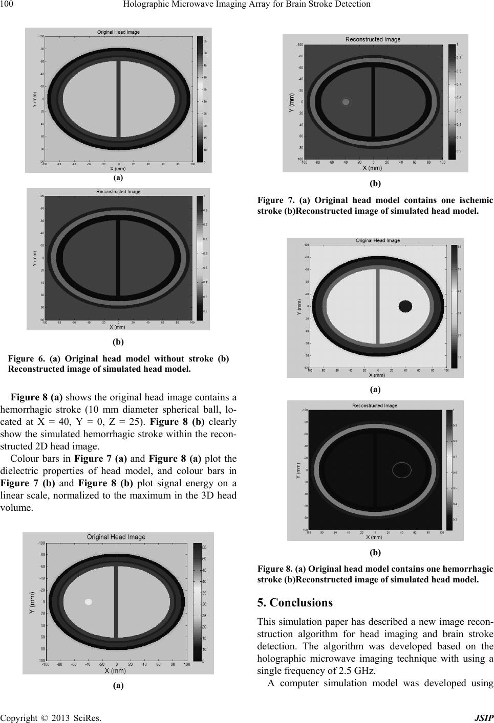
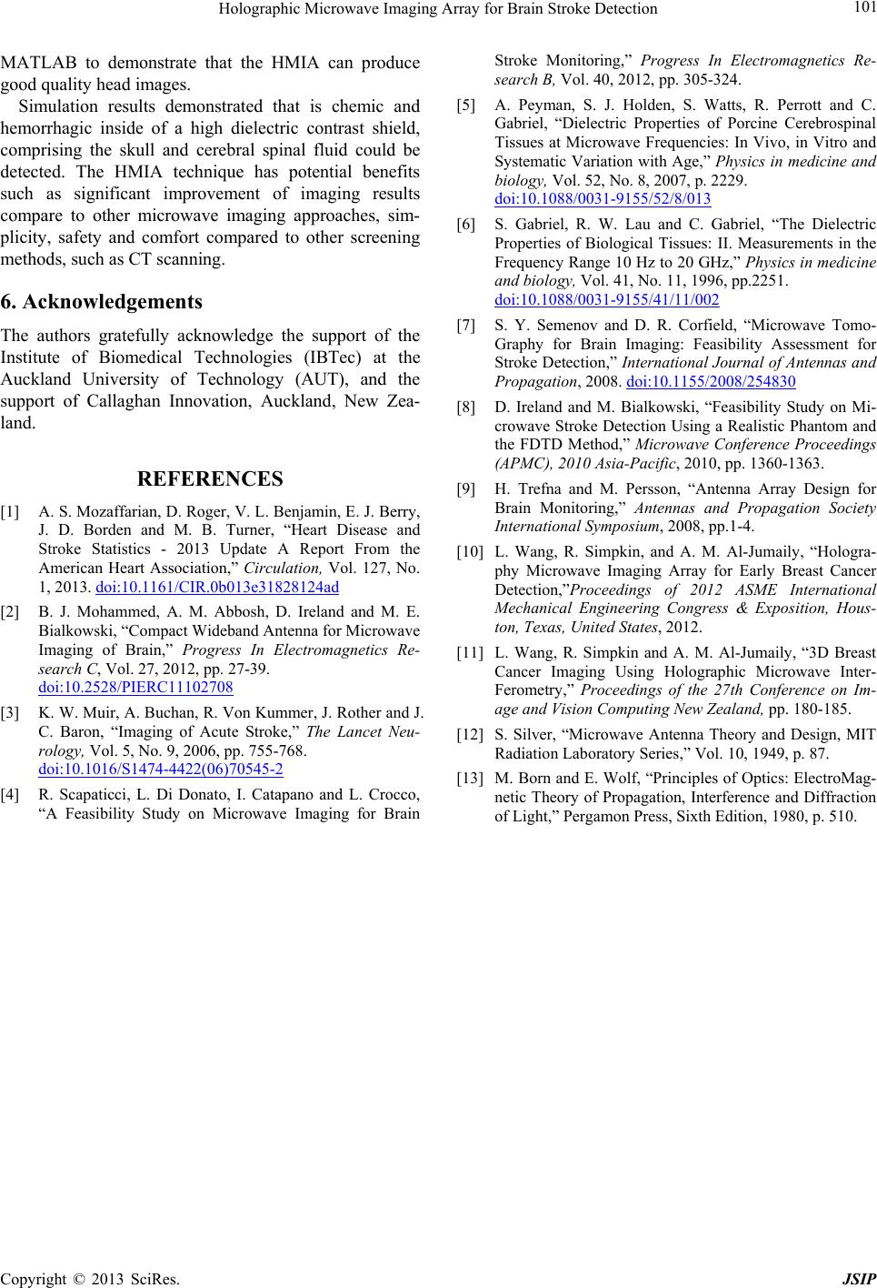
MATLAB to demonstrate that the HMIA can produce
good quality head images.
Simulation results demonstrated that is chemic and
hemorrhagic inside of a high dielectric contrast shield,
comprising the skull and cerebral spinal fluid could be
detected. The HMIA technique has potential benefits
such as significant improvement of imaging results
compare to other microwave imaging approaches, sim-
plicity, safety and comfort compared to other screening
methods, such as CT scanning.
6. Acknowledgements
The authors gratefully acknowledge the support of the
Institute of Biomedical Technologies (IBTec) at the
Auckland University of Technology (AUT), and the
support of Callaghan Innovation, Auckland, New Zea-
land.
REFERENCES
[1] A. S. Mozaffarian, D. Roger, V. L. Benjamin, E. J. Berry,
J. D. Borden and M. B. Turner, “Heart Disease and
Stroke Statistics - 2013 Update A Report From the
American Heart Association,” Circulation, Vol. 127, No.
1, 2013. doi:10.1161/CIR.0b013e31828124ad
[2] B. J. Mohammed, A. M. Abbosh, D. Ireland and M. E.
Bialkowski, “Compact Wideband Antenna for Microwave
Imaging of Brain,” Progress In Electromagnetics Re-
search C, Vol. 27, 2012, pp. 27-39.
doi:10.2528/PIERC11102708
[3] K. W. Muir, A. Buchan, R. Von Kummer, J. Rother and J.
C. Baron, “Imaging of Acute Stroke,” The Lancet Neu-
rology, Vol. 5, No. 9, 2006, pp. 755-768.
doi:10.1016/S1474-4422(06)70545-2
[4] R. Scapaticci, L. Di Donato, I. Catapano and L. Crocco,
“A Feasibility Study on Microwave Imaging for Brain
Stroke Monitoring,” Progress In Electromagnetics Re-
search B, Vol. 40, 2012, pp. 305-324.
[5] A. Peyman, S. J. Holden, S. Watts, R. Perrott and C.
Gabriel, “Dielectric Properties of Porcine Cerebrospinal
Tissues at Microwave Frequencies: In Vivo, in Vitro and
Systematic Variation with Age,” Physics in medicine and
biology, Vol. 52, No. 8, 2007, p. 2229.
doi:10.1088/0031-9155/52/8/013
[6] S. Gabriel, R. W. Lau and C. Gabriel, “The Dielectric
Properties of Biological Tissues: II. Measurements in the
Frequency Range 10 Hz to 20 GHz,” Physics in medicine
and biology, Vol. 41, No. 11, 1996, pp.2251.
doi:10.1088/0031-9155/41/11/002
[7] S. Y. Semenov and D. R. Corfield, “Microwave Tomo-
Graphy for Brain Imaging: Feasibility Assessment for
Stroke Detection,” International Journal of Antennas and
Propagation, 2008. doi:10.1155/2008/254830
[8] D. Ireland and M. Bialkowski, “Feasibility Study on Mi-
crowave Stroke Detection Using a Realistic Phantom and
the FDTD Method,” Microwave Conference Proceedings
(APMC), 2010 Asia-Pacific, 2010, pp. 1360-1363.
[9] H. Trefna and M. Persson, “Antenna Array Design for
Brain Monitoring,” Antennas and Propagation Society
International Symposium, 2008, pp.1-4.
[10] L. Wang, R. Simpkin, and A. M. Al-Jumaily, “Hologra-
phy Microwave Imaging Array for Early Breast Cancer
Detection,”Proceedings of 2012 ASME International
Mechanical Engineering Congress & Exposition, Hous-
ton, Texas, United States, 2012.
[11] L. Wang, R. Simpkin and A. M. Al-Jumaily, “3D Breast
Cancer Imaging Using Holographic Microwave Inter-
Ferometry,” Proceedings of the 27th Conference on Im-
age and Vision Computing New Zealand, pp. 180-185.
[12] S. Silver, “Microwave Antenna Theory and Design, MIT
Radiation Laboratory Series,” Vol. 10, 1949, p. 87.
[13] M. Born and E. Wolf, “Principles of Optics: ElectroMag-
netic Theory of Propagation, Interference and Diffraction
of Light,” Pergamon Press, Sixth Edition, 1980, p. 510.