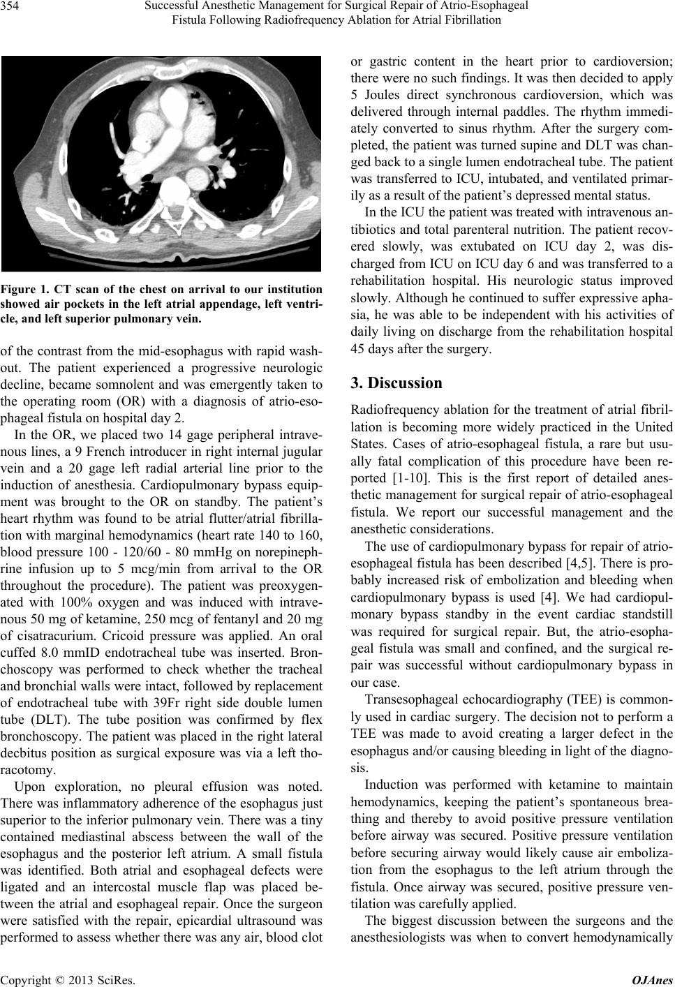
Successful Anesthetic Management for Surgical Repair of Atrio-Esophageal
Fistula Following Radiofrequency Ablation for Atrial Fibrillation
Copyright © 2013 SciRes. OJAnes
355
unstable atrial fibrillation to sinus rhythm. The patient
became hemodynamically unstable shortly after arrival to
the OR. According to ACLS protocol, hemodynamically
unstable atrial fibrillation should be cardioverted. But in
this specific case, atrial fibrillation kept atrial pressure
low enough to avoid hemorrhage from the atria to the
chest or esophagus. Also converting to sinus rhythm in-
creases the likelihood of embolization of air, blood clot,
gastric contents in the left atrium into the brain or other
organs. And also the patient had had atrial fibrillatio n for
at least several days and the patient had very high risk for
thrombus formation due to factor V Leiden deficiency
and DVT. We did not know whether there was thrombus
or air in the left atria or how big it would be if thrombus
was present at this point. Cardioversion likely could have
ended up causing massive bleeding or embolization.
Hemodynamics was managed instead by infusing pheny-
lephrine and norepinephrine to maintain a systolic blood
pressure around 100 mmHg. Only once air in the atria
was suctioned, and no other air, blood clots or gastric
contents were detected by direct epicardial ultrasound
and the fistula was surgically repaired, caridoversion was
performed by directly applying paddles to the heart.
The patient was kept intubated and transferred to the
ICU due to the hemodyn amic instab ility and n eurolog ical
deterioration shown preoperatively which could impair
airway protection postoperatively. The patient was treat-
ed for sepsis with broad spectrum of antibiotics initially
to cover the likely bacterial species inh abiting esophagus.
The role of esophageal stent was considered intraope-
ratively. There was a successful case of esophageal stent
for atrio-esophageal fistula, using esophagoscopy [7],
even though most of the literature did recommend avoid-
ance of esophagoscopy [4-6,9]. But more data is needed
to determine which cases are indicated for esophageal
stent and esophagoscopy.
The key to managing these patients’ safety is the pre-
vention of massive bleeding and multiple air emboliza-
tion during the surgical repair and then later the postop-
erative management of sepsis and multiple organ failure,
which are the main causes of death from this complica-
tion.
4. Conclusion
We had a successful case in anesthetic management for
surgical repair of atrio-esophageal fistula with avoidance
of positive pressure ventilation before securing airway
and careful control of hemodynamics by delaying car-
dioversion to prevent massive bleeding as well as multi-
ple air embolization through the fistula.
REFERENCES
[1] A. M. Gillinov, G. Pettersson and T. W. Rice, “Esophag-
eal Injury during Radiofrequency Ablation for Atrial Fib-
rillation,” The Journal of Thoracic and Cardiovascular
Surgery, Vol. 122, No. 6, 2001, pp. 1239-1240.
http://dx.doi.org/10.1067/mtc.2001.118041
[2] N. Doll, M. A. Borger, A. Fabricius, S. Stephan, J. Gum-
mert, F. W. Mohr, et al., “Esophageal Perforation during
Left Atrial Radiofrequency Ablation: Is the Risk Too
High?” The Journal of Thoracic and Cardiovascular Sur-
gery, Vol. 125, No. 4, 2003, pp. 836-842.
http://dx.doi.org/10.1067/mtc.2003.165
[3] G. Hindricks and H. Kottkamp, “Potential Benefits, Risks,
and Complications of Catheter Ablation of Atrial Fibrilla-
tion: More Questions Than Answers,” Journal of Cardio-
vascular Electrophysi ology, Vol. 13, No. 8, 2002, pp. 768-
769. http://dx.doi.org/10.1046/j.1540-8167.2002.00768.x
[4] B. Sonmez, E. Demirsoy, N. Yagan, M. Unal, H. Arbatli,
D. Sener, et al., “A Fatal Complication Due to Radiofre-
quency Ablation for Atrial Fibrillation: Atrio-Esophageal
Fistula,” The Annals of Thoracic Surgery, Vol. 76, No. 1,
2003, pp. 281-283.
http://dx.doi.org/10.1016/S0003-4975(03)00006-7
[5] C. Pappone, H. Oral, V. Santinelli, G. Vicedomini, C. C.
Lang, F. Manguso, et al., “Atrio-Esophageal Fistula as a
Complication of Percutaneous Transcatheter Ablation of
Atrial Fibrillation,” Circulation, Vol. 109, No. 22, 2004,
pp. 2724-2726.
http://dx.doi.org/10.1161/01.CIR.0000131866.44650.46
[6] M. I. Scanavacca, A. D’avila, J. Parga and E. Sosa, “Left
Atrial-Esophageal Fistula Following Radiofrequency Ca-
theter Ablation of Atrial Fibrillation,” Journal of Cardio-
vascular Electrophysi ology, Vol. 15, No. 8, 2004, pp. 960-
962. http://dx.doi.org/10.1046/j.1540-8167.2004.04083.x
[7] T. J. Bunch, J. Nelson, T. Foley, S. Allison, B. G. Cran-
dall, J. S. Osborn, et al., “Temporary Esophageal Stenting
Allows Healing of Esophageal Perforations Following
Atrial Fibrillation Ablation Procedures,” Journal of Car-
diovascular Electrophysiology, Vol. 17, No. 4, 2006, pp.
435-439.
http://dx.doi.org/10.1111/j.1540-8167.2006.00464.x
[8] P. Schley, H. Gu¨lker and M. Horlitz, “Atrio-Oesophageal
Fistula Following Circumferential Pulmonary Vein Abla-
tion: Verification of Diagnosis with Multislice Computed
Tomography,” Europace, Vol. 8, No. 3, 2006, pp. 189-
190. http://dx.doi.org/10.1093/europace/euj050
[9] J. E. Cummings, R. A. Schweikert, W. I. Saliba, J. D.
Burkhardt, F. Kilikaslan, E. Saad, et al., “Brief Commu-
nication: Atrial-Esophageal Fistulas after Radiofrequency
Ablation,” Annals of Internal Medicine, Vol. 144, No. 8,
2006, pp. 572-574.
http://dx.doi.org/10.7326/0003-4819-144-8-200604180-0
0007
[10] A. Takahashi, T. Kuwahara and Y. Takahashi, “Compli-
cations in the Catheter Ablation of Atrial Fibrillation: In-
cidence and Management,” Circulation Journal, Vol. 73,
No. 2, 2009, pp. 221-226.
http://dx.doi.org/10.1253/circj.CJ-08-1097.