Open Journal of Anesthesiology, 2013, 3, 345-348
http://dx.doi.org/10.4236/ojanes.2013.38073 Published Online October 2013 (http://www.scirp.org/journal/ojanes)
345
Pneumothorax Complicating Port-a-Cath and Groshong
Catheter Positioning in Children: Our Experience before
Routine Ultrasound-Guided Puncture*
Silvia Guenzani1,2#, Paola Previtali2, Federico Piccioni2, Maria Chiara Allemano2, Serena Catania3,
Martin Langer1,2
1Department of Pathophysiology and Transplantation, University of Milan, Milan, Italy; 2Department of Anesthesia, Intensive Care
and Palliative Care, Fondazione IRCCS Istituto Nazionale dei Tumori, Milan, Italy; 3Pediatric Oncology Unit, Fondazione IRCCS
Istituto Nazionale dei Tumori, Milan, Italy.
Email: #silvia.guenzani@gmail.com
Received June 18th, 2013; revised July 20th, 2013; accepted August 19th, 2013
Copyright © 2013 Silvia Guenzani et al. This is an open access article distributed under the Creative Commons Attribution License,
which permits unrestricted use, distribution, and reproduction in any medium, provided the original work is properly cited.
ABSTRACT
Objective: To study incidence and management of long term central venous catheter (CVC) placement related pneu-
mothorax (PNX) in children. Aim: To construct a baseline value before the introduction of systematic use of ultrasound
guidance, which requires specific training and equipment. Background: Anesthesia Service and Pediatric Oncology of
the Italian National Cancer Center; patients were children (age 18 years) with solid tumors, needing long-term central
venous catheters (Groshong or Port-a-Cath). Materials/Methods: Catheter placement was performed, mostly under
general anesthesia, utilizing a micropuncture 5 - 7 Fr needle and fluoroscopy. In the study period ultrasound was used
only in case of previously failed attempts. Relevant data were collected retrospectively. Results: From August 2008 to
December 2011, 452 catheters were implanted to our patients. The prevalent approach was from subclavian vein (left
85.7%, right 9.7%); in few cases internal jugular vein was chosen (right 2.4%, left 2.2%). Pneumothorax occurred in 14
patients (3.1%; 95%CI 1.9 - 5.1). In 4/14 children the PNX was considered minimal and not treated. In 10 patients the
PNX was drained. In 7 cases a traditional, surgical thoracostomy was performed, while in 3 children a 14-Ga polyure-
thane catheter (Arrow International®) was inserted over a wire guide in the pleural space by anaesthetists. Conclusions:
In our centre rates of PNX are the same as those described in literature and are expected to lower when ultrasound guid-
ance of the puncture will be routinely applied. Percutaneous drainage of PNX seems as effective as surgically placed
thoracostomy catheter, but less invasive.
Keywords: Pneumothorax; Central Venous Catheter; Groshong; Port-a-Cath; Children; Drainage
1. Introduction
Long term venous access through totally implantable
venous access devices (TIVAD), partially implantable
tunneled cuffed catheters, or more recently peripherically
inserted central venous catheters (PICC) are essential for
administering chemotherapy also to young oncologic
patients. Positioning of these catheters is reported as a
relatively safe procedure with little associated morbidity
and nearly no mortality, where this risk is outweighed by
the benefit obtained [1].
An implanted port is preferred when its use is inter-
mittent (the skin protects the device from infections and
rupture) and can remain in situ for years, while an exter-
nal catheter is commonly used for continuous infusion
during a shorter period of time. Percutaneous positioning
of catheters can be achieved by various central accesses:
through the subclavian vein (SCV), the internal jugular
vein (IJV), or, in special cases, the femoral vein, while
the brachial vein is preferred for PICC insertion.
Pneumothorax (PNX) is a typical early, mechanical
complication of the puncture of the subclavian vein. The
SCV approach is more frequently used than the IJV ac-
cess in small children because the exit site is placed at
proper distance from the mouth, it is easy to protect and
*Important Note: Main results of this study have been presented as oral
communication at SMART CONFERENCE on May 2012, this is why
you can find a similar abstract at this link:
http://www.altrimedia.biz/smart-cd2012/Comunicazioni_Libere/10_TE
RAPIA_INTENSIVA_PEDIATRICA/54_Guenzani.pdf.
#Corresponding author.
Copyright © 2013 SciRes. OJAnes
Pneumothorax Complicating Port-a-Cath and Groshong Catheter Positioning in
Children: Our Experience before Routine Ultrasound-Guided Puncture
346
tunneling of the catheter is rather simple and linear. The
infraclavicular, landmark based insertion for long term
central venous catheters (CVCs) in children is a widely
used approach, but ultrasound (US) guidance is difficult.
Also in our center catheter placement in pediatric patients
under sonographic guidance is still uncommon, and anes-
thetists usually perform a landmark based puncture of the
SCV, with the approach through the IJV as second choice.
In this retrospective study we try to assess the risk of
PNX in our experience compared to the literature, in or-
der to discuss/revise our catheter placement policy in
children.
2. Materials and Methods
Two options for elective long term venous accesses are
available at our Hospital: Port-a-Cath (Titanium Low-
Profile Implantable Port-Bard Access Systems or others),
a totally implantable venous access device (TIVAD), and
Groshong catheter (Bard Access Systems), a tunneled
external silicon-rubber device with a three-position pres-
sure-sensitive valve near the distal tip.
Long term CVC placement in pediatric patients is
proposed by pediatric oncologists to the anesthesia ser-
vice. The preoperative evaluation includes medical his-
tory, body examination to evaluate their physical habitus
and anatomic pitfalls (obesity, chest tumors or adeno-
pathies of the mediastinum), laboratory exams (com-
plete blood and platelet count, coagulation screening)
and a chest X-ray. Cannulation is performed percutane-
ously in an operating room where anesthesia facilities
and C-arm X-ray machine are available.
Left infraclavicular SCV cannulation is the access of
first choice. If more then one attempt fails, other sites
(IJV) are tried. A micropuncture set (Micropuncture In-
troducer Set 5.0 - 7.0 Fr-Cook) is used for the puncture
of the vein and catheters are inserted under fluoroscopic
control. In nearly all pediatric patients the procedure is
performed under general, total intravenous anesthesia,
mechanical ventilation and standard monitoring. There is
no dedicated catheter-team and all staff anesthetists and
many residents are actively involved.
A chest X-ray is performed 6 - 12 hrs after catheter
placement to document the location of catheter tip and to
rule out mechanical complications like PNX, and hemo-
or hydrothorax.
In case of evidence of PNX the need for drainage is
discussed with a thoracic surgeon and/or a staff-anesthe-
tist, taking into account the clinical symptoms, size and
progression of the pneumothorax. Drainage of PNX is
performed by the thoracic surgeon with a 16Ch chest
tube drainage, or by the anesthetist with a single-lumen
14Ga venous catheter (Arrow® International-central ve-
nous catheterization set) over a wire guide, after percu-
taneous puncture of the pleural space. The choice be-
tween chest tube drainage or percutaneous catheter is not
standardized yet, but we are getting used to utilize in-
creasingly the less invasive option.
Data analysis was performed using IBM SPSS Statis-
tics v19. Chi-quare test was adopted to compare discrete
variables. Normality distribution of continuous variables
was verified with Shapiro-Wilk test. T-test for inde-
pendent groups was used to compare parametric data. A
p value of less than 0.05 was considered as statistically
significant.
3. Results
From August 2008 to December 2011, 452 long term
vascular access devices were implanted in pediatric pa-
tients (median age 8 years, range 3 months - 18 years) at
our Institute.
In 159 (35.2%) children a Groshong catheter was cho-
sen and in 293 (64.8%) a TIVAD. Infraclavicular SCV
cannulation was in all cases the access of first choice,
followed by puncture of IJV when more than one attempt
failed. The procedure was performed in 93% of cases
under general anesthesia, and in 7% of cases with local
anesthesia. The final catheterization site was primarly the
subclavian vein (left SCV 85.7%, right SCV 9.7%, right
IJV 2.4%, left IJV 2.2%). Anatomic landmark oriented
puncture was the first choice technique in all patients, US
guidance after initial failed attempts was performed in
2% of the procedures.
PNX was diagnosed at the control chest X-ray in 14
cases (3.1%; 95%CI 1.9 - 5.1) always after several failed
punctures of the SCV. See Table 1 for patients’ demo-
graphic data. Ten of these patients underwent PNX drai-
nage, in 4 cases the PNX was minimal, not requiring any
treatment. Seven patients underwent a 16Ch tube thora-
costomy, while in 3 cases the pleural air was drained by a
single-lumen 14Ga venous catheter (Arrow® Interna-
tional-central venous catheterization set). In the latter
approach air was aspired by a syringe once or twice a day
with the catheter locked during the intervals. Chest tubes
and “chest catheters” remained in situ for a variable pe-
riod between 2 and 12 days (average: catheters for 4.3
days, chest tubes for 5.6 days; p = 0.633). No other early
complications or long term consequences were recorded.
4. Discussion
In our study the rate of pneumothorax (3.1%; 95%CI 1.9
- 5.1) after a central venous catheter insertion by punc-
ture of the SCV (infraclavicular approach) was in the
higher range, compared to the pooled data (Table 2 and
Figure 1) from 7 pertinent studies in 2839 children
(1.7%; 95%CI 1.3 - 2.3) [28]. The 1.4% difference is -
Copyright © 2013 SciRes. OJAnes
Pneumothorax Complicating Port-a-Cath and Groshong Catheter Positioning in
Children: Our Experience before Routine Ultrasound-Guided Puncture
Copyright © 2013 SciRes. OJAnes
347
Table 1. Demographic data of patients with PNX (DT, days of thoracostomy; US, ultrasound; SB, spontaneous breathing and
local anesthesia; MV, mechanical ventilation and general anesthesia; SCV, subclavian vein; PNET, primitive neuroectoder-
mal tumor; MPNST, malignal peripheral nerve sheath tumor; NHL, non Hodgkin lymphoma).
PT N˚ Sex Age Diagnosis DT Site of catheterizationUS SB or MV Procedure
1 M 16 Hepatoblastoma 4 Left SCV NO MV Tube thoracostomy
2 M 12 Medulloblastoma / Left SCV NO MV None
3 M 10 Glioblastoma 5 Left SCV NO MV One-lumen 14Ga Arrow
4 M 14 Lynfoprolipherative disease / Left SCV NO MV None
5 F 14 Desmoid fibromatosis 12 Left SCV NO SB Tube thoracostomy
6 M 11 Glioblastoma 6 Left SCV YES MV Tube thoracostomy
7 F 14 Intracranic fibrosarcoma 3 Left SCV NO MV Tube thoracostomy
8 M 17 PNET 2 Left SCV NO MV Tube thoracostomy
9 F 3 Neuroblastoma 2 Left SCV NO MV Tube thoracostomy
10 M 15 MPNST / Left SCV NO MV None
11 M 18 Mediastinal sarcoma / Left SCV NO MV None
12 F 1 Rabdomiosarcoma 2 Left SCV YES MV One-lumen 14Ga Arrow
13 M 16 Troncoencephalic glioma 10 Left SCV NO SB Tube thoracostomy
14 M 9 NHL 6 Left SCV NO MV One-lumen 14Ga Arrow
Table 2. Incidence data from the literature concerning pneumothorax (PNX) after central venous catheter (CVC) positioning
through the SCV. The 95%CI are calculated ex post.
N˚ subclavian CVCs N˚ PNX % PNX 95%CI
Karapinar et al. 2007 [2] 119 3 2.5 0.9 - 7.2
Araujo et al. 2007 [3] 197 4 2.0 0.8 - 5.1
Citak et al. 2002 [4] 148 2 1.4 0.4 - 4.8
Casado-Flores 2001 [5] 235 6 2.6 1.2 - 5.5
Lefrant et al. 2002 [6] 707 22 3.1 2.1 - 4.7
Casado-Flores 1991 [7] 322 6 1.9 0.9 - 4.0
Johnson et al. 1998 [8] 1180 6 0.5 0.2 - 1.1
Guenzani et al. 2013 452 14 3.1 1.9 - 5.1
Overall 3360 63 1.87 1.5 - 2.4
remarkable but statistically not significant (p = 0.061).
Excluding the outstanding good results in the large series
of Johnson et al. (0.5%; 95%CI 0.2 - 1.1), the mean in-
cidence of PNX during cannulation of SCV in 2180 chil-
dren, from the remaining 6 studies, is 2.5% (95%CI 1.9 -
3.3).
Pneumothorax complicating landmark-oriented punc-
ture of the SCV remains therefore a real risk of CVC
placement in children. The standard treatment of PNX is
the chest tube insertion and connection to a chest drain
canister. Of course this is a cumbersome maneuver for
sick children, requiring mostly general anesthesia or deep
sedation, more or less postoperative pain and bed rest.
The attempt to drain pleural air in spontaneously breath-
ing children with a “minimally invasive” drain (a stan-
dard central venous catheter, inserted over a wire-guide)
was successful when adopted, leading to rapid resolution,
also without catheter connection to the drain canister.
One or two syringe aspirations per day allowed removing
air and checking for new air accumulation. When
Pneumothorax Complicating Port-a-Cath and Groshong Catheter Positioning in
Children: Our Experience before Routine Ultrasound-Guided Puncture
348
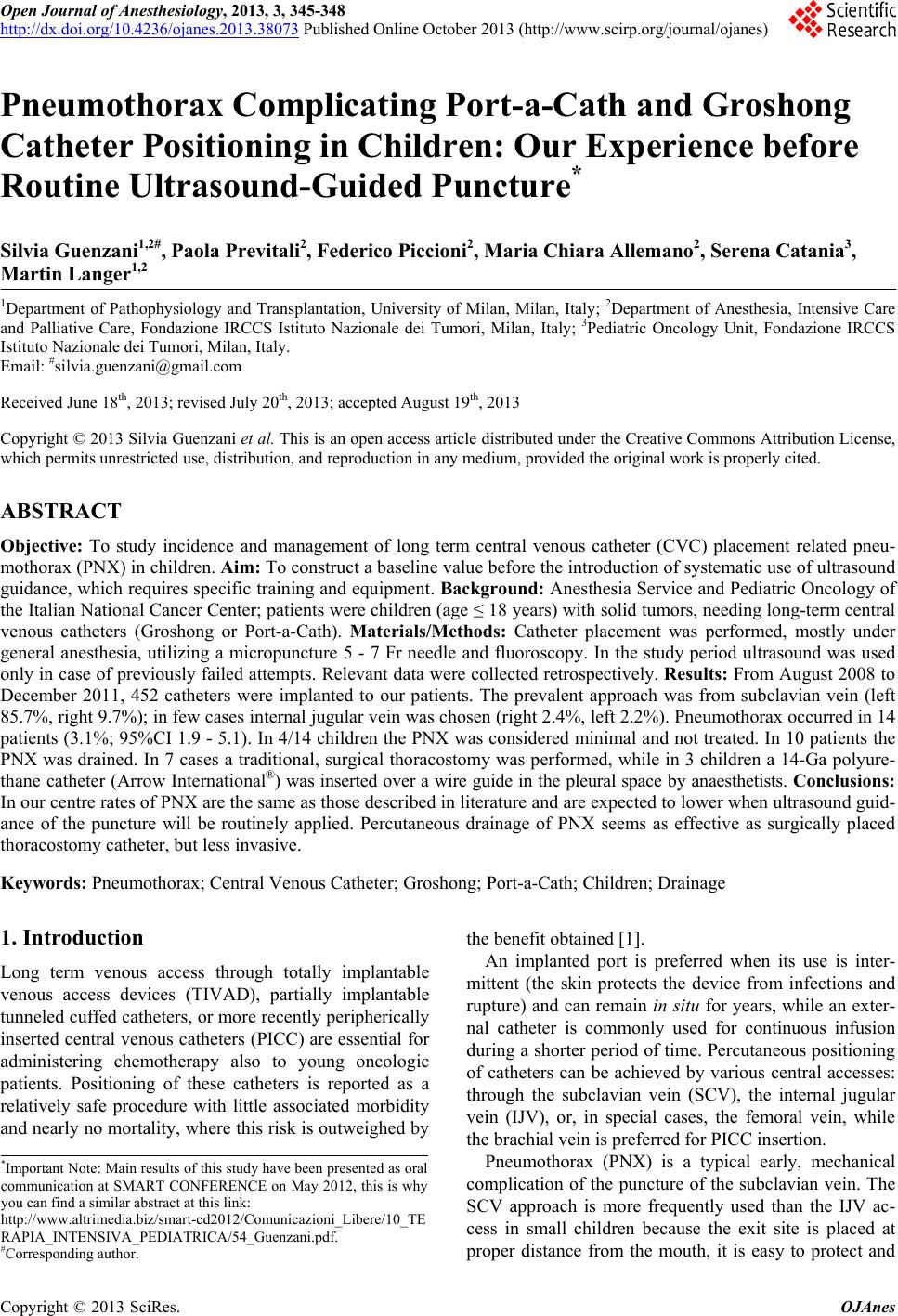
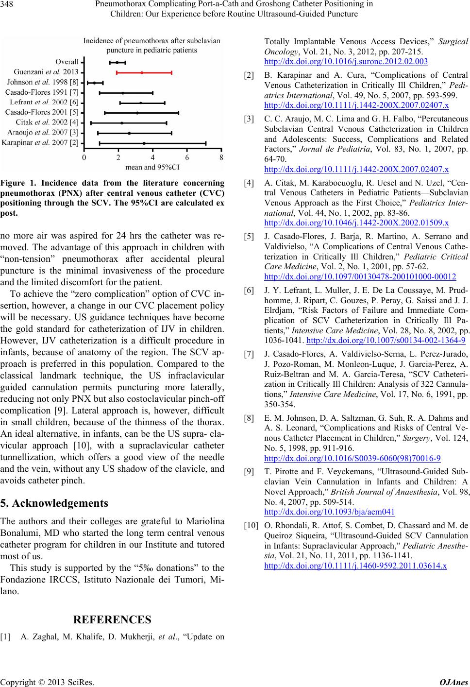
Figure 1. Incidence data from the literature concerning
pneumothorax (PNX) after central venous catheter (CVC)
positioning through the SCV. The 95%CI are calculated ex
post.
no more air was aspired for 24 hrs the catheter was re-
moved. The advantage of this approach in children with
“non-tension” pneumothorax after accidental pleural
puncture is the minimal invasiveness of the procedure
and the limited discomfort for the patient.
To achieve the “zero complication” option of CVC in-
sertion, however, a change in our CVC placement policy
will be necessary. US guidance techniques have become
the gold standard for catheterization of IJV in children.
However, IJV catheterization is a difficult procedure in
infants, because of anatomy of the region. The SCV ap-
proach is preferred in this population. Compared to the
classical landmark technique, the US infraclavicular
guided cannulation permits puncturing more laterally,
reducing not only PNX but also costoclavicular pinch-off
complication [9]. Lateral approach is, however, difficult
in small children, because of the thinness of the thorax.
An ideal alternative, in infants, can be the US supra- cla-
vicular approach [10], with a supraclavicular catheter
tunnellization, which offers a good view of the needle
and the vein, without any US shadow of the clavicle, and
avoids catheter pinch.
5. Acknowledgements
The authors and their colleges are grateful to Mariolina
Bonalumi, MD who started the long term central venous
catheter program for children in our Institute and tutored
most of us.
This study is supported by the “5‰ donations” to the
Fondazione IRCCS, Istituto Nazionale dei Tumori, Mi-
lano.
REFERENCES
[1] A. Zaghal, M. Khalife, D. Mukherji, et al., “Update on
Totally Implantable Venous Access Devices,” Surgical
Oncology, Vol. 21, No. 3, 2012, pp. 207-215.
http://dx.doi.org/10.1016/j.suronc.2012.02.003
[2] B. Karapinar and A. Cura, “Complications of Central
Venous Catheterization in Critically Ill Children,” Pedi-
atrics International, Vol. 49, No. 5, 2007, pp. 593-599.
http://dx.doi.org/10.1111/j.1442-200X.2007.02407.x
[3] C. C. Araujo, M. C. Lima and G. H. Falbo, “Percutaneous
Subclavian Central Venous Catheterization in Children
and Adolescents: Success, Complications and Related
Factors,” Jornal de Pediatria, Vol. 83, No. 1, 2007, pp.
64-70.
http://dx.doi.org/10.1111/j.1442-200X.2007.02407.x
[4] A. Citak, M. Karabocuoglu, R. Ucsel and N. Uzel, “Cen-
tral Venous Catheters in Pediatric Patients—Subclavian
Venous Approach as the First Choice,” Pediatrics Inter-
national, Vol. 44, No. 1, 2002, pp. 83-86.
http://dx.doi.org/10.1046/j.1442-200X.2002.01509.x
[5] J. Casado-Flores, J. Barja, R. Martino, A. Serrano and
Valdivielso, “A Complications of Central Venous Cathe-
terization in Critically Ill Children,” Pediatric Critical
Care Medicine, Vol. 2, No. 1, 2001, pp. 57-62.
http://dx.doi.org/10.1097/00130478-200101000-00012
[6] J. Y. Lefrant, L. Muller, J. E. De La Coussaye, M. Prud-
homme, J. Ripart, C. Gouzes, P. Peray, G. Saissi and J. J.
Elrdjam, “Risk Factors of Failure and Immediate Com-
plication of SCV Catheterization in Critically Ill Pa-
tients,” Intensive Care Medicine, Vol. 28, No. 8, 2002, pp.
1036-1041. http://dx.doi.org/10.1007/s00134-002-1364-9
[7] J. Casado-Flores, A. Valdivielso-Serna, L. Perez-Jurado,
J. Pozo-Roman, M. Monleon-Luque, J. Garcia-Perez, A.
Ruiz-Beltran and M. A. Garcia-Teresa, “SCV Catheteri-
zation in Critically Ill Children: Analysis of 322 Cannula-
tions,” Intensive Care Medicine, Vol. 17, No. 6, 1991, pp.
350-354.
[8] E. M. Johnson, D. A. Saltzman, G. Suh, R. A. Dahms and
A. S. Leonard, “Complications and Risks of Central Ve-
nous Catheter Placement in Children,” Surgery, Vol. 124,
No. 5, 1998, pp. 911-916.
http://dx.doi.org/10.1016/S0039-6060(98)70016-9
[9] T. Pirotte and F. Veyckemans, “Ultrasound-Guided Sub-
clavian Vein Cannulation in Infants and Children: A
Novel Approach,” British Journal of Anaesthesia, Vol. 98,
No. 4, 2007, pp. 509-514.
http://dx.doi.org/10.1093/bja/aem041
[10] O. Rhondali, R. Attof, S. Combet, D. Chassard and M. de
Queiroz Siqueira, “Ultrasound-Guided SCV Cannulation
in Infants: Supraclavicular Approach,” Pediatric Anesthe-
sia, Vol. 21, No. 11, 2011, pp. 1136-1141.
http://dx.doi.org/10.1111/j.1460-9592.2011.03614.x
Copyright © 2013 SciRes. OJAnes