World Journal of Cardiovascular Diseases, 2013, 3, 448-453 WJCD
http://dx.doi.org/10.4236/wjcd.2013.37070 Published Online October 2013 (http://www.scirp.org/journal/wjcd/)
Syntax score: The fallacies and remedies—A perspective
Pravin K. Goel*, Roopali Khanna, Aditya Batra, Sanjeev K. Syal
Department of Cardiology, Sanjay Gandhi Postgraduate Institute of Medical Sciences, Lucknow, India
Email: *golf_pgi@yahoo.co.in
Received 27 July 2013; revised 29 August 2013; accepted 12 September 2013
Copyright © 2013 Pravin K. Goel et al. This is an open access article distributed under the Creative Commons Attribution License,
which permits unrestricted use, distribution, and reproduction in any medium, provided the original work is properly cited.
ABSTRACT
The syntax score devised as a tool to grade complexity
of coronary artery disease with a view to its suitabil-
ity for PCI or otherwise on deeper introspection is
fraught with some inherent limitations and fallacies
largely as a result of the variability in coronary ana-
tomy and the fixed nomenclature of the devised scor-
ing system. We, in this special report, bring out the
limitations and fallacies in the same and possible re-
medies presented as a viewpoint which may be useful
to the interventional community at large.
Keywords: Syntax Score; PCI
1. INTRODUCTION
With advancements in percutaneous coronary interven-
tion techniques and availability of reliable drug eluting
stents, more and more patients with multivessel disease
are being offered percutaneous coronary interventions
(PCI). However, the best management strategy for an
individual patient with multivessel disease, (i.e. either
PCI or bypass surgery) has always been an issue of de-
bate [1].
The SYNTAX score [2-4] was devised as an an-
giographic tool to grade the complexity of coronary ar-
tery disease (CAD) with a view to its suitability for PCI
or otherwise. This score incorporates a combination of
the effect of the area subtended by a diseased vessel/
segment in question, the severity of stenosis and the ana-
tomical complexity of the lesion/lesions. It uses a com-
bination of the AHA classification for coronary segments
as modified for the ARTS study [5] and for the anatomi-
cal coronary segments, the Leaman score for weighted con-
tribution of the stenosis to total myocardial jeopardy [6],
the total occlusion classification system of the European
Total Surveillance study [7] for classification of occlu-
sions and the Duke and ICPS classification systems for
bifurcation lesions [8,9]. Although the syntax study was
not meant to segregate patients based upon the Syntax
score, it delivered an indirect message to the practising
clinician and interventionists that if one could ascertain
the syntax score in day to day practice and use the same
in stratifying patients into those suitable for PCI or
coronary artery bypass grafts (CABG), it may help in ob-
taining better clinical outcomes from the revasculariza-
tion procedure.
Introspecting into the score, we have made some ob-
servations which we would like to bring to the attention
of the cardiology community at large. These observations
summarise the inherent fallacies/difficulties with the
score and possible remedies, if any. The fallacies are
largely a result of the high inter individual variability of
the coronary anatomy and not uncommonly this variabil-
ity may be so significant that the prefixed arterial no-
menclature based scoring, as used by syntax, would not
allow the flexibility needed for such wide variations in
coronary anatomy. More so to have a computer generated
software which would cover for all such variability
would certainly not be easy. We have tried to work out
the fallacies in some detail and the possible remedies in
principle which could help make the score more precise
and still maintain reproducibility.
2. FALLACIES IN SYNTAX SCORE
2.1. Right Coronary Artery (RCA) vs Left
Coronary Artery (LCA) Dominance Issue
Incompletely Addressed
The syntax scoring system accounts for right or left
dominance based on Posterior Descending Artery (PDA)
origin alone i.e. PDA from RCA or Circumflex (Cx).
However in real life extremes of dominance are not un-
common with a just dominant right system having PDA
as the only off-shoot of RCA at one extreme (Figure
1(A)) and on the other the entire posterior and inferior
Left Ventricular (LV) including the adjacent Obtuse Mar-
ginal (OM) territory could be supplied by super domi-
*Corresponding author.
Published Online October 2013 in SciRes. http://www.scirp.org/journal/wjcd
P. K. Goel et al. / World Journal of Cardiovascular Diseases 3 (2013) 448-453 449
nant RCA (Figure 1(B)) while the usual average domi-
nant RCA would encompass a PDA and at least one ma-
jor adjacent posterior inferior LV branch (Figure 1(C)).
It goes without saying then that all these three types of
right dominance would call for the same myocardial
weight of 1.0 (16% of LV) in the syntax scoring system
but in reality a lesion in each of these three different
RCA’s of same complexity should actually have a dif-
ferent clinical impact on total myocardial supply of the
heart if left non revascularised. Similar logic could stand
for variations in size of CX system (Figures 2(A) and
(B)) independent of RCA dominance.
2.2. LAD Length Issue Not Addressed at All
The left anterior descending is known to vary in its
length to a large extent and it is not uncommon to see
patients with short LAD which does not even reach the
apex (Type I) (Figure 3(A)) while there are others where
the LAD is so long that it not only crosses the apex but
also takes over almost the complete supply of PDA (Type
III) and in these cases the PDA is invariably rather small
Figure 1. Dominant RCA of three varying sizes: (A) Just domi-
nant RCA giving rise to PDA only; (B) Super-dominant RCA
with PDA and 2/3 PLVs reaching the OM territory; (C) Average
dominant RCA with PDA and one PLV.
Figure 2. Both the angiograms show left injections from right
dominant systems as per Syntax definitions with stark differ-
ence in size of LCX ((A): Small, (B): large) which can not be
equated.
Figure 3. (A) LAD falling short of apex; (B) LAD wrapping
around the apex and extending more than half the length into
the posterior interventricular groove.
and of much smaller myocardial value (Figure 3(B)). It
would then be quite unwise to give the same score to a
lesion in mid or distal LAD in either of these two situa-
tions as advocated by the Syntax scoring system.
2.3. Myocardial Weight of Proximal Segment
Independent of the Distal Ramification of
the Vessel
If one takes the example of a RCA giving rise to a PDA
(score 1.0) and two or three PLV’s each being scored 0.5
respectively as per the syntax system then the total myo-
cardial weight of this RCA becomes 2.5 but a lesion in
the proximal RCA would continue to be weighed by a
multiplication factor of 1.0 which is the fixed segmental
weight of a dominant RCA main stem which does not
sound to logic (Figure 4(A)). In the same context a
proximal Cx in a usual right dominant circulation would
continue to the weighed by a factor of 1.5 irrespective of
the area of its distal supply with one, two or more OM’s.
Each of these individual OM’s would add to the total
weight of the vessel but a lesion in proximal or mid seg-
ment of the CX would continue to be weighted as per the
fixed myocardial weight of proximal Cx of 1.5 as this is
independent of the distal ramification of the vessel in the
current syntax scoring system which seems incorrect.
2.4. Score Dependent on the Nomenclature of the
Vessel Rather than Its Anatomical Size
The syntax score prefixes the myocardial weight of each
individual branch of a coronary artery based on its no-
menclature which by and large is reasonable but there are
several situations involving vessels like the Ramus in-
termedius, OM and Diagonal (Dx) which at times are so
huge that one single branch e.g. Ramus would cover the
entire territorial supply (Figure 5(A)) of adjacent Dx and
OM territories but would be equated to be of similar sig-
nificance as any average size or even small ramus inter-
medius (Figure 5(B)) which by no means could equate
to above. The only alternative is to exclude a particular
Copyright © 2013 SciRes. WJCD
P. K. Goel et al. / World Journal of Cardiovascular Diseases 3 (2013) 448-453
450
Figure 4. A-RCA has severe disease (white arrow) in proximal
part which gets a value of only 1.0 in a right dominant system
as per SYNTAX score but additional disease in PLV and PDA
(black arrows) keeps adding a value of 0.5 each (total 1.5)
making total weight of the vessel 2.5 but the proximal RCA
lesion shall have myocardial value only 1.0.
Figure 5. Two Ramus intermedius vessels ((A): small, (B):
large) of contrastingly different sizes but getting the same value
in SYNTAX scoring system since both are at least 1.5 mm in
diameter.
vessel from accounting provided it is less than 1.5 mm in
diameter.
2.5. Overloading of the Syntax Score with
Multiple Branches within Same Territory
(Dx, PLV, OM)
Cases are not uncommon where a standard size Cx vessel
supplying the lateral LV wall has multiple OM branches
of similar size (>2 - 3) each being individually weighed
with a factor of 0.5 as per the syntax scoring system
(Figure 6(A)) and complex lesions like ostial stenosis or
ostial occlusion in one or more of these individual
branches would put an extra load on the total score which
athough in reality may not have much significance as far
Figure 6. (A) An average sized LCX with three average sized
OMs each more than 3 mm in diameter. An ostial lesion in all
three OMs with heavily overload the SYNTAX score even
though the territory supplied by these collectively remains rela-
tively small; (B) Multiple diagonals which can overload the
SYNTAX score if any of the stems had important stenosis.
as completeness and complexity of revascularization is
concerned because even if this entire chunk is left un-
touched it might matter little as far as final out comes are
concerned as these would depend more upon the propor-
tion of entire myocardium left incompletely revascular-
ised rather than the number of stems left non revascular-
ised. Similar logic could stand for multiple Dx (Figure
6(B)) and multiple PLV’s from RCA.
2.6. Inappropriateness of 1.5 mm Calibre as the
Only Criteria for a Vessel Being Considered
Significant Enough to Be Accounted for
A 1.5 mm diameter criteria alone may not be the most
appropriate criteria for considering a vessel significant.
The absolute diameter of the vessel in any patient may
relate more to the total arterial texture or size of the vas-
cular tree in general just as an individual’s built or habi-
tus. Also atherosclerosis being a generalised process, it is
not uncommon in diffuse disease situations to have none
of the vessels being truly 1.5 mm in diameter on meas-
urement on angiogram. Does it mean that none of these
vessels would be accounted for in Syntax calculations
(Figure 7). The real size of a vessel or a stem is its actual
myocardial value which depends upon the area of myo-
cardium subtended by it or it is supplying in a way i.e. its
myocardial weight in other terms, or so called proportion
of left ventricular myocardium supplied by the individual
stem irrespective of its diameter and the need for inter-
vention must be based upon its importance thus estab-
lished rather than its diameter.
3. REMEDIES
The Greenlane system [10,11] of coronary artery report-
ing gives the flexibility of covering for all the anatomical
variations and provides segment wise myocardial score
to each individual vessel/stem which makes it an accu-
Copyright © 2013 SciRes. WJCD
P. K. Goel et al. / World Journal of Cardiovascular Diseases 3 (2013) 448-453
Copyright © 2013 SciRes.
451
rate and reproducible system. However, the division of
myocardial segments based on this system is time con-
suming and tedious and requires expertise and training
which would be difficult to reproduce on a computer based
system. However, based on the above system we have
worked out a myocardial segmental scoring where in we
have devised a prefixed segmental weightage, based on
the basic coronary anatomy of an individual case, which
takes care of most if not all of the variations and fallacies
in the Syntax score highlighted by us (Table 1).
Figure 7. All vessels including LAD are of thin caibre (less than 1.5 mm, thinner than 5F catheter) but still
remain important for myocardial perfusion.
Table 1. Myocardial value of various coronary segments in different anatomical subsets of coronary anatomy.
Cx. Dominant System Dominant RCA (PDA only)Dominant RCA (Average)Super Dominant RCA (super)
LAD size Vessel
Name Short AverageLong Short AverageLongShort AverageLongShort AverageLong
LM 15 15 15 12 13 14 10.5 11.5 12.59 (7.5) 10 (8.5)11 (9.5)
Prox. Lad 6 7 8 6 7 8 6 7 8 6 7 8
Mid. LAD 4 5 6 4 5 6 4 5 6 4 5 6
Distal LAD 2 3 4 2 3 4 2 3 4 2 3 4
Major Dx 1.5 1.5 1.5 1.5 1.5 1.5 1.5 1.5 1.51.5 1.5 1.5
Small Dx 0.75 0.75 0.75 0.75 0.75 0.75 0.75 0.75 0.750.75 0.75 0.75
Prox. Cx 9 8 7 6 6 6 4.5 4.5 4.53 (1.5) 3 (1.5)3 (1.5)
Ramus/OM1 1.5 1.5 1.5 1.5 1.5 1.5 1.5 1.5 1.51.5 1.5 1.5
Major OM/O M2 1.5 1.5 1.5 1.5 1.5 1.51.5 1.5 1.51.5 (0) 1.5 (0)1.5 (0)
Distal Cx./OM3 1.5 1.5 1.5 1.5 1.5 1.5 1.5 1.5 1.50 0 0
Distal Cx./PLV 1.5 1.5 1.5 1.5 1.5 1.50 0 0 0 0 0
Prox./Mid RCA 0 0 0 3 2 1 4.5 3.5 2.56 (7.5) 5 (6.5)4 (5.5)
PDA (RCA) 0 0 0 3 2 1 3 2 1 3 2 1
PDA (off Cx.) 3 2 1 0 0 0 0 0 0 0 0 0
PLV 1 (RCA) 0 0 0 0 0 0 1.5 1.5 1.51.5 1.5 1.5
PLV 2 (RCA) 0 0 0 0 0 0 0 0 0 1.5 1.5 1.5
PLV 3 (RCA) 0 0 0 0 0 0 0 0 0 0 (1.5) 0 (1.5)0 (1.5)
Non-Dominant
RCA (No PDA) 0 0 0 0 0 0 0 0 0 0 0 0
*Values in parenthesis relate to a situation where there are at least three major PLV’s from the RCA in a super dominant right system.
WJCD
P. K. Goel et al. / World Journal of Cardiovascular Diseases 3 (2013) 448-453
452
1) The issue of arterial dominance—The circulation
needs to be first classified into right dominant (average),
right dominant (super), left dominant (PDA from LCX)
and co-dominant (PDA from RCA but PLVs from LCX)
instead of right/left dominant alone. The average right
dominant RCA has been given the weight of a RCA with
PDA and one major PLV while standard left dominant
system has entire LV supply from left circulation alone
and none from RCA while a co-dominant circulation, has
a RCA with myocardial weightage of only PDA from the
RCA and rest of the LV supply is accomplished from Cx.
A right super-dominant system is given the myocardial
weight of PDA plus 2 PLVs for RCA and only one or
two OMs from Cx as the case may be.
2) Similarly, a type I, II or III LAD can be pre classi-
fied and the score of the entire left tree is proportionately
increased or decreased based on the length of the LAD in
the system.
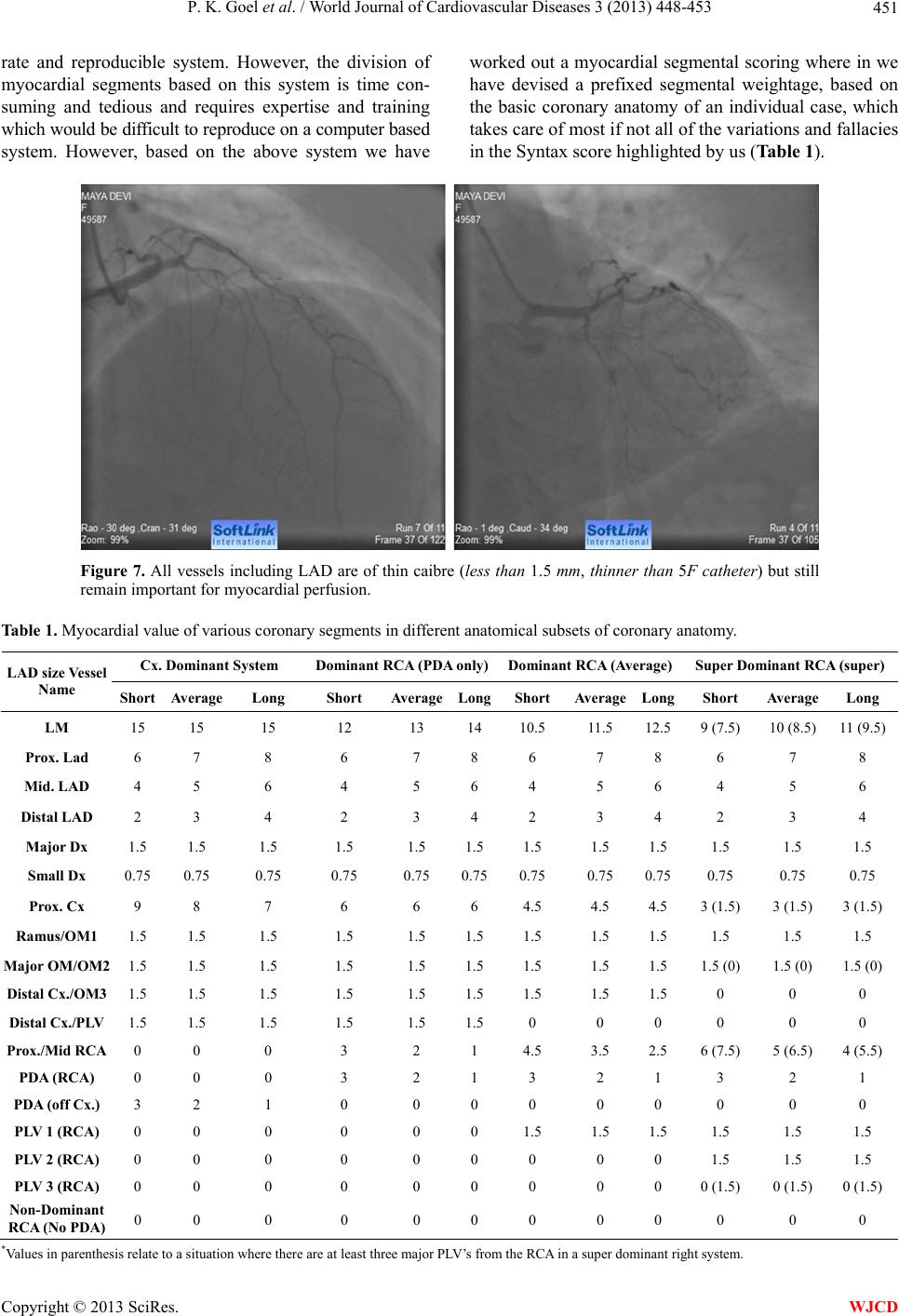
3) The total myocardial score in the Syntax system is
six with 5.0 (84% of LV) to LCA and 1.0 (16%) to PDA
while in the Greenlane system it being 15, the different
coronary segments get a proportionate score as a division
of 15. A conceptual division of myocardial score for dif-
ferent vessel segments based on the type of circulation
(Right dominant (average)/Right dominant (super)/left
dominant/co-dominant) and type I/II/III LAD is shown in
table attached in detail (Table 1 ). As an example an av-
erage RCA would contribute a score of 3.5/15 with 2/15
from PDA and 1.5/15 from PLV, which makes about 23%
of the LV myocardium as against 16% in the syntax sys-
tem and so on.
4) The Greenlane scoring system which was formu-
lated during the non interventional era of cardiology
gives a simple understanding to intervene in any vessel
when it has a myocardial value of 1.5 which in a way is a
vessel supplying 10% of the entire LV myocardial supply
and seems logically more correct than sticking to the 1.5
mm calibre criteria which has its own limitation as ex-
plained in the text above. This accounts for revasculari-
sation of PLV/OMs/Dx done based on the size of vessel
which in individual operator’s mind accounts for a myo-
cardial value of 1.5 or 10% of the entire supply of the
myocardium irrespective of its anatomical diameter.
5) A complexity factor based on LV function also can
be added which is the bottom line of all revasculariza-
tion procedural outcomes.
4. CONCLUSION
We have tried to express a viewpoint on possible
fallacies or limitations inherent in the syntax score and
present an outline of a modified remedial system and
strongly feel that a combination of the modified Green-
lane system of myocardial segment weight age as sug-
gested by us with assessment of lesion complexity as per
the syntax protocol could possibly give a complexity as-
sessment of the coronary anatomy which would be more
realistic, practical and even more precise than the current
syntax score without loss of ease for use. It would how-
ever need a large prospective study similar to the syntax
trial to adjudicate the same and such a score would then
be very close to actual decision making on PCI vs CABG
in an individual patient with multivessel disease.
REFERENCES
[1] Hannan, E.L., Wu, C., Walford, G., et al. (2008) Drug-
eluting stents vs. coronary-artery bypass grafting in mul-
tivessel coronary disease. New England Journal of Medi-
cine, 358, 331-341.
http://dx.doi.org/10.1056/NEJMoa071804
[2] Sianos, G., Morel, M.A., Kappetein, A.P., Morice, M.C.,
Colombo, A., Dawkins, K., van den Brand, M., Van
Dyck, N., Russell, M.E., Mohr, F.W. and Serruys, P.W.
(2005) The SYNTAX Score: An angiographic tool grad-
ing the complexity of coronary artery disease. EuroInter-
vention, 1, 219-227.
[3] Kappetein, A.P., Feldman, T.E., Colombo, A., et al.
(2011) Comparison of coronary bypass surgery with
drug-eluting stenting for the treatment of left main and/or
three-vessel disease: 30-year follow-up of the SYNTAX
trial. European Heart Journal, 32, 2125-2134.
http://dx.doi.org/10.1093/eurheartj/ehr213
[4] Garg, S., Sarno, G., Garcia-Garcia, H.M., et al. (2010) A
new tool for the risk stratification of patients with com-
plex coronary artery disease: The clinical SYNTAX score.
Circ. Cardiovascular Interventions, 3, 317-326.
http://dx.doi.org/10.1161/CIRCINTERVENTIONS.109.9
14051
[5] Serruys, P.W., Unger, F., van Hout, B.A., van den Brand,
M.J., van Herwerden, L.A., van Es, G.A., Bonnier, J.J.,
Simon, R., Cremer, J., Colombo, A., Santoli, C., Van-
dormael, M., Marshall, P.R., Madonna, O., Firth, B.G.,
Breeman, A., Morel, M.A. and Hugenholtz, P.G. (1999)
The ARTS study (Arterial Revascularization Therapies
Study). Seminars in Interventional Cardiology, 4, 209-
219.
[6] Leaman, D.M., Brower, R.W., Meester, G.T., Serruys, P.
and van den Brand, M. (1981) Coronary artery athero-
sclerosis: Severity of the disease, severity of angina pec-
toris and compromised left ventricular function. Circula-
tion, 63, 285-299.
http://dx.doi.org/10.1161/01.CIR.63.2.285
[7] Hamburger, J.N., Serruys, P.W., Scabra-Gomes, R.,
Simon, R., Koolen, J.J., Fleck, E., Mathey, D., Sievert, H.,
Rutsch, W., Buchwald, A., Marco, J., Al-Kasab, S.M.,
Pizulli, L., Hamm, C., Corcos, T., Reifart, N., Hanrath, P.
and Taeymans, Y. (1997) Recanalization of total coro-
nary occlusions using a laser guidewire (the European
TOTAL Surveillance Study). American Journal of Car-
diology, 80, 1419-1423.
http://dx.doi.org/10.1016/S0002-9149(97)00704-2
[8] Topol, E.J. (1998) Textbook of interventional cardiology,
Copyright © 2013 SciRes. WJCD
P. K. Goel et al. / World Journal of Cardiovascular Diseases 3 (2013) 448-453 453
3rd Edition, WB Saunders Co., Philadelphia, p. 728.
[9] Lefevre, T., Louvard, Y., Morice, M.C., Dumas, P., Lou-
beyre, C., Benslimane, A., Premchand, R.K., Guillard, N.
and Piechaud, J.F. (2000) Stenting of bifurcation lesions:
Classification, treatments, and results. Catheterization and
Cardiovascular Interventions, 49, 274-283.
http://dx.doi.org/10.1002/(SICI)1522-726X(200003)49:3
<274::AID-CCD11>3.0.CO;2-N
[10] Brandt, P.W.T., Partridge, J.B. and White, W.J. (1977)
Coronary arteriography: Methods of presentation of the
arteriogram report and scoring system. Clinical Radiol-
ogy, 28, 361-365.
http://dx.doi.org/10.1016/S0009-9260(77)80140-2
[11] Goel, P.K. (1997) A relook at the reporting of coronary
angiograms in the interventional era: A perspective. In-
dian Heart Journal, 49, 323-326.
Copyright © 2013 SciRes. WJCD