Paper Menu >>
Journal Menu >>
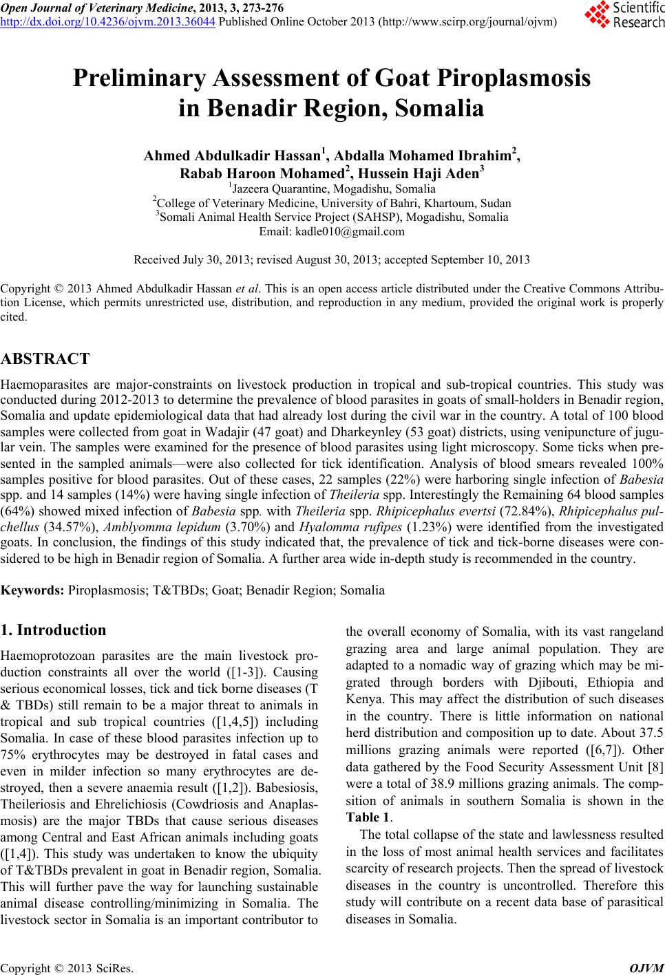 Open Journal of Veterinary Medicine, 2013, 3, 273-276 http://dx.doi.org/10.4236/ojvm.2013.36044 Published Online October 2013 (http://www.scirp.org/journal/ojvm) Preliminary Assessment of Goat Piroplasmosis in Benadir Region, Somalia Ahmed Abdulkadir Hassan1, Abdalla Mohamed Ibrahim2, Rabab Haroon Mohamed2, Hussein Haji Aden3 1Jazeera Quarantine, Mogadishu, Somalia 2College of Veterinary Medicine, University of Bahri, Khartoum, Sudan 3Somali Animal Health Service Project (SAHSP), Mogadishu, Somalia Email: kadle010@gmail.com Received July 30, 2013; revised August 30, 2013; accepted September 10, 2013 Copyright © 2013 Ahmed Abdulkadir Hassan et al. This is an open access article distributed under the Creative Commons Attribu- tion License, which permits unrestricted use, distribution, and reproduction in any medium, provided the original work is properly cited. ABSTRACT Haemoparasites are major-constraints on livestock production in tropical and sub-tropical countries. This study was conducted during 2012-2013 to determine the prevalence of blood parasites in goats of small-holders in Benadir region, Somalia and update epidemiological data that had already lost during the civil war in the country. A total of 100 blood samples were collected from goat in Wadajir (47 goat) and Dharkeynley (53 goat) districts, using venipuncture of jugu- lar vein. The samples were examined for the presence of blood parasites using light microscopy. Some ticks when pre- sented in the sampled animals—were also collected for tick identification. Analysis of blood smears revealed 100% samples positive for blood parasites. Out of these cases, 22 samples (22%) were harboring single infection of Babesia spp. and 14 samples (14%) were having single infection of Theileria spp. Interestingly the Remaining 64 blood samples (64%) showed mixed infection of Babesia sp p. with Th eileria spp. Rhipicephalus evertsi (72.84%), Rhipicephalus pul- chellus (34.57%), Amblyomma lepidum (3.70%) and Hyalomma rufipes (1.23%) were identified from the investigated goats. In conclusion, the findings of this study indicated that, the prevalence of tick and tick-borne diseases were con- sidered to be high in Benadir region of Somalia. A further area wide in-depth study is recommended in the country. Keywords: Piroplasmosis; T&TBDs; Goat; Benadir Region; Somalia 1. Introduction Haemoprotozoan parasites are the main livestock pro- duction constraints all over the world ([1-3]). Causing serious economical losses, tick and tick borne diseases (T & TBDs) still remain to be a major threat to animals in tropical and sub tropical countries ([1,4,5]) including Somalia. In case of these blood parasites infection up to 75% erythrocytes may be destroyed in fatal cases and even in milder infection so many erythrocytes are de- stroyed, then a severe anaemia result ([1,2]). Babesiosis, Theileriosis and Ehrelichiosis (Cowdriosis and Anaplas- mosis) are the major TBDs that cause serious diseases among Central and East African animals including goats ([1,4]). This study was undertaken to know the ubiquity of T&TBDs prevalent in goat in Benadir region, Somalia. This will further pave the way for launching sustainable animal disease controlling/minimizing in Somalia. The livestock sector in Somalia is an important contributor to the overall economy of Somalia, with its vast rangeland grazing area and large animal population. They are adapted to a nomadic way of grazing which may be mi- grated through borders with Djibouti, Ethiopia and Kenya. This may affect the distribution of such diseases in the country. There is little information on national herd distribution and composition up to date. About 37.5 millions grazing animals were reported ([6,7]). Other data gathered by the Food Security Assessment Unit [8] were a total of 38.9 millions grazing animals. The comp- sition of animals in southern Somalia is shown in the Table 1. The total collapse of the state and lawlessness resulted in the loss of most animal health services and facilitates scarcity of research projects. Then the spread of livestock diseases in the country is uncontrolled. Therefore this study will contribute on a recent data base of parasitical diseases in Somalia. C opyright © 2013 SciRes. OJVM 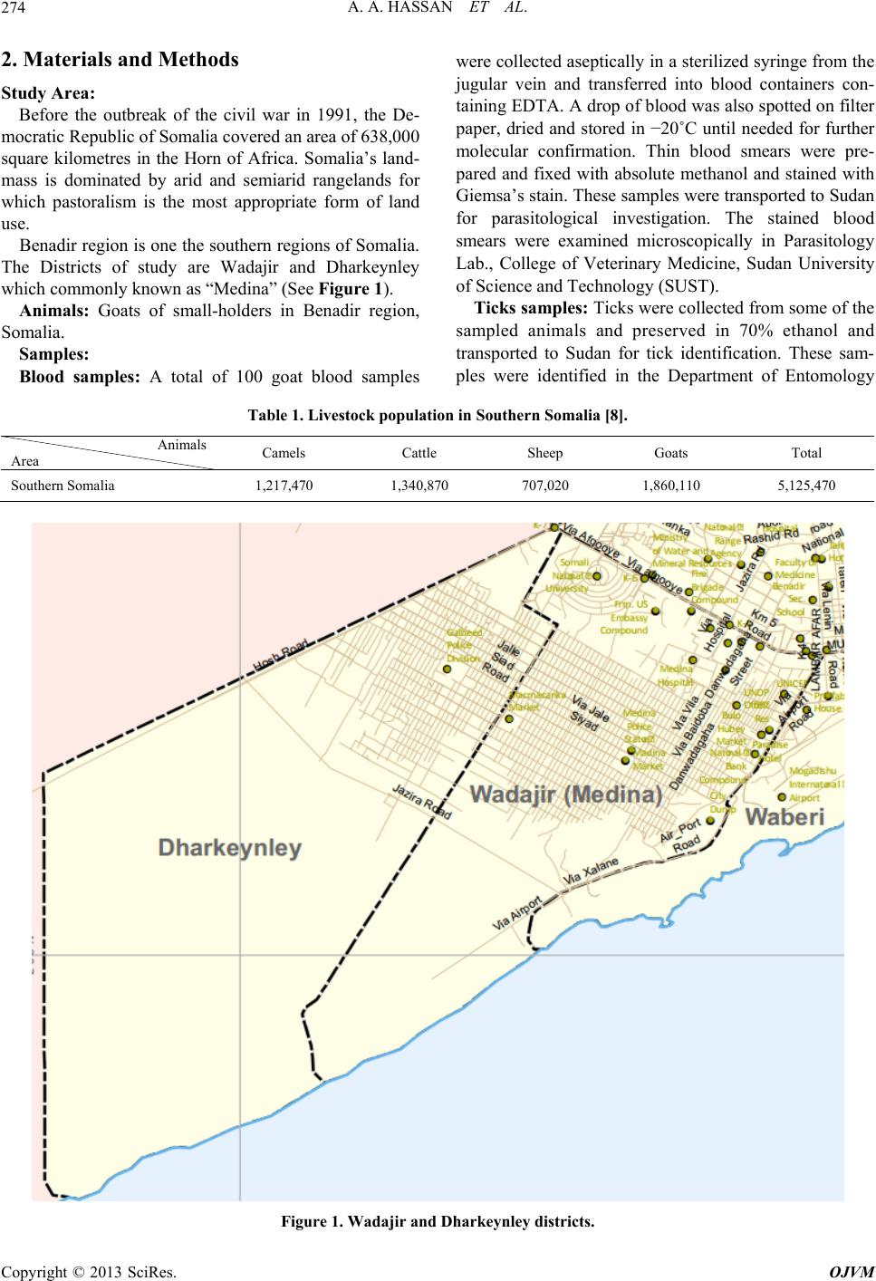 A. A. HASSAN ET AL. 274 2. Materials and Methods Study Area: Before the outbreak of the civil war in 1991, the De- mocratic Republic of Somalia covered an area of 638,000 square kilometres in the Horn of Africa. Somalia’s land- mass is dominated by arid and semiarid rangelands for which pastoralism is the most appropriate form of land use. Benadir region is one the southern regions of Somalia. The Districts of study are Wadajir and Dharkeynley which commonly known as “Medina” (See Figure 1). Animals: Goats of small-holders in Benadir region, Somalia. Samples: Blood samples: A total of 100 goat blood samples were collected aseptically in a sterilized syringe from the jugular vein and transferred into blood containers con- taining EDTA. A drop of blood was also spotted on filter paper, dried and stored in −20˚C until needed for further molecular confirmation. Thin blood smears were pre- pared and fixed with absolute methanol and stained with Giemsa’s stain. These samples were transported to Sudan for parasitological investigation. The stained blood smears were examined microscopically in Parasitology Lab., College of Veterinary Medicine, Sudan University of Science and Technology (SUST). Ticks samples: Ticks were collected from some of the sampled animals and preserved in 70% ethanol and transported to Sudan for tick identification. These sam- ples were identified in the Department of Entomology Table 1. Livestock population in Southern Somalia [8]. Animals Area Camels Cattle Sheep Goats Total Southern Somalia 1,217,470 1,340,870 707,020 1,860,110 5,125,470 Figure 1. Wadajir and Dharkeynley districts. Copyright © 2013 SciRes. OJVM 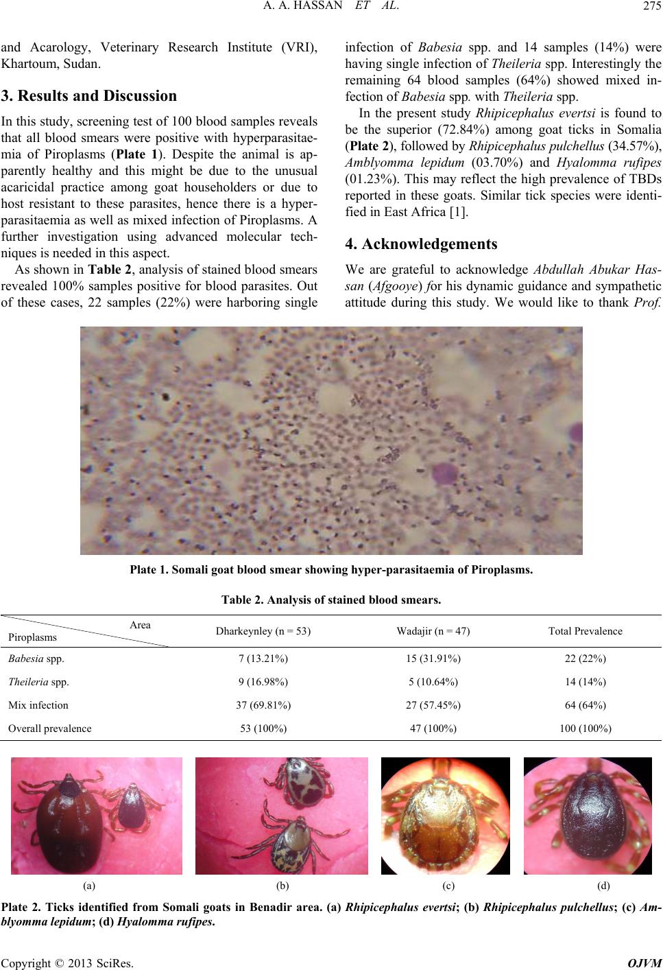 A. A. HASSAN ET AL. 275 and Acarology, Veterinary Research Institute (VRI), Khartoum, Sudan. 3. Results and Discussion In this study, screening test of 100 blood samples reveals that all blood smears were positive with hyperparasitae- mia of Piroplasms (Plate 1). Despite the animal is ap- parently healthy and this might be due to the unusual acaricidal practice among goat householders or due to host resistant to these parasites, hence there is a hyper- parasitaemia as well as mixed infection of Piroplasms. A further investigation using advanced molecular tech- niques is needed in this aspect. As shown in Table 2, analysis of stained blood smears revealed 100% samples positive for blood parasites. Out of these cases, 22 samples (22%) were harboring single infection of Babesia spp. and 14 samples (14%) were having single infection of Theileria spp. Interestingly the remaining 64 blood samples (64%) showed mixed in- fection of Babesia sp p. with Theileria spp. In the present study Rhipicephalus evertsi is found to be the superior (72.84%) among goat ticks in Somalia (Plate 2), followed by Rhipicephalus pulchellus (34.57%), Amblyomma lepidum (03.70%) and Hyalomma rufipes (01.23%). This may reflect the high prevalence of TBDs reported in these goats. Similar tick species were identi- fied in East Africa [1]. 4. Acknowledgements We are grateful to acknowledge Abdullah Abukar Has- san (Afgooye) for his dynamic guidance and sympathetic attitude during this study. We would like to thank Prof. Plate 1. Somali goat blood smear showing hyper-parasitaemia of Piroplasms. Table 2. Analysis of stained blood smears. Area Piroplasms Dharkeynley (n = 53) Wadajir (n = 47) Total Prevalence Babesia spp. 7 (13.21%) 15 (31.91%) 22 (22%) Theileria spp. 9 (16.98%) 5 (10.64%) 14 (14%) Mix infection 37 (69.81%) 27 (57.45%) 64 (64%) Overall prevalence 53 (100%) 47 (100%) 100 (100%) (a) (b) (c) (d) Plate 2. Ticks identified from Somali goats in Benadir area. (a) Rhipicephalus evertsi; (b) Rhipicephalus pulchellus; (c) Am- lyomma lepidum; (d) Hyalomma rufipes. b Copyright © 2013 SciRes. OJVM  A. A. HASSAN ET AL. 276 Ahmed A. Ismail, SUST and VRI for their technical sup- port. REFERENCES [1] E. J. L. Soulsby, “Helminths, Arthropods & Protozoa of Domesticated Animals,” 7th Edition, Bailliere Tindall, London, 1986. [2] G. M. Urquhart, J. Armour, J. L. Duncan, A. M. Dunn and F. W. Jennings, “Veterinary Parasitology,” 2nd Edi- tion, B-Blackwell Science, 1996. [3] K. T. Friedhoff, “Transmission of Babesia,” In: M. Ristic, Ed., Babesiosis of Domestic Animals and Man, CRC Press, Boca Raton, 1988, pp. 23-52. [4] A. M. El Hussein, A. M. Majid and S. M. Hassan, “The Present Status of Tick-Borne Diseases in the Sudan,” Ar- chives de l’Institut Pasteur de Tunis, Vol. 81, No. 5, 2004, pp. 31-34. [5] S. M. Hassan and D. A. Salih, “Bibliography with Ab- stracts, Ticks and Tick-borne Diseases in the Sudan,” 1st Edition, Central Laboratory, Ministry of Science and technology, Khartoum, 2009. [6] FAO/World Bank Cooperative Programme, the World Bank, European Union Report No. 04/001 IC-SOM, 2004. [7] Food and Agriculture Organization (FAO) of the United Nations investment centre division, Somalia, “Somalia towards a Livestock Sector Strategy,” Final Report, 1998. [8] Food Security Assessment Unit (FSAU), 1999. Copyright © 2013 SciRes. OJVM |