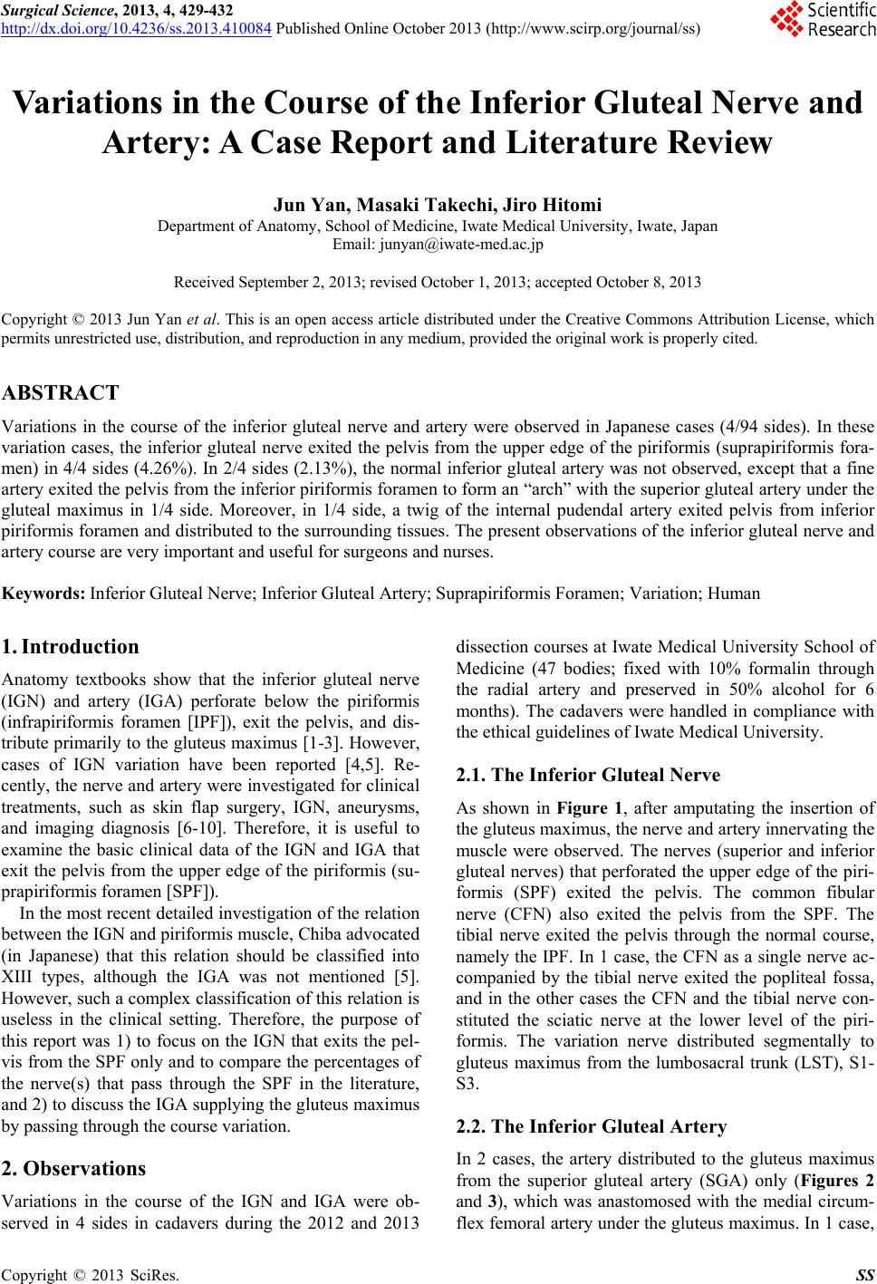
Surgical Science, 2013, 4, 429-432
http://dx.doi.org/10.4236/ss.2013.410084 Published Online October 2013 (http://www.scirp.org/journal/ss)
Variations in the Course of the Inferior Gluteal Nerve and
Artery: A Case Report and Literature Review
Jun Yan, Masaki Takechi, Jiro Hitomi
Department of Anatomy, School of Medicine, Iwate Medical University, Iwate, Japan
Email: junyan@iwate-med.ac.jp
Received September 2, 2013; revised October 1, 2013; accepted October 8, 2013
Copyright © 2013 Jun Yan et al. This is an open access article distributed under the Creative Commons Attribution License, which
permits unrestricted use, distribution, and reproduction in any medium, provided the original work is properly cited.
ABSTRACT
Variations in the course of the inferior gluteal nerve and artery were observed in Japanese cases (4/94 sides). In these
variation cases, the inferior gluteal nerve exited the pelvis from the upper edge of the piriformis (suprapiriformis fora-
men) in 4/4 sides (4.26%). In 2/4 sides (2.13%), the normal inferior gluteal artery was not observed, except that a fine
artery exited the pelvis from the inferior piriformis foramen to form an “arch” with the superior gluteal artery under the
gluteal maximus in 1/4 side. Moreover, in 1/4 side, a twig of the internal pudendal artery exited pelvis from inferior
piriformis foramen and distributed to the surrounding tissues. The present observations of the inferior gluteal nerve and
artery course are very important and useful for surgeons and nurses.
Keywords: Inferior Gluteal Nerve; Inferior Gluteal Artery; Suprapiriformis Foramen; Variation; Human
1. Introduction
Anatomy textbooks show that the inferior gluteal nerve
(IGN) and artery (IGA) perforate below the piriformis
(infrapiriformis foramen [IPF]), exit the pelvis, and dis-
tribute primarily to the gluteus maximus [1-3]. However,
cases of IGN variation have been reported [4,5]. Re-
cently, the nerve and artery were investigated for clinical
treatments, such as skin flap surgery, IGN, aneurysms,
and imaging diagnosis [6-10]. Therefore, it is useful to
examine the basic clinical data of the IGN and IGA that
exit the pelvis from the upper edge of the piriformis (su-
prapiriformis foramen [SPF]).
In the most recent detailed investigation of the relation
between the IGN and piriformis muscle, Chiba advocated
(in Japanese) that this relation should be classified into
XIII types, although the IGA was not mentioned [5].
However, such a complex classification of this relation is
useless in the clinical setting. Therefore, the purpose of
this report was 1) to focus on the IGN that exits the pel-
vis from the SPF only and to compare the percentages of
the nerve(s) that pass through the SPF in the literature,
and 2) to discuss the IGA supplying the gluteus maximus
by passing through the course variation.
2. Observations
Variations in the course of the IGN and IGA were ob-
served in 4 sides in cadavers during the 2012 and 2013
dissection courses at Iwate Medical University School of
Medicine (47 bodies; fixed with 10% formalin through
the radial artery and preserved in 50% alcohol for 6
months). The cadavers were handled in compliance with
the ethical guidelines of Iwate Medical University.
2.1. The Inferior Gluteal Nerve
As shown in Figure 1, after amputating the insertion of
the gluteus maximus, the nerve and artery innervating the
muscle were observed. The nerves (superior and inferior
gluteal nerves) that perforated the upper edge of the piri-
formis (SPF) exited the pelvis. The common fibular
nerve (CFN) also exited the pelvis from the SPF. The
tibial nerve exited the pelvis through the normal course,
namely the IPF. In 1 case, the CFN as a single nerve ac-
companied by the tibial nerve exited the popliteal fossa,
and in the other cases the CFN and the tibial nerve con-
stituted the sciatic nerve at the lower level of the piri-
formis. The variation nerve distributed segmentally to
gluteus maximus from the lumbosacral trunk (LST), S1-
S3.
2.2. The Inferior Gluteal Artery
In 2 cases, the artery distributed to the gluteus maximus
from the superior gluteal artery (SGA) only (Figures 2
and 3), which was anastomosed with the medial circum-
flex femoral artery under the gluteus maximus. In 1 case,
C
opyright © 2013 SciRes. SS
J. YAN ET AL.
430
Figure 1. A photo (posterior view) showing the inferior
gluteal nerve (IGN) exited the pelvic from upper edge of the
piriformis muscle (PM) with the common fibula nerve
(CFN). The tibia nerve (TN) was from normal course exited
the pelvic and the posterior femoral cutaneous nerve
(PFCN) was from both upper and low edge of piriformis
muscle. The superior gluteal artery (SGA) was the main
artery supplying the gluteus maximus musc le .
Figure 2. A sketch (posterior view) showing the nerve and
artery supplying gluteal maximus muscle exited the pelvic
from upper edge of piriformis muscle, and the artery anas-
tomosis with the medial circulation femoral artery to form
an “arch” under the gluteal maximus (Red arrow). Note the
normal inferior gluteal artery was absence. GM: gluteal
maximus; GMe: gluteal medius; GMi: gluteal minimus; SN:
sciatic nerve; Red star mark: the nerves innervating gluteal
maximus exited the pelvic from upper edge of pirifomis
muscle.
Figure 3. A sketch showing the form of the sacral plexus
(posterior view). The nerves innervating gluteal maximus
exited the pelvic from upper edge of pirifomis (Red star
mark). Under the gluteal maximus, an “artery arch” (Red
arrow) was formed by the superior gluteal artery and a
twig from internal pudendal artery (IP A). The normal infe-
rior gluteal artery was absence.
the IGA was very fine and anastomosed with the SGA
under the gluteus maximus to form an “arch”. In another
case, a fine branch exited the pelvis from the IPF, but
was a twig of the internal pudendal artery; this twig
branched into the surrounding tissue.
3. Discussion
The course of the IGN and IGA is important for clinical
treatment, such as in plastic surgery, grafting surgery,
and clinical nursing [11-14]. However, many variations
in the courses of the IGN and IGA have been observed
and discussed [15-18], although the IGA was not dis-
cussed [5,19, 2 0] .
In the literature, the relative frequencies of the IGN
exiting the pelvis from the upper edge of the p iriformis in
human adults were 1.4% (8/579) [15]; 0.4% (1/246) [16];
0.4% (1/284) [21]; 2.1% (3/144) [22]; 1.0% (3/306) [23];
3.3% (4/120) [24]; 4.4% (14/320) [17]; 1.9% (2/106)
[25]; 2.0% (4/200) [26]; 0.9% (1/112) [27]; and 0.2%
(1/257) [5]. This frequency in fetuses was 0.4% (1/240)
[28]; 1.2% (2/164) [29]; 1.1% (4/364) [30]; and 2.0%
(4/200) [31]. In our series, this frequency in adults was
4.26% (4/94), which was closer to that reported by Ha-
simoto and Toyama (1942). Variations in the course of
the IGN are closely related with the generation of the
piriformis muscle [5], thus it is likely that the IGN fibers
Copyright © 2013 SciRes. SS
J. YAN ET AL. 431
changed course and transferred it with the SGN to exit
the pelvis from the SPF.
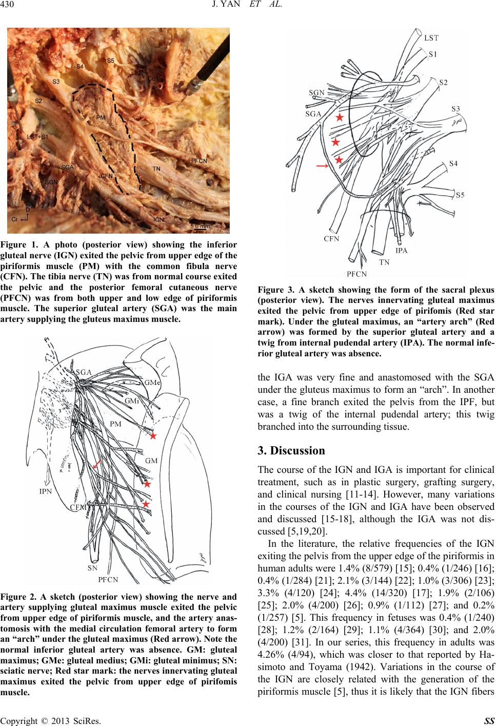
Generally, the IGA branches from the internal iliac ar-
tery, passes through the lower edge of the piriformis, and
distributes to the lower part of the gluteus maximus; the
arteries supplying the gluteus maximus are derived from
both the SGA and IGA. In the literature, the IGA has
been indicated to originate from the internal pudenda
artery (IPA) or further from the obturator artery [4], and
the absence of the IGA has been reported in 1 case [32].
Recently, superior and inferior gluteal artery perforator
flaps (SGAP and IGAP) have been used for transplanta-
tion surgery. On the other hand, it has been mentioned
that a descending branch of the IGA is an important ves-
sel for the flap and was present in 91% of patients; how-
ever, the authors did not mention whether the IGA was
absent or exited the pelvis from the SPF [7]. Gabrielli et
al. investigated this artery in 80 sides in humans and re-
ported that none exited the pelvis from the upper edge of
the piriformis [6]. The present observation indicated that
the artery supplying the gluteus maximus muscle could
originate from the SGA only and/or from other branches,
in agreement with the individual results of Bergman and
Reddy [4,32]. Therefore, the variation in the course of
the artery and the rare occurrence of the IGA being a
twig to form an “arch” under the gluteus maximus are
clinically important.
Moreover, present variation cases show that the varia-
tion of inferior gluteal nerve accompanies with the varia-
tion of the artery in all cases is effected by unknown
mechanism in early stage of generation. Therefore, fur-
ther investigations of the embryological changes in the
nerves and arteries distributed to gluteus muscle group
are necessary.
4. Conclusions
The relative frequency of the inferior gluteal nerve exit-
ing the pelvis from the upper edge of the piriformis
ranges from 0.2% to 4.4% in human adults and 0.4% to
3.2% in fetuses. In our series, this frequency was 4.26%
in Japanese adults.
The inferior gluteal artery could be absent or present
as a very fine anastomosis twig that forms an “arch” with
the superior glut eal artery un der the glut eus maximus.
5. Acknowledgements
We thank Mr. S. Takahashi and Mr. N. Sasaki (Iwate
Medical University) for their technical advice. This work
was supported financially by the Advanced Medical Sci-
ence Center of Iwate Medical University.
REFERENCES
[1] P. E. Celli, “Sulla Morphologie del M. Piriformis,”
Anatomischer Anzeiger, Vol. 41, 1913, pp. 551-560.
http://www.biodiversitylibrary.org/item/43338#page/579/
mode/1up
[2] G. Gabella, “Gray’s Anatomy,” In: P. L. Williams, Ed.,
Pelvic Girdle and Lower Limb, 38th Edition, Churchill
Livingstone, New York, 1995, pp. 1545-1564.
[3] V. Mahadevan, “Gray’s Anatomy,” In: S. Standing, Ed.,
Pelvic Girdle and Lower Limb, 40th Edition, Churchill
Livingstone, New York, 2008, pp. 1329-1390.
[4] R. A. Bergman, S. A. Thomson, A. K. Afifi and F. A.
Saadesh, “Compendium of Human Anatomic Variations,”
Urban & Schwarzenberg, Baltimore, Munich, 1988, pp.
84-85.
[5] S. Chiba, “Multiple Positional Relationships of Nerves
Arising from the Sacral Plexus to the Piriformis Muscle
in Human,” Journal of Anatomy, Vol. 67, No. 6, 1992, pp.
691-724. (in Japanese)
[6] C. Gabrielli, E. Olave, A. Sarmento, C. Mizusaki and J. C.
Prates, “Abnormal Extrapelvic Course of the Inferior
Gluteal Artery,” Surgical and Radiologic Anatomy, Vol.
19, No. 3, 1997, pp. 139-142.
http://dx.doi.org/10.1007/BF01627962
[7] C. Windhofer, E. Brenner, B. Moriggl and C. Papp, “Re-
lationship between the Descending Branch of the Inferior
Gluteal Artery and the Posterior femoral Cutaneous
Nerve Applicable to Flap Surgery,” Surgical and Ra-
diologic Anatomy, Vol. 24, No. 5, 2002, pp. 253-257.
http://dx.doi.org/10.1007/s00276-002-0064-z
[8] Z. X. Ling and V. P. Kumar, “The Course of the Inferior
Gluteal Nerve in the Posterior Approach to the Hip,” The
Bone & Joint Journal, Vol. 88-B, No. 12, 2006, pp. 1580-
1583. http://dx.doi.org/10.1302/0301-620X.88B12.18182
[9] F. S. Anthony, F. M. Michael, W. Gary and B. Kath,
“Relationship of Inferior Gluteal Nerve and Vessels:
Target for Application of Stimulation Devices for the
Prevention of Pressure Ulcers in Spinal Cord Injury”,
Surgical and Radiologic Anatomy, Vol. 30, No. 1, 2008,
pp. 41-45. http://dx.doi.org/10.1007/s00276-007-0282-5
[10] S. Mariano and D. M. Gilda, “Exposure of the Sciatic
Nerve in the Gluteal Region without Sectioning the Glu-
teus Maximus: Analysis of a Series of 18 Cases,” Surgi-
cal Neurology International, Vol. 3, No. 1, 2012, pp. 15-
20. http://dx.doi.org/10.4103/2152-7806.92929
[11] G. C. Cormack and B. G. H. Lamberty, “The Blood Sup-
ply of Thigh Skin,” Plastic & Reconstructive Surgery,
Vol. 75, No. 3, 1985, pp. 342-354.
http://dx.doi.org/10.1097/00006534-198503000-00008
[12] A. Frick, R. G. H. Baumeister and B. Wiebecke, “Mi-
crovasculature of the Inferior Gluteal Flap,” European
Journal of Plastic Surgery, Vol. 16, No. 1, 1993, pp. 30-
32. http://dx.doi.org/10.1007/BF00192703
[13] M. Tarek, “Superior Gluteal Artery Perforator Flap for
Closure of Large Sacral Defects,” Egyptian Journal of
Plastic and Reconstructive Surgery, Vol. 28, No. 2, 2004,
pp. 175-179.
[14] J. A. Robert, M. L. Maria and W. G. Jay, “Inferior
Gluteal Perforator Flaps for Breast Reconstruction,” Se-
minars in Plastic Surgery, Vol. 20, No. 2, 2006, pp. 89-94.
Copyright © 2013 SciRes. SS
J. YAN ET AL.
Copyright © 2013 SciRes. SS
432
http://dx.doi.org/10.1055/s-2006-941715
[15] B. Adachi, “Anatomische Untersuchungen an Japanern,”
Zeitschrift für Morphologie und Anthropologie, Vol. 2,
No. 2, 1900, pp. 198-222.
[16] C. R. Bardeen and A. W. Elting, “A Statistical Study of
the Variations in the Formation and Position of the Lum-
bosacral Plexus in Man,” Anatomical Record, Vol. 19, No.
1, 1901, pp. 209-232.
[17] M. Hasimoto and K. Toyama, “The Relationship of the
Sacral Nerve and Piriformis Muscle in Chinese,” Acta
Anatomica Nipponica, Vol. 19, No. 2, 1942, pp. 171-184.
(in Japanese)
[18] G. Odajima and T. Kurihara, “Supplementary Findings to
the Morphology of M. Piriformis,” Journal of Showa
Medical Society, Vol. 20, No. 3. 1960, pp. 1-4. (in Japa-
nese)
[19] M. E. Tarek and H. A. Abdelghany, “Inferior Gluteal
Nerve and Artery: An Anatomical Study,” Alexandria
Journal of Medicine, Vol. 45, No. 2, 2009, pp. 1-17.
[20] P. S. Sharadkumar, “The Anatomical Study of Division of
the Sciatic Nerve Proximal to its Exit,” Mad Science, Vol.
2, No. 1, 2013, pp. 169-171.
[21] Y. Koganei, S. Arai and S. Shikinami, “The Statistics of
the Variation of the Muscles,” Journal of Tokyo Medical
Society, Vol. 17, No. 2, 1903, pp. 127-131. (in Japanese)
[22] S. Igarashi, “The Relationship of the N. Ischiadicus and
M. Piriformis,” Journal of Kanazawa Medical University,
Vol. 19, No. 1, 1935, pp. 123-153. (in Japanese)
[23] N. Fukumoto, “The M. Piriformis and N. Ischiadicus in
Japanese,” Journal of Fukuoka Medical University, Vol.
28, No. 3, pp. 756-763. (in Japanese)
[24] L. E. Beaton and B. J. Anson, “The Relation of the Sci-
atic Nerve and Its Subdivisions to the Piriformis Muscle,”
The Anatomical Record, Vol. 70, No. 1, 1938, pp. 1-5.
http://dx.doi.org/10.1002/ar.1090700102
[25] T. Hayama, “The Relationship of Sciatic Nerve and Piri-
formis Muscle,” Journal of Medical University of Japan,
Vol. 13, No. 3, 1954, pp. 1583-1588. (in Japanese)
[26] C. Nizankowski, J. Stociak and J. Szybejko, “Odimany
Przebiegu Nerwu Kulszwogo u Cztowieka,” Folia Mor-
phologica, Vol. 31, 1972, pp. 507-513.
[27] B. Tillmann, “Verlaufsvarianten des N. Gluteus Inferior,”
Anatomical Record, Vol. 145, No. 3, 1979, pp. 293-302.
[28] T. Kusida, “The Lumbosacral Plexus in Japanese Fetus:
Part III,” Acta Anatomica Nipponica, Vol. 16, No. 1,
1941, pp. 28-47. (in Japanese)
[29] J. Sugiyama, “The Relationship of the Sciatic Nerve and
Piriformis Muscle in Japanese Fetal Twins,” Anatomical
Study of the Fetal Twins, Vol. 4, 1943, pp. 1-26. (in
Japanese)
[30] K. Kubota, I. Noguchi and T. Nakao, “Rare Types of
Relation between the Sciatic Nerve and the Piriformis
Muscle,” Okajimas Folia Anatomica Japonica, Vol. 36,
No. 2, 1960, pp. 329-344.
[31] K. Honma, “The Anatomical Study of Sciatic Nerve in
Japanese Fetus,” Journal of Tokyo Medical University,
Vol. 17, No. 4, 1959, pp. 1955-1993. (in Japanese)
[32] R. Ausencia, R. V. Venkata and R. Mohandas, “Absence
of Inferior Gluteal Artery: A Rare Observ ation,” Interna-
tional Journal of Morphology, Vol. 25, No. 1, 2007, pp.
95-98.