International Journal of Geosciences, 2013, 4, 60-63
http://dx.doi.org/10.4236/ijg.2013.45B010 Published Online September 2013 (http://www.scirp.org/journal/ijg)
Copyright © 2013 SciRes. IJG
Micr o c ystin Accumulation in Nile Ti lapia, Oreochromis
niloticus and Giant Freshwater P r awns, Macrobrachium
rosenbergii in Green Water Sys tem Cultivation
Khomsan Ruangrit1, Yuwadee Peerapornpisal1, Jeeraporn Pekkoh1, Niwooti Whangchai2*
1Department of Biology, Faculty of Science, Chiang Mai University, Chiang Mai, Thailand
2Faculty of Fisheries Technology and Aquatic Resources, Maejo University, Chiang Mai, Thailand
Email: *niwooti@hotmail.co.th
Received July 2013
ABSTRACT
Phytoplankton including blue-green algal or cyanobacterial blooms frequently occurred in aquaculture ponds. Some
cyanobacteria produced cyanotoxins that may accumulate in the food web and eventually in the aquacult ure products. I n
this study, accumulatation of microcystins in Nile tilapia (Oreochromis niloticus) and giant freshwater prawn (Macro-
brachium rosenbergii) cultured in green water system was investigated. Nile tilapia was cultured in green water sy stem
and fish food; green water system with Microcystis aeruginosa Kützing and fish food and green water system with M.
aeruginosa. Giant freshwater prawn was cultured: in green water systems with and without toxic M. aeruginosa. Mi-
crocystins of 8.32 ± 0.76 and 9.35 ± 1.45 µg·kg1 d.w. were detected in fish cultured in green water system with M.
aeruginosa and fish food and in green water system with M. aeruginosa, respectively. Microcystins of 14.42 ± 1.63
µg·kg1 was found in prawn samples. It implied that aquaculture products were likely to be contaminated with micro-
cystins. This finding is useful for aquaculture in terms of food safety.
Keywords: Microcystis aeruginosa Kützing; Microcystins; Aquaculture; Green Wa t e r System
1. Introduction
Thailand is the fourth ranking Nile tilapia (Oreochromis
niloticus) producer in the world since 2000. Its produc-
tion has increased almost exponentially [1]. It is con-
sumed and exported to other countries. Nile tilapia is
mostly raised in earthen ponds using manures and other
recyclable wastes, as low cost commercial pellet feeds
are not necessary for growing tilapia and the traditional
cultivation, nutrient-enriched water, “green water”, pro-
duced by the addition of animal manure or fertilizer is
sufficient to achieve a marketable fish [2] as well as the
prawns [3]. There are many aquaculture ponds through-
out Thailand where giant freshwater prawns, Macrobra-
chium rosenbergii, are cultured. In fact, the prawns can
be grown in all freshwater bodies [3]. They are commer-
cially important because they are widely used for human
consumption. Domestic consumption was 70 % of total
production [1]. Green water systems can cause eutrophi-
cation of surface water, resulting in increased occurrence
of toxic cyanobacterial bloom, especially Microcystis [3].
The occurrence can create a significant water quality
problem, including their ability to produce toxins, name-
ly microcystins (MCs). The toxins accumulate in aquatic
organisms and are transferred to higher trophic levels. It
involves the risk for human exposure through the con-
sumption of contaminated aquatic organisms [4-6]. In
Thailand, cyanobacterial genera with known toxin- pro-
ducing taxa occurred in many reservoirs in all regions.
Microcystis aeruginosa is the most frequently blooms
[7-9]. Ruangrit et al. [10] found high amount of M. aeru-
ginosa in prawn pond and microcystins were detected in
prawn. Therefore, it is needed to clarify whether MCs are
able to accumulate in aquatic organism cultured with
traditional method. The data would be useful for food
safety aspect and public health to avoid the damaging
effect of cyanobacteria and their toxins.
2. Materials and Methods
2.1. Culturing of Nile Tilapia and Giant
Freshwater Prawn
Nile tilapia about 5 cm in size and giant freshwater
prawn about 5 - 7 cm in size were obtained from the Fa-
culty of Fisheries Technology and Aquatic Resources,
Maejo University, Chiang Mai, Thailand. The fish were
cultured in 3 cement ponds, 1.5 m × 1.5 m and the depth
of 0.50 m containing green water 0.30 m deep, 30 fish in
*Corresponding a uthor.
K. RUANGRIT ET AL.
Copyright © 2013 SciRes. IJG
61
each pond. Feeding treatments were; 1) Green water sys-
tem (Tr. 1). 2) Green water system with 18 - 30 × 106
cells·L1 M. aeruginosa from natural pond and combined
with commercial pellet feed (Tr. 2). 3) Green water sys-
tem with 18 – 30 × 106 cells·L1 M. ae ruginosa (Tr. 3).
The prawns were cultured in two cement ponds of
similar size, 30 prawns were in the pen (0.45 m × 0.45 m
with water depth of 0.30 m) made of blue net (mezh size
2 mm) attached to the pond, 30 prawns were outside the
pen. Feeding treatments were: Green water system (Tr. 1)
and green water system with M. aeruginosa combined
with commercial pellet feed (Tr. 2).
Completely randomize design (CRD) with duplicate
treatments were carried out. Both fish and prawn were
cultured for 2 months. Water samples were collected
every two weeks to determine the amount of M. aerugi-
nosa, phytoplankton and microcystins.
2.2. Identification and Enumeration of M.
aeruginosa and Phytoplankton
Morphological classification of Microcystis spp. and phy-
toplankton were done under compound microscope (Olym-
pus model CH30RF200) using related texts such as
Komárek and Komáková-Legnerová [12] and Hindak [13].
Cells of M. aeruginosa we re counted on a haem acytometer .
2.3. Analysis of Microcystins
2.3.1. Extraction of Microcystins
Microcystins were extracted after Kankaanpää et al. [4]
with modification. Fish and prawn tissues were dissected
and freeze-dried at 20˚C for 24 - 72 hours before ex-
traction and ELISA analysis. One mL of 100% methanol
was added into 2 g fish and prawn tissues for extraction
overnight. The extracts were centrifuged at 12,000 rpm
for 30 min and the supernatants were concentrated to 150
µl with a heat blo ck at 50 ˚C, overnight and centrifuged at
12,000 rpm for 30 min before ELISA analysis.
2.3.2. Microcystin Analysis by ELISA Assay
ELISA Microcystin Plate Kit (Catalog No. EP022), EN-
VIROLOGIX INC was used and performed in accor-
dance with the manufacturer’s instructions. A standard
curve was constructed using three calibrations 0.16, 0.5
and 2.5 µg·L1 supplied with the kit. The absorbance at
450 nm was measured with a microplate reader (Spectra
MR, DYNEX Technologies). The microcystin concentra-
tion in each extract was expressed as MC-LR equivalent.
3. Results and Discussion
3.1. Identification and Enumeration of M.
aeruginosa and Phytoplankton in Fish Ponds
Dominant species of phytoplankton excluding M. aeru-
ginosa were found to belong to 5 divisions i.e. Divisions
Chlorophyta, Cyanophyta, Euglenophyta, Bacillariophyta
and Pyrrhophyta. Dominant species were Chlorophyta
(green algae) such as Scencedesmus spp . and Pediastrum
spp. The species composition was similar in each treat-
ment except Tr. 2, Microcystis wesenbergii was found as
dominant species.
Tr. 1 had lowest amounts of phytoplankton. Whereas
the highest amounts of phytoplankton were found in Tr.
3 and Tr. 2, respectively (Figures 1 and 2) .
3.2. Microcystin Contents in Fish Samples
High microcystin contents in fish samples were detected.
Microcystin contents of 8.32 ± 0.76 and 9.35 ± 1.45
µg·kg1 d.w. were found in the fish of Tr. 2 and Tr. 3,
respectively. Whereas microcystins in the corresponding
Tr. 2 and T r. 3 pond s w ere 2 0.08 ± 0 .24 and 1 9.52 ± 0.49
µg·L1, respectively.
Microcystins were released from dead Microcystis
cells causing high content of microcystin in the water.
However, microcystin in the fish was obtained from in-
gestio n of Microcystis cells by the fish [14,15].
Figure 1. Amounts of phytoplankton and M. aeruginosa in
each treatment (Replicate I fish pond).
Figure 2. Amounts of phytoplankton and M. aeruginosa in
each treatment (Replicate II fish pond).
K. RUANGRIT ET AL.
Copyright © 2013 SciRes. IJG
62
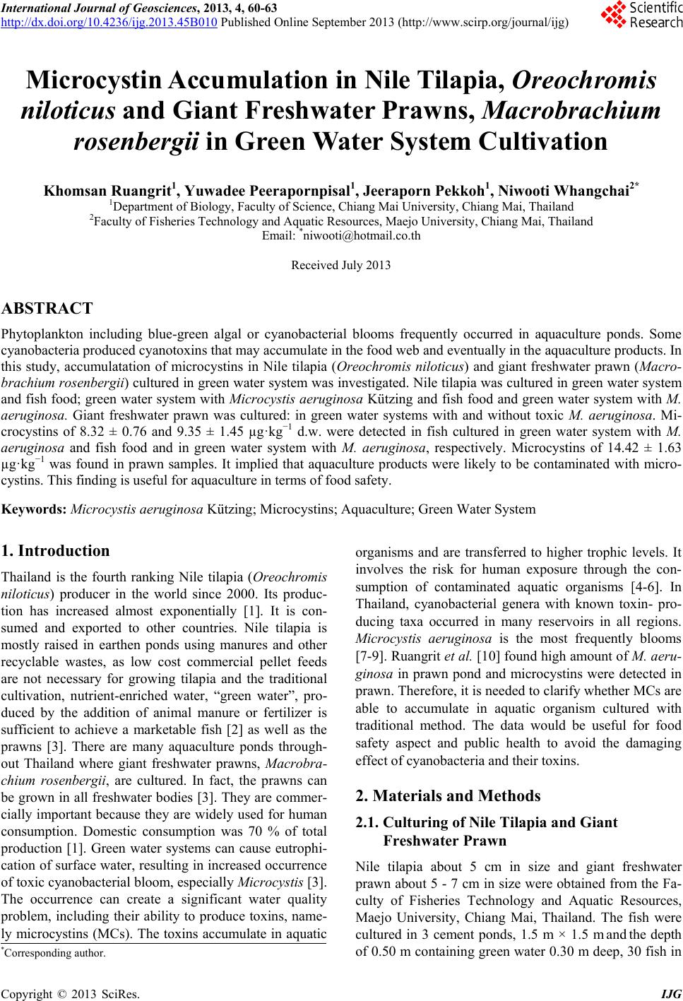
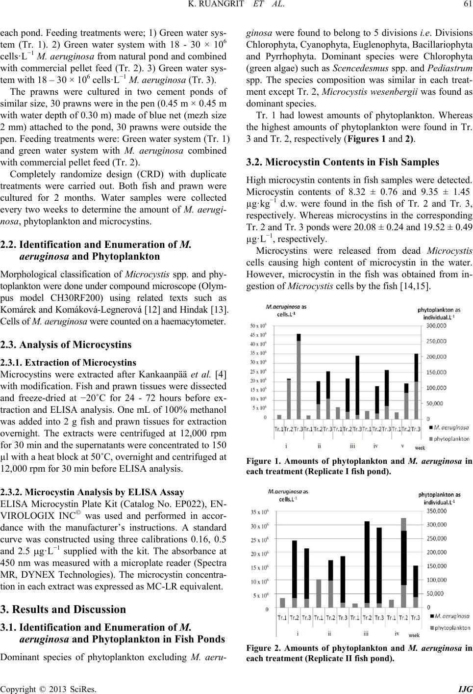
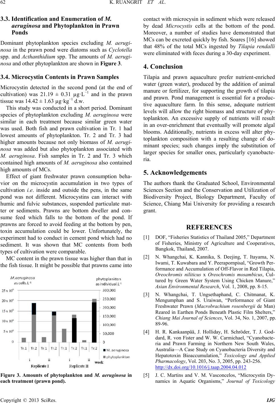
3.3. Identification and Enumeration of M.
aeruginosa and Phytoplankton in Prawn
Ponds
Dominant phytoplankton species excluding M. aerugi-
nosa in the prawn pond were diatoms such as Cyclotella
spp. and Acthanthidium spp. The amounts of M. aerugi-
nosa and other phyto plankton a re s hown in Figure 3.
3.4. Microcystin Contents in Prawn Samples
Microcystin detected in the second pond (at the end of
cultivation) was 21.19 ± 0.31 µg·L1 and in the prawn
tissue was 14.42 ± 1.63 µg·kg1 d.w.
This study was conducted in a short period. Dominant
species of phytoplankton excluding M. aeruginosa were
similar in each treatment because similar green water
was used. Both fish and prawn cultivation in Tr. 1 had
lowest amounts of phytoplankton. Tr. 2 and Tr. 3 had
higher amounts because not only biomass of M. aerugi-
nosa was added but also phytoplankton associated with
M. aeruginosa. Fish samples in Tr. 2 and Tr. 3 which
contained high amounts of M. aeruginosa also contained
high amounts of MCs.
Effect of giant freshwater prawn consumption beha-
vior on the microcystin accumulation in two types of
cultivation i.e. inside and outside the pens, in the same
pond was not different. Microcystins can interact with
humic and fulvic substances, suspended particulate mat-
ter or sediments. Prawns are bottom dweller and con-
sume feed which falls to the bottom of the pond. If
prawns are forced to avoid feeding at the bottom by pen,
toxin accumulation could be lower. Unfortunately, the
experiment had to condu ct in cement pond which had no
sediment. It was shown that MC contents from both
types of cultivation were comparable.
MC content in the prawn tissue was higher than that in
the fish tissue. It might be possible that prawns came into
Figure 3. Amounts of phytoplankton and M. aeruginosa in
each treatment (prawn pond).
contact with microcysin in sediment which were released
by dead Microcystis cells at the bottom of the pond.
Moreover, a number of studies have demonstrated that
MCs can be excreted quickly by fish. Soares [16] showed
that 48% of the total MCs ingested by Tilapia rendalli
were eliminated with feces during a 30-day experiment.
4. Conclusion
Tilapia and prawn aquaculture prefer nutrient-enriched
water (green water), produced by the addition of animal
manure or fertilizer, for supporting the growth of tilapia
and prawn. Pond management is essential for a produc-
tive aquaculture farm. In this sense, adequate nutrient
levels will allow the right biomass and structure of phy-
toplankton. An excessive supply of nutrients will result
in an over-enrichment that eventually will promote algal
blooms. Additionally, nutrients in excess will alter phy-
toplankton composition with a resulting change of do-
minant species; such changes imply the substitution of
larger species for smaller ones, particularly cyanobacte-
ria.
5. Acknowledgements
The authors thank the Graduated School, Environmental
Sciences Section and the Conservation and Utilization of
Biodiversity Project, Biology Department, Faculty of
Science, Chiang Mai University for providing a research
grant.
REFERENCES
[1] DOF, “Fisheries Statistics of Thailand 2005,” Department
of Fisheries, Ministry of Agriculture and Cooperatives,
Bangkok, Thailand, 2007.
[2] N. Whangchai, K. Kannika, S. Deejing, T. Itayama, N.
Iwami, T. Kuwabara and Y. Peerapornpisal, “Growth Per-
formance and Accumulation of Of f-Fl avor in Red Tilapia,
Oreochromis niliticus x Oreochromis mosambicus, Cul-
tured by Green Water System Using Chicken Manure,”
Asian Environmental Research, Vol. 1, 2008, pp. 8-15.
[3] N. Whangchai, T. Ungsethaphand, C. Chitmanat, K.
Mengumphan and S. Uraiwan, “Performance of Giant
Freshwater Prawn (Macrobrachium rosenbergii de Man)
Reared in Earthen Ponds Beneath Plastic Film Shelters,”
Chiang Mai Journal of Sciences, Vol. 34, No. 1, 2007, pp.
89-96.
[4] H. R. Kankaanpää, J. Holliday, H. Schröder, T. J. God-
dard, R. von Fister and W. W. Carmichael, “Cyanobacte-
ria and Prawn Farming in Northern New South Wales,
Australia—A Case Study on Cyanoba cteria Di versity and
Hepatotoxin Bioaccumulation,” Toxicology and Applied
Pharmacology, Vol. 203, No. 3, 2005, pp. 243-256.
http://dx.doi.org/10.1016/j.taap.2004.04.012
[5] J. C. Martins and V. M. Vasconcelos, “Microcystin Dy-
namics in Aquatic Organisms,” Journal of Toxicology
K. RUANGRIT ET AL.
Copyright © 2013 SciRes. IJG
63
and Environmental Health, Part B, Vol. 12, 2009, pp.
65–82. http://dx.doi.org/10.1080/10937400802545151
[6] T. Papadimitriou, I. Kagalou, V. Bacopoulos and I. D.
Leonardos, “Accumulation of Microcystins in Water and
Fish Tissues: An Estimation of Risks Associated with
Microcystins in Most of the Greek Lakes,” Environmen-
tal Toxicology, Vol. 25, No. 4, 2010, pp. 418-427.
http://dx.doi.org/10.1002/tox.20513
[7] W. Yongmanitchai, P. Intachot and D. Chonudomkul,
“Survey of Cyanobacterial Diversity in Thailand,” In:
Workshop on Eutrophication and Toxic Cyanobacteria in
Reservoirs, Water Research Center, Chiang Mai, 2001.
[8] Y. Peerapornpisal, W. Sonthichai, M. Sukchotiratana, S.
Lipigorngoson, W. Ruangyuttikarn, K. Ruangrit, J. Pek-
koh, R. Prommana, N. Panuvanitchakorn, N. Ngearnpat,
S. Kiatpradub and S. Promkutkaew, “Survey and Moni-
toring of Toxic Cyanobacteria in Water Supplied and Fi-
sheries in Thailand,” Chiang Mai Journal of Sciences,
Vol. 29, No. 2, 2002. pp. 71-79.
[9] J. Pekkoh, “Diversity and Cyanotoxins of Toxic Cyano-
bacteria in Some Water Resources of Thailand,” Ph.D.
Thesis, Chiang Mai University, Chiang Mai, 2008.
[10] K. Ruangrit, N. Whangchai, W. Ruangyuttikarn, J. Pek-
koh and Y. Peerapornpisal, “First Report on Microcystins
Contamination in Giant Freshwater Prawn (Macrobra-
chium rosenbergii) and Nile Tilapia (Tilapia nilotica)
Cultured in Earthen Ponds,” International Journal of
Agriculture & Biology, Vol. 13, No. 6, 2011, pp. 1025-
1028.
[11] FAO, “Farming Freshwater Prawns: A Manual for the
Culture of the Giant River Prawn (Macrobrachium ro-
senbergii),” Fisheries Technical Paper 428, 2002.
[12] J. Komárek and J. Komáková-Legnerová, “Review of
European Microcystis-morphospecies (Cyanoprokaryotes)
from Nature,” Czech Phycology, Olomouc, Vol. 2, 2002,
pp. 1-22.
[13] F. Hindak, “Colour Atlas of Cyanophytes,” VEDA, Pub-
lishing House of the Slovak Academy of Sciences, 2008.
[14] V. F. Magalhães, R. M. Soares and S. M. F. O. Azevedo,
“Microcystin Contamination in Fish from the Jacrepaqua
Lagoon (Rio de Janeiro, Brazil): Ecological Implication
and Human Health Risk,” Toxicon, Vol. 39, 2001, pp.
1077-1085.
http://dx.doi.org/10.1016/S0041-0101(00)00251-8
[15] V. F. Magalhães, M. M. Marinho, P. Domingos; A. C.
Oliveira, S. M. Costa, L. O. Azevedo and S. M. F. O.
Azevedo, “Microcystins (Cyanobacteria hepatotoxins)
Bioaccumulation in Fish and Crustaceans from Sepetiba
Bay (Brasil, RJ),” Toxicon, Vol. 42, 2003, pp. 289-295.
http://dx.doi.org/10.1016/S0041-0101(03)00144-2
[16] R. M. Soares, V. F. Magalhaes and S. M. F. O. Azevedo,
“Accumulation and Depuration of Microcystins (Cyano-
bacteria hepatotoxins) in Tilapia rendalli (Cichlidae) un-
der Laboratory Conditions,” Aquatic Toxicology, Vol. 70,
2004, pp. 1-10.
http://dx.doi.org/10.1016/j.aquatox.2004.06.013