D. V. Singh et al. / J. Biomedical Science and Engineering 4 (2011) 29-33
Copyright © 2011 SciRes. JBiSE
30
μg/ml was prepared by dissolving 1 ml of stock solution
in 199 ml of normal saline. Endotoxin was I/v infused in
the animals at the rate of 5 μg/kg BW/ hr for 3 hrs was
followed immediately with infusion of flunixin meglu-
mine at the rate of 1.1 mg/kg BW in group-I and with
hypertonic saline solution (7.2%Nacl acq.) at the rate of
4 ml/Kg. BW followed by flunixin meglumine at the rate
of 1.1 mg/kg BW in group-II as one time infusion.
The animals were casted in right lateral recumbency
on the operation table. Before endotoxin infusion, an
area over the jugular furrow was shaved and disinfected
with savlon. The local anaesthetic lignocaine (2%) at the
rate of 90 ml was injected subcutanaeously and intra-
muscularly before catheterization of the carotid artery
and jugular vein to alleviate pain. The skin was incised
to expose and catheterize the carotid artery and jugular
vein. Siliconized polyethylene catheter was inserted into
the carotid artery and was connected to mercury ma-
nometer through a 3-way cannula with stop cork for the
record of arterial blood pressure. The jugular vein was
catherterized and attached to the saline manometer
(Ramson’s scientific and surgical India Pvt. Ltd,
Agra-India) for the record of CVP and administration of
endotoxin and flunixin meglumine.
Packed cell volume was estimated by microhaemotocrit
method while Hb was measured by cyannomethaemoglo-
bin method by the use of spectrophotometer by colorimet-
ric method at 540 mm [4]. Body temperature was re-
corded by using standard clinical thermometer from the
rectum of the animal. Thermometer was in touch with the
mucosa for one minute during every observation.
The data were pooled and analyzed using Completely
Randomized Design ANOVA and t-test [5]. All the val-
ues obtained were compared with the pre-infusion nor-
mal values within the group.
3. RESULTS AND DISCUSSION
The I.V. infusion of endotoxin in animals led to the de-
velopment of clinical symptoms of restlessness, respira-
tory distress characterized by labored and abdominal
respiration, diarrhea and profuse salivation. The animals
closed their eyes and struggled intermittently with the
progression of endotoxin infusion. On i.v. infusion of
hypertonic saline solution and flunixin meglumine, all
the animals opened their eyes and were alert. A profuse
urination was observed one hour after hypertonic saline
solution infusion in group-II animals.
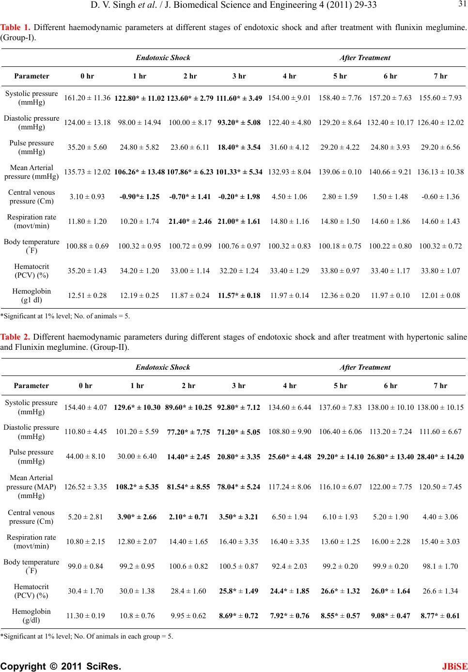
The normal mean systolic pressure was observed to be
161.2 ± 11.36 mmHg (Table 1) and 154.40 ± 4.07
mmHg (Table 2) which is slightly higher than 145.60 ±
17.3 to 146.60 ± 2.78 mmHg as reported in buffalo
calves [3]. The mean systolic pressure decreased imme-
diately after endotoxin infusion. An increase in systolic
pressure was seen on treatment with flunixin meglumine
(FM) and it remained non-significantly below the nor-
mal values (Table 1, Figure 1). Similar results have
been reported by Singh et al., 2005 [6].
The normal mean diastolic pressure was 124.00 ±
13.18 mmHg (Table 1) and 110.80 ± 4.45 mmHg. (Ta-
ble 2) which is close to 118.0 ± 7.80 to 122.40 ± 7.4
mmHg as reported. [3]. The diastolic pressure was sig-
nificantly (P < 0.01) lower at 3rd hour of start of en-
dotoxin infusion (Table 1). Similar results have been
reported in buffalo calves [3,6]. After flunixin meglu-
mine treatment, the diastolic pressure reached slightly
above normal value at the end of the experiment i.e. 7th
hour of observation (Table 1).
The normal pulse pressure was 35.20 ± 5.60 mmHg
(Table 1) and 44.0 + 8.10 mmHg (Table 2). Apart from
a general decline in pulse pressure, a significantly (P <
0.01) lower pulse pressure was observed at 3 hour of
start of endotoxin which after treatment increased and
was non-significantly lower than the normal pre-infusion
level throughout the period of observation in group-1
while in group-2 pulse pressure was significantly below
normal pre-infusion values throughout the observation
period. Endotoxin infusion lowered the pulse pressure
and treatment with flunixin meglumine led to an increase
in pulse pressure, yet it was still lower then the normal
value at the end of the experiment (Table 1).
The normal MAP (Mean arterial pressure) was found
to be 135.73 ± 12.02 (table 1) and 126.52 ± 3.35 mmHg
which is similar to 130.00 ± 6.4 mm Hg [6] but lower
than 153.88 ± 2.00 mmHg [7]. The fall in MAP
throughout endotoxin infusion was significant i.e., upto
3rd hour and after infusion of Flunixin meglumine, it was
slightly higher than the normal value at the end of the
experiment (Table 1). The fall in MAP during endotoxin
infusion may be due to the release of 6 -Keto pros-
taglandin-F1-α [8]. The rise in MAP after HSS infusion
may be due to the fact that HSS infusion increases the
plasma osmolality and osmiotically draws intracellular
and interstitial water into vascular space. The consequent
plasma volume expansion is 3 ml for every 1 ml of hy-
pertonic saline solution infused [9]. This rapid plasma
volume expansion increases the cardiac output and the
mean arterial pressure. Hypertonic saline may also elicit
a beneficial effect through reduction of endothelial
swelling which results in narrowed vessel diameter with
increased hydraulic resistance making perfusion of tis-
sues more difficult. According to Olson et al. (1995)
[10], in response to endotoxin, through the action of a
membrane bound enzyme prostaglandin synthase, ara-
chidonic acid is converted to cyclicendoperoxidases i.e.,
PGG2 and PGH2 which are rapidly converted into
ThromboxaneA2 (TXA2) and PGI2. PGI2 is a potent sys-
temic vasodilator which could contribute to endotoxin-
induced systemic hypotension and lethality. The rise in