 Open Journal of Urology, 2013, 3, 232-245 http://dx.doi.org/10.4236/oju.2013.36044 Published Online October 2013 (http://www.scirp.org/journal/oju) MG289 in Mycoplasma genitalium Enhances Microbial Invasion and Bacterial Persistence in Benign Human Prostate Cells* Wasia Rizwani1, Leticia Reyes2, Jeongsoon Kim1, Steve Goodison1, Charles J. Rosser1# 1Cancer Research Institute, MD Anderson Cancer Center Orlando, Orlando, USA 2Department of Infectious Disease & Pathology, College of Veterinary Medicine, University of Florida, Gainesville, USA Email: wasia.rizwani@gmail.com, LREYES@dental.ufl.edu, Jeongsoon.Kim@orlandohealth.com, steven.goodison@orlandohealth.com, #deacdoc@aol.com Received August 9, 2013; revised September 5, 2013; accepted September 12, 2013 Copyright © 2013 Wasia Rizwani et al. This is an open access article distributed under the Creative Commons Attribution License, which permits unrestricted use, distribution, and reproduction in any medium, provided the original work is properly cited. ABSTRACT Introduction: Recent studies suggest that infectious organisms may facilitate initiation and metastasis of many human cancers. One infectious organism of interest is Mycoplasma genitalium (Mg), a prevalent organism in humans known to cause sexually transmitted infection, as well as urethritis and prostatitis. Previous studies have demonstrated that benign, non-tumorigenic human prostate cells (BPH-1) chronically exposed to M. genitalium led to the malignant transforma- tion of these cells as demonstrated in in vitro and in vivo models. Based on work from our laboratory, we felt this ma- lignant transformation revolved around a specific M. genitalium’s ABC transporter (MG289) with homology to M. hy- orhinis’ ABC transporter, p37. In this study, differences in M. genitalium’s ability to infect and induce a unique pro- teome conducive to tumoral growth were studied with engineered M. genitalium in which the p37 protein was silent. Materials and Methods: Wild-type M. genitalium (strain 431c, designated as M. genitalium WT) and MG289 deficient M. genitalium mutant (strain 260_3, designated as Mg260_3) were used for this study. We studied the infectivity poten- tial between M. genitalium WT and Mg260_3 upon exposure to BPH-1 cells. Furthermore, we set out to identify a unique proteome in BPH-1 cells exposed to M. genitalium WT that could explain its ability to induce malignant trans- formation of benign cells. Validation of selected proteomic targets was carried out by Western blot analysis. Results: Both M. genitalium WT and Mg260_3 strains showed somewhat similar growth curve when absorbance at 450nm was matched at day 0. Colony forming units (CFUs) were similar for both strains at the same absorbance. However, the ability to infect BPH-1 cells was greatly reduced in Mg260_3 compared to the M. genitalium WT (p < 0.001). This was evident by infectivity assays and confocal microscopy. Proteomic analysis of BPH-1 cells infected with either M. geni- talium WT or Mg260_3 for 8 hr, 24 hr and 6 days demonstrated a considerable shift in protein expression over unin- fected BPH-1 cells at each time point. The preponderance of perturbed proteins regulated protein synthesis and protein processing, triggering endoplasmic reticulum stress. Conclusions: In summary, we demonstrate that Mg260_3, which is deficient of the phosphonate ABC transporter substrate-binding protein; MG289 (homologue to M. hyorhinis p37), is less effective in invading and maintaining an intracellular persistence in benign human prostate cells. In addition, dele- tion of MG289 resulted in altered BPH-1 responses to M. genitalium infection as evidenced by differential proteome profiling of BPH-1 infected cells. Keywords: Cancer; ER Stress Proteins; Infection; Mycoplasma genitalium; Prostate; Proteomics 1. Introduction Mycoplasma genitalium (Mg) is the smallest known self-replicating microorganism capable of living in para- sitic association with host cells, thus obtaining most of their nutrients that they cannot synthesize from the host for survival. M. genitalium can grow in close relationship with mammalian cells, often without any apparent pa- thology or symptoms for extended periods of time [1]. M. genitalium has been implicated in urogenital infections of both men and women around the world and it is recog- nized as an emerging cause of sexually transmitted infec- *The authors declare that they have no competing interests. #Corresponding author. C opyright © 2013 SciRes. OJU 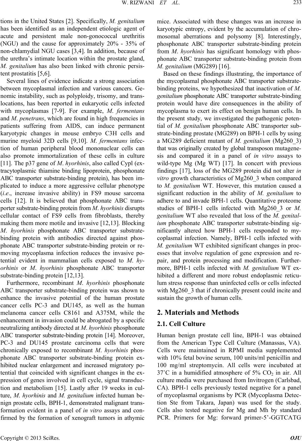 W. RIZWANI ET AL. 233 tions in the United States [2]. Specifically, M. genitalium has been identified as an independent etiologic agent of acute and persistent male non-gonococcal urethritis (NGU) and the cause for approximately 20% - 35% of non-chlamydial NGU cases [3,4]. In addition, because of the urethra’s intimate location within the prostate gland, M. genitalium has also been linked with chronic persis- tent prostatitis [5,6]. Several lines of evidence indicate a strong association between mycoplasmal infection and various cancers. Ge- nomic instability, such as polyploidy, trisomy, and trans- locations, has been reported in eukaryotic cells infected with mycoplasmas [7-9]. For example, M. fermentans and M. penetrans, which are found in high frequencies in patients suffering from AIDS, can induce permanent karyotypic changes in mouse embryo C3H cells and murine myeloid 32D cells [9,10]. M. fermentans infec- tion of human peripheral blood mononuclear cells can also promote immortalization of these cells in culture [11]. The p37 gene of M. hyorhinis, also called Cypl (ex- tracytoplasmic thiamine binding lipoprotein, phosphonate ABC transporter substrate-binding protein), has been im- plicated to induce a more aggressive cellular phenotype (i.e., increase invasive ability) in FS9 mouse sarcoma cells [12]. It is believed that phosphonate ABC trans- porter substrate-binding protein from M. hyorhinis disrupts cellular contact of FS9 cells from fibroblasts, thereby making them more motile and invasive [12,13]. Blocking M. hyorhinis phosphonate ABC transporter substrate- binding protein with antibodies directed against phos- phonate ABC transporter substrate-binding protein or re- moving mycoplasma infection reduces the invasive po- tential evident in mammalian cells exposed to M. hy- orhinis or M. hyorhinis phosphonate ABC transporter substrate-binding protein [12,13]. Furthermore, recombinant M. hyorhinis phosphonate ABC transporter substrate-binding protein was shown to enhance the invasive potential of the human prostate cancer cells PC-3 and DU145, as well as the human melanoma cancer cells C8161 and A375M, while the enhancement in invasion could be abrogated by a specific neutralizing antibody directed at M. hyorhinis phosphonate ABC transporter substrate-binding protein [14]. Moreover, PC-3 and DU145 prostate carcinoma cells that were chronically exposed to recombinant M. hyorhinis phos- phonate ABC transporter substrate-binding protein ex- hibited nuclear enlargement and increased migratory po- tential that coincided with significant changes in the ex- pression of genes involved in cell cycle, signal transduc- tion and metabolism [15]. Lastly after 19 weeks in cul- ture, M. hyorhinis and M. genitalium infected human be- nign prostate cells, BPH-1, demonstrated malignant trans- formation evident in a panel of in vitro assays and con- firmed by the formation of xenograft tumors in athymic mice. Associated with these changes was an increase in karyotypic entropy, evident by the accumulation of chro- mosomal aberrations and polysomy [8]. Interestingly, phosphonate ABC transporter substrate-binding protein from M. hyorhinis has significant homology with phos- phonate ABC transporter substrate-binding protein from M. genitalium (MG289) [16]. Based on these findings illustrating, the importance of the mycoplasmal phosphonate ABC transporter substrate- binding proteins, we hypothesized that inactivation of M. genitalium phosphonate ABC transporter substrate-binding protein would have dire consequences in the ability of mycoplasma to exert its effect on benign human cells. In the present study, we investigated the pathogenic poten- tial of M. genitalium phosphonate ABC transporter sub- strate-binding prostate (MG289) on BPH-1 cells by using a MG289 deficient mutant of M. genitalium (Mg260_3) that was originally created by global transposon mutagene- sis and compared it in a panel of in vitro assays to wild-type Mg (Mg WT) [17]. In concert with previous findings [17], loss of the MG289 protein did not alter in vitro growth characteristics of Mg260_3 when compared to M. genitalium WT. However, this mutation caused a significant reduction in the ability of M. genitalium to adhere to and invade BPH-1 cells. Quantitative proteome studies of BPH-1 cells infected with Mg260_3 or M. genitalium WT also revealed that loss of the M. genital- ium phosphonate ABC transporter substrate-binding sig- nificantly altered how BPH-1 cells responded to my- coplasmal infection. Namely, BPH-1 cells infected with M. genitalium WT exhibited significant changes in proc- esses that involve regulation of gene expression and re- pair, and protein processing and modification. Further- more, BPH-1 cells infected with M. genitalium WT ex- hibited a different and more robust endoplasmic reticu- lum stress response than uninfected cells or cells infected with Mg260_3 that if chronically present could incite and sustain the growth of human cells. 2. Materials and Methods 2.1. Cell Culture Human benign prostate cell line, BPH-1 was obtained from the American Type Cell Culture (Manassas, VA). Cells were maintained in RPMI media supplemented with 10% fetal bovine serum, 100 units/ml penicillin and 100 mg/ml streptomycin. All cells were incubated at 37˚C in a humidified atmosphere of 5% CO2 in air. All culture media were purchased from Invitrogen (Carlsbad, CA). BPH-1 cells previously tested negative for a panel of mycoplasmal organisms by PCR (Mycoplasma Detec- tion Ste from Takara, Japan) was used for the study. Cells also tested negative for Mg and Mh by standard PCR. Primers for Mg: forward primer-5’-GGTCATG Copyright © 2013 SciRes. OJU  W. RIZWANI ET AL. 234 GACAAACAAATTCAGCTTCAC-3’; reverse primer-5’- CAATACCAATGTCATACAAAAGCGGATCA-3’ and Mh: forward primer-5’-CGATCCAACTGTTCAAGGT- AATTTTTA-3’; reverse primer-5’-CACTTTTTCTAAA- CACTCCAACATCAT-3’ were used. PCR was carried out using 20 μl reaction volume containing 200 ng of DNA and 200 nM of primers (Promega PCR mix). The amplification conditions of PCR consisted of initial de- naturation at 95˚C for 2 min followed by 40 cycles of denaturation at 95˚C for 45 sec, annealing at 55˚C for 45 sec and elongation at 72˚C for 45 sec min, followed by 1 cycle at 72˚C for 10 min. 2.2. Mycoplasma Preparation and Culture Stock solutions of both M. genitalium strains were pre- pared by growing the microorganisms to early log phase in SP4 broth, and stored frozen at −80˚C. Both strains of M. genitalium were grown in SP4 media under standard conditions [8]. 2.3. Growth Curve Cultures for colony forming unit (CFU) determination of inoculum were serially diluted 10-fold to 10-8 in SP4 broth then 20 ml from each sample and its corresponding dilutions were plated in SP4 broth and Agar plates. Broth cultures were incubated at 37˚C in ambient air, while Agar plates were incubated at 37˚C in 5% CO2. Broth tubes were checked daily for a color change, and the re- ciprocal of the last dilution to show growth was deemed the color-changing unit (CCU). In order to confirm growth of M. genitalium WT and Mg260_3, CCU, which provide a relative indication of the amount of microbes present, were determined. Samples were serially diluted tenfold to 10-8 in SP4 broth in duplicate. Broth tubes were checked daily for color change, and the CCU was recorded as the reciprocal of the last dilution to show growth at 14 days. Agar cultures were incubated for at least 14 days before colonies were counted. Log-phase cultures of M. genitalium WT and Mg260_3 with the same absorbances (i.e., same number of colonies) at 450 nm were diluted to 20 ml to give zero absorbance at 450 nm compared to SP4 blank on day 0 and growth was observed over a period of 14 days. Absorbance was read every 24 hrs and plotted graphically. The same absorb- ances gave same CFUs for both M. genitalium WT and Mg260_3 cultures. The experiment was carried out in duplicate in three separate experiments and p value of 0.05 was considered significant. 2.4. Infectivity Assay BPH-1 cells (105) were inoculated with log-phase cul- tures (3 × 10⁴ CFU) of M. genitalium WT or Mg260_3 to determine whether the mycoplasmal protein MG289 al- tered its ability to attach to and infect human cells [18]. To quantify intracellular M. genitalium load, the inocu- lum was removed following 3 hrs incubation for cellular attachment and entry and replaced with medium con- taining gentamicin (200 μg/ml, Sigma) for 2 hrs. Infected cells were washed thoroughly and fresh RPMI media added to the wells. The ability for M. genitalium to per- sist in culture media and not be transported into human cells was also quantified from culture supernatants in separate wells. Cell fractions or culture supernatants were collected at 8 hrs, 24 hrs and 6 days post-infection fol- lowing removal of the inoculums for quantification of bacterial loads using both CCU and CFU assays. In every case, intracellular M. genitalium was quantitated by Real- Time PCR (RT-PCR) and standard PCR using MG289 and p110 primers. Extracellular M. genitalium was ob- served and quantitated using CFU assay (p < 0.05; Stu- dent’s t-test). Each assay was performed in duplicate. 2.5. Real-Time PCR/ PCR Following infections, DNA was extracted from cells us- ing DNA isolation kit (Qiagen) as per manufacturer’s instructions. Quantitative reverse transcriptase (RT)-PCR was carried out using ABI 7300 Real-Time PCR System (Life Technologies) in a 20 μl reaction volume contain- ing 1 μl of the DNA, 1 μM of gene-specific TaqMan primer and probe mix. The primers used were purchased from Integrated DNA Technologies. Mg p110: Probe-5’-/ 56-FAM/AGCGAGCAGCATGACCAACTTGTT/36-Ta mSp/-3’; Primer 1-5’-TGAACCGGAAAGTGCAACA- AGTGC-3’ and Primer 2-5’-AAACTTCCCTGCATTG- CTGTTCCC-3’. Human GAPDH: Probe-/56-FAM/AA- AGCCAGTCCCCAGAACCCC/36-TAMSp/-3’; Primer 1-5’-CTCCCACCTTTCTCATCCAAG-3’ and Primer 2-5’-ACATCACCCCTCTACCTCC-3’. Relative fold chan- ges in DNA levels were calculated after normalization to GAPDH using the comparative Ct method (27). PCR was also carried out using 20 μl reaction volumes containing 200 ng of DNA, 1 μM of gene-specific TaqMan primer and probe mix for semi-quantitative analysis. Standard DNA PCR was carried out for M. genitalium MG289 using primers described earlier and Mg p110 primers-for- ward primers-5’-AAGGGCAAAACGCAAAGG GA-3’ and p110-reverse primers-5’-TCCCCCTCCAGAAACA CTGG-3’ for validation of M. genitalium WT and Mg260_3. 2.6. iTRAQ/LC-MS/MS BPH-1 cells (106) were plated and allowed to attach and grow for a day. Next, cells were infected with log-phase M. genitalium WT or Mg260_3 for 3 hrs (initial infection time, 3 × 10⁵ bacteria per infection), followed by 2 hrs Copyright © 2013 SciRes. OJU  W. RIZWANI ET AL. 235 incubation with 200 μg/ml gentamicin in fresh media. Cells were then washed three times with PBS to remove unbound or non-invading mycoplasma. Fresh media was added and the cells were grown for a total of 8 hrs, 24 hrs or 6 days post initial infection time. Cells were washed with PBS again and displaced by versene, collected and centrifugation at 1500 × g for 5 min at 4˚C. Infection of all BPH-1 cell pellets was confirmed by PCR using MG289 and p110 primers as already described. Cell pel- lets were stored at −80˚C until processing for proteome studies. Protein lysates from cell pellets were prepared using mammalian proteome extraction kit (Calbiochem, Billerica, MA) according to the manufacturer’s protocol. Extracted protein samples were purified and concen- trated using the Ultrafree®-0.5 centrifugal filter device (5 kDa cut-off from Millipore). Protein supernatants were dissolved in 1 M Urea/0.5M triethylammonium bicar- bonate buffer and were assayed using CBX kit (from G- Biosciences). A total of three separate iTRAQ labelling reactions per infection were processed according to the manufacturer’s instructions (AB Sciex) and as described by Alvarez et al. [19]. Briefly, the protein samples were reduced/alkylated and then digested with trypsin. Pep- tides were then labelled with iTRAQ reagents as follows; Uninfected 8 hrs, Uninfected 24 hrs and Uninfected 6 days with isobaric tag 114; M. genitalium WT 8 hrs, M. genitalium WT 24 hrs, M. genitalium WT 6 days with isobaric tag 115; Mg260_3 8 hrs, Mg260_3 24 hrs, Mg260_3 6 days with isobaric tag 116. For each iTRAQ experiment, peptides labelled with one of the three labels (114 - 116) were pooled before separation using strong cation exchange (SCX). The samples were dried and the peptides were separated using SCX as described by Al- varez [19]. Thirteen peptide fractions were collected, dried, and analyzed once by LC-MS/MS using nanoLC- ESI-LTQ Orbitrap Velos (Thermo Scientific) as previ- ously outlined in [20]. The data acquired for the 13 frac- tions were processed and merged for each biological rep- licate independently. The raw protein files were processed for peak detec- tion using Mascot Distiller v2.4 (Matrix Science Inc., Boston, MA, USA). The MS/MS settings include single peak window selection from m/z 113.5 to m/z 117.5. The protein peak was identified and quantified with Mascot Daemon v2.3 (Matrix Science Inc.) using NCBInr (Jan 2012) entries for Bacteria (7,982,362 sequences) and Mammalia (811,620 sequences). The following settings were used for the search: trypsin as cleavage enzyme; two missed cleavages; methylthio modification of cys- teines, iTRAQ (N terminal), iTRAQ (K) were fixed modi- fications; and methionine oxidation, deamidation (N, Q), and iTRAQ (Y) were selected as variable modifications. The mass error tolerance for precursor ions was set to 15 ppm and 0.08 Da for fragment ions. An automatic decoy database search was also performed to assess the false positive rate of protein identification. Automatic isotope correction was carried out using the values supplied with the AB Sciex reagents. For reporting quantification re- sults, the protein ratio type used was the “weighted” geo- metric mean, normalization method was “summed inten- sities”, outlier removal was “automatic” (Dixon’s method up to 25 data points, Rosner’s method above 25 data points), the peptide threshold was “above homology” (i.e., peptide score does not exceed absolute threshold but is an outlier from the quasi-normal distribution of random scores), and the minimum number of peptides for protein identification and quantification was two with an ex- pected value <0.05. The intensity values of the assigned peptides were summed and the protein ratio calculated from the summed values. 2.7. Lysate Preparation and Western Blotting Corresponding protein lysates from uninfected BPH-1 cells and cells infected with M. genitalium WT and Mg260_3 were prepared using mammalian proteome ex- traction kit (Calbiochem) as described earlier and 10 μg of protein was run on polyacrylamide-SDS gel. The pro- teins were transferred to a nitrocellulose membrane and immunoblotted with antibodies corresponding to various proteins identified from the proteomics. Blots were also probed for ER stress proteins using the ER stress Anti- body kit (cat# 9956, Cell Signaling Technology, Danvers, MA). Mouse monoclonal antibody to β-Actin was pur- chased from Sigma-Aldrich, St. Louis, MO and used as a loading control. 2.8. Immunofluorescence and Confocal Microscopy BPH-1 cells were plated onto cover slips (2 × 10⁴ cells per cover slip) and were infected with log phase M. genitalium WT or Mg260_3 for 3 hrs followed by 200 μg/ml gentamicin treatment for 2 hrs. Cells were washed with PBS twice and then fixed with 10% buffered-for- malin for 15 min. Uninfected cells were used as control. Immunofluorescence was performed as per the protocol published previously [21]. The primary M. genitalium antibody (polyclonal anti-rabbit Mg obtained from Dr. Mary B. Brown, University of Florida, College of Vet- erinary Medicine) was used at 1:1000 dilution. Secon- dary antibody was mouse anti-rabbit Alexa Fluor-488 (Molecular Probes, Carlsbad, CA). Control experiments (i.e., no primary M. genitalium antibody) demonstrated no cross-reactivity between mouse anti-rabbit secondary antibody as evident by no detectable staining by secon- dary antibody alone (data not shown). DAPI (Vector labs, Burlingame, CA) was used to stain the nuclei. Cells were visualized with a DM16000 inverted Zeiss scanning con- Copyright © 2013 SciRes. OJU  W. RIZWANI ET AL. 236 focal microscope with a 63X/1.40NA oil immersion ob- jective. 2.9. Data Analysis M. genitalium’s ability to invade human cells was ana- lyzed by ANOVA followed by Tukey’s test. A value of p ≤ 0.05 was considered significant. Cluster analysis of iTRAQ data was performed as previously described [22] using JMP Genomics software (SAS, Cary, NC). Briefly, only protein ratios that were deemed to be significantly different from uninfected cells as determined by Pro GroupTM algorithm were used in the analysis. Protein ratios (M. genitalium infected/uninfected control) from each time point were normalized prior to clustering and clustering was performed by the method of Ward. 3. Results 3.1. Growth of Mycoplasma genitalium—Wild Type and Mutant Strains Mycoplasma genitalium MG289 mutant clone (Mg260_3, M. hyorhinis p37 homologue deleted) was created by TN4001 transposon insertion in the MG289 gene; the transposon is located 570 bases into the 1158 bp MG289 gene [17]. Disruption of MG289 gene was checked by PCR (Figure 1(A)). M. genitalium WT was positive for both MG289 and p110 whereas Mg260_3 was positive for only p110 gene confirming that MG289 gene in Mg260_3 is disrupted. In vitro growth properties of M. genitalium WT and Mg260_3 were assessed in SP4 growth media by CCU method and optical density measurements taken at 450 nm (Figure 1(B)). During stationary phase, both M. genitalium WT and Mg260_3 attained similar microbial numbers. However, Mg260_3 did not attain stationary growth until day 7 of culture whereas M. geni- talium WT achieved stationary phase by day 6 of culture (Figure 1(B)). Live bacteria were confirmed by CFU assay (data not shown). 3.2. Mycoplasma genitalium MG289 Facilitates Microbial Invasion and Persistence in BPH-1 Previous studies have shown that M. genitalium can in- vade and persist in endocervical cells [23]. Therefore, we evaluated the ability of M. genitalium WT and Mg260_3 to invade and persist in BPH-1 cells at 2 hrs, 8 hrs, 24 hrs, and 6 days post-inoculation. BPH-1 cell lysates collected at 2 hrs post-inoculation confirmed that bacteria attached and invaded BPH-1 cells. In follow-up assays assessing internalized bacteria, these BPH-1 cells exposed to M. genitalium WT and Mg260_3 and subsequently treated to remove extracellular M. genitalium had its DNA isolated and subjected to semi-quantitative PCR using a PCR Figure 1. Growth curves of M. genitalium (M. genitalium WT and Mg260_3). (A) PCR confirming the depletion of MG289 (homologue to M. hyorhinis p37) in the mutant strain, Mg260_3; (B) Growth curve shows that M. genital- ium WT reaches stationary phase earlier than Mg260_3 (p < 0.05). Values represent the mean and standard error of one experiment performed in triplicate. Each experiment was conducted three times. primer/probe set specific for the p110 gene, which is present in both M. genitalium WT and Mg260_3 (Figure 2(A)). At 40 cycles, M. genitalium WT DNA was de- tected with equivalent intensity at all post-inoculation time points. This is in contrast to Mg260_3 in which less PCR product was detected at 2, 8, and 24 hrs post-in- oculation. There was no amplified DNA detected in Mg260_3 infected cell lysates obtained from 6 day old cultures (Figure 2(A)). We next quantified internalized M. genitalium bacteria by RT-PCR in BPH-1 cell RNA lysates obtained from a separate infectivity assay (Figure 2(B)). As shown in Figure 2(B), significantly more M. genitalium WT were retrieved from BPH-1 cells at all post-infection time points compared to Mg260_3 (p < 0.001). Although there was a significant decline in the DNA copy number of both M. genitalium WT and Mg260_3 over the course of the ex- periment, the decline was more pronounced in Mg260_3 suggesting that deletion of MG289 may impact both mi- crobial adherence and invasion. Viable bacteria were re- trieved from both M. genitalium WT and Mg260_3 in- fected cultures (Figure 2(C)) confirming that bacteria that invaded BPH-1 cells were viable and that M. genitalium Copyright © 2013 SciRes. OJU  W. RIZWANI ET AL. 237 Figure 2. MG289 (homologue to M. hyorhinis p37) gene facilitates adherence and invasion of M. genitalium in BPH-1 cells. (A) PCR showing presence of M. genitalium within BPH-1 cells. M. genitalium WT could infect cells with higher efficiency than Mg260_3; (B) DNA copy number as determined by RT-PCR. M. genitaliumg DNA was normal- ized to BPH-1 GAPDH DNA. **Indicates a significant dif- ference between M. genitalium WT and Mg260_3 (p < 0.05); (C) Extracellular M. genitalium as determined by culture. Values represent the mean ± SD log CFU of triplicate. Each experiment was conducted three times. may be trafficking through BPH-1 cells in a similar fash- ion as human endocervical cells [23]. Infection of BPH-1 cells with M. genitalium WT and Mg260_3 was further examined by confocal microscopy (Figure 3). For infection experiments, all M. genitalium cultures were adjusted so that 105 BPH-1 cells were in- oculated with 3 × 10⁴ CFU of M. genitalium WT or Mg260_3 that were harvested during log phase growth. At 5 hrs post-inoculation, both internalized M. genitalium WT and Mg260_3 were found within the perinuclear region of BPH-1 cells by microscopy (Figure 3(A)), which is consistent with previous studies [24]. However, con- Figure 3. Intracellular location of M. genitalium within BPH-1 cells. (A) Representative confocal microscopic im- ages of uninfected and M. genitalium infected (M. genital- ium WT and Mg260_3) BPH-1 cells at 7 hrs post-inocula- tion. Images were captured using 63X/1.40NA oil immer- sion objective with DM16000 inverted Zeiss scanning con- focal microscope, magnification −120 μm; (B) Images are 3 dimensional representation of infected cells, magnification −120 μm. siderably more aggregates of M. genitalium WT were found within BPH-1 cells than Mg260_3. The staining pattern for M. genitalium was specific since neither anti- body controls, nor uninfected BPH-1 cells were positive for M. genitalium immunofluorescence (Figure 3(A), first panel). Figure 3(B) is a 3 dimensional representation of M. genitalium WT and Mg260_3 in BPH-1 cells. 3.3. BPH-1 Cells Infected with Mycoplasma genitalium Exhibit an Altered Proteome Profile In order to identify the host cell processes that are per- turbed by M. genitalium infection, we used quantitative proteome profiling of uninfected, M. genitalium WT in- fected and Mg260_3 infected BPH-1 whole cell extracts. The BPH-1 infection and cell harvest scheme is summa- rized in Figure 4. Proteins from BPH-1 cell extracts that were harvested at 8 hr, 24 hr, and 6 days post-inoculation were identified and quantified using Mascot v2.3. The Venn diagram (Figure 5(A)) illustrates the number of proteins that significantly changed in abundance in at least one of the ratios (M. genitalium WT vs . uninfected control, Mg260_3 vs. uninfected control or M. genitalium WT vs. Mg 260_3). Next, we examined the BPH-1 proteome for proteins that may be affected during microbial invasion such as the endosome pathways [25,26], ubiquination and pro- teasome degradation pathways [27] and proteins involved Copyright © 2013 SciRes. OJU  W. RIZWANI ET AL. 238 Figure 4. Diagram summarizing the proteome experimental design. BPH-1 cells were subjected to Mg infection (M. geni- talium WT and Mg260_3) for an initial period of 3hr, fol- lowed by removal of the inoculum and gentamicin treat- ment (200 µg/ml) for 2hr to eliminate any remaining bacte- ria that were not internalized in BPH-1 cells. Infected BPH-1 cells were then harvested at 8 hr, 24 hr and 6 days post-inoculation and analyzed by PCR—to confirm pres- ence of Mg, iTRAQ analysis—to identify proteome, and Western blot—to confirm targets from proteome. in SRP-dependent co-translational protein targeting [28]. BPH-1 cells infected with M. genitalium WT exhibited a significant increase in endosome specific proteins and proteins that regulate endosome formation and traffick- ing. In contrast, the majority of these proteins were de- creased in Mg 260_3 infected cells. M. genitalium infec- tion also perturbed proteins involved in the proteasome degradation pathway and the SRP-dependent co-transla- tional protein targeting to membrane pathway, which may have an impact on endoplasmic reticulum function [29]. The majority of proteins associated with these processes were significantly increased in BPH-1 cells infected with M. genitalium WT, but decreased in cells infected with Mg 260_3. Two approaches were used to profile the effect of M. genitalium WT and Mg260_3 infection on BPH-1 cells. In the first analysis, protein ratios (M. genitalium WT/ control and Mg260_3/control) that reflected a significant change (p ≤ 0.05), and were accurately identified in all infected groups were used in this analysis (Figure 5(B)). Thus, proteins that did not conform to these criteria were removed before analysis. At 8 hrs post-inoculation, clus- ter groups 4 and 9 (demarcated with arrows) displayed the most dramatic differences. Proteins found within clus- ter group 4 were markedly reduced in M. genitalium WT infected cells when compared to cells infected with Mg260_3. Assigned functions of these proteins include oxidative reduction, perturbation induction of apoptosis, angiogenesis/cytokine activity/glycolysis, and negative regulation of cell proliferation. In contrast proteins in clus- ter group 9 were markedly increased in M. genitalium WT infected cells and their assigned functions include virus infectious cycle, cellular protein metabolic process, transcriptional regulation, and regulation of apoptosis. Cluster analysis of cell cultures harvested at 24 hrs post-inoculation, revealed three clusters that were dramati- cally different between M. genitalium WT and Mg260_3 infected cells (Figure 5(B)). Proteins in clusters 4 and 6 were markedly increased in M. genitalium WT infected cells, and their assigned functions include apoptosis, cel- lular response to reactive oxygen species, biosynthetic processes, cell differentiation, regulation of microtubule function, protein import into the nucleus, mRNA proc- essing, and inhibition of transforming growth factor re- ceptor signaling pathway. Conversely, proteins in cluster 9 were markedly reduced in Mg WT infected cells, and these proteins are involved in transcription, translation, and inhibition of DNA damage response or signal trans- duction by p53 class mediator. Cluster analysis of infected cultures harvested on Day 6 identified three markedly different cluster groups be- tween M. genitalium WT and Mg260_3 infected cells (Figure 5(B)). BPH-1 proteins in cluster group 6 were markedly reduced compared to cells infected with Mg260_3, and these proteins are involved in translation, regulation of defense response, telomere maintenance, RNA processing, and steroid hormone mediated signal- ing. Proteins in clusters groups 9 and 10 were markedly increased in M. genitalium WT infected cells, and their assigned functions include regulation of cytokinesis, cell- cell signaling, vesicle-mediated transport, translation, and long-chain fatty acid biosynthetic process. The biological response of BPH-1 cells to M. genital- ium WT and Mg260_3 was also evaluated by determin- ing the proportion of biological functions or cellular proc- esses that were significantly perturbed by M. genitalium infection (Figure 6). Protein ratios that reflected a signifi- cant difference between M. genitalium WT and Mg260_3 infected cells (p ≤ 0.05) were grouped according to their assigned biological process based on the blast2go gene ontology database (http://www.blast2go.com) [30]. Most notable changes were observed in proteins over the course of the assay involved in genetic processes such as regula- tion of transcription, RNA processing and DNA repair (20% - 22%). Similarly, another 20% of proteins that were significantly altered between Mg WT and Mg260_3 infected cells are involved in protein processing such as post-translational modification, enzyme activation, un- folded protein response, and protein targeting. Though the infectivity is suppressed in Mg260_3, absence of MG289 has produced significant changes in the proteome of infected BPH-1 cells (Figure 6). 3.4. Endoplasmic Reticulum (ER) Stress Response Increases with Mycoplasma genitalium Infection It is believed that microbial pathogens may manipulate the ER stress response as a means of prolonging their intracellular survival within host cells [31]. Since Mg infection resulted in significant perturbations in the pro- teasome pathway, which can affect the ER stress response Copyright © 2013 SciRes. OJU  W. RIZWANI ET AL. Copyright © 2013 SciRes. OJU 239 Figure 5. Proteomic analysis reveals a significant number of proteins changing in expression with M. genitalium WT infec- tion/uninfected control and Mg260_3 infection/uninfected control. (A) Venn diagram illustrates the number of proteins that significantly changed in abundance in at least 1 of the ratios between comparisons (M. genitalium WT vs. uninfected control, Mg260_3 vs. uninfected and M. genitalium WT vs. Mg 260_3); (B) Heat maps were generated with protein ratios (M. genital- ium WT/control and Mg260_3/control) that reflected a significant change (p ≤ 0.05), and were accurately identified in all in- fected groups used in this analysis. or unfolded protein response [32], we assessed the ER stress response in uninfected and M. genitalium infected BPH-1 cells by measuring a panel of ER associated pro- teins by Western blot analysis. We did not observe any changes in resident ER proteins such as calnexin, BiP, protein disulfide isomerase, and endoplasmic oxidore- ductin-1 (data not shown). However, we did observe changes in proteins that regulate UPR [reviewed in Mar- tin Hausmann, International Journal of Inflammation 2010] such as IRE1 (Inositol-requiring enzyme-1), PERK (pro- tein kinase RNA-like endoplasmic reticulum kinase), and CHOP/GADD153 (CCAAT/-enhancer-binding protein homologous protein) (Figures 7(A) and (B)). At 8hr and 24 hr post-infection, both M. genitalium WT and Mg260_3 infected cells exhibited similar responses. Namely, both M. genitalium WT and Mg260_3 infection induced an increase in PERK with no appreciable change in the lev- els of IRE-1 and CHOP. However, by 6 days post-in- oculation, M. genitalium WT infected cells had a marked increase in CHOP, whereas cells infected with Mg260_3 displayed a reduction in all three UPR proteins (Figures 7(A) and (B)). 4. Discussion M. genitalium is an obligate parasite of the human uro-  W. RIZWANI ET AL. 240 Figure 6. Functional distribution of protein ratios that were significantly different between M. genitalium WT and Mg260_3 infected cells at 8hr (A), 24 hr (B), and 6 d (C) post-infection. Gene ontology designations were obtained from the Blast2go database. genital tract [33] that has been implicated in pelvic in- flammatory disease [34], acute and persistent male non- gonococcal urethritis (NGU) [3,4], and chronic persistent prostatitis [5,6]. Furthermore, M. genitalium may also play a role in cancer since our group has previously demonstrated that chronic exposure of human BPH-1 cells to M. genitalium resulted in malignant transformation of these benign cells as evident by in vitro and in vivo as- says [8]. Despite the various reports indicating an asso- ciation between Mg infection and human diseases, the means by which M. genitalium can persist in the host and thus contribute to the pathobiology of these diseases has not been determined. In an attempt to identify the minimal essential genes for life, Glass et al. [17] established that the phosphonate ABC transporter substrate-binding protein, MG289 (homo- logue to Mh p37), in M. genitalium was not necessary for the survival of this microbe in cell free culture media. However, given that M. genitalium is an obligate parasite, retention of MG289 suggests that this gene serves a critical function during natural infection. Another inter- esting feature of MG289 is its similarity to the M. hy- orhinis p37 [16], which has been shown to enhance ma- lignancy in various cells lines [12-15]. Based on these characteristics, we postulated that MG289 serves an im- portant role in the virulence of M. genitalium. In this report we have demonstrated that MG289 enhances mi- crobial attachment, invasion and intracellular persistence in BPH-1 cells. Further, by using quantitative proteome profiling, we have been able to identify M. genitalium WT induced perturbations in BPH-1 cells that pertain to DNA repair, protein synthesis and protein processing, which could impact endoplasmic reticulum function. Overall, our studies with M. genitalium WT were con- sistent with previous reports [17,18,24]. Although we observed a slight delay in the exponential growth phase of Mg260_3 in SP4 media, this mutant did attain a simi- lar microbial density as M. genitalium WT at stationary phase, reaffirming that MG289 is not essential for growth of mycoplasma in cell free culture media [17]. We also noted that both M. genitalium WT and Mg260_3 could invade BPH-1 cells, and also exist outside of these cells. Furthermore, both internalized M. genitalium WT and Mg260_3 were in association with the nucleus of BPH-1 cells as well as in the cytoplasm. However, deletion of MG289 affected the efficiency with which M. genitalium could invade BPH-1 cells, and this may have also im- pacted the ability of the mutant to maintain an intracellu- lar existence. For example, the temporal decline in the number of internalized Mg260_3 was more pronounced than with Mg WT, but the numbers of extracellular bac- teria found within BPH-1 cultures were similar for both strains at all examined time points. M. genitalium WT and Mg260_3 also induced different proteome profiles pertaining to intracellular trafficking. For instance, BPH-1 cells infected with M. genitalium WT exhibited greater concentrations of proteins involved in endosome recy- cling and late endosome/lysosome pathways. Since we examine BPH-1 whole cell lysates, these differences in Copyright © 2013 SciRes. OJU 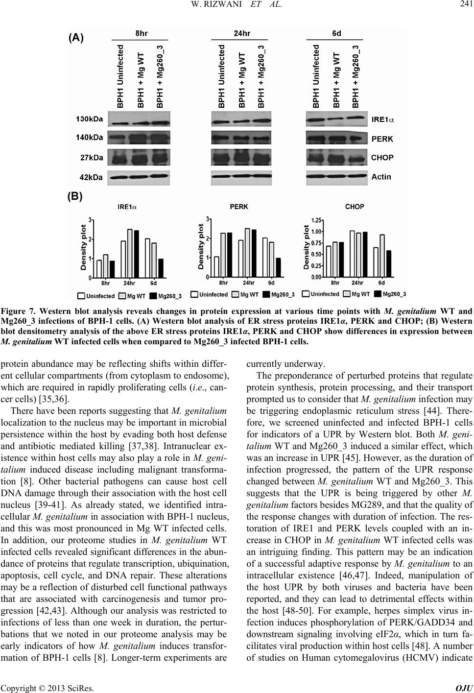 W. RIZWANI ET AL. Copyright © 2013 SciRes. OJU 241 Figure 7. Western blot analysis reveals changes in protein expression at various time points with M. genitalium WT and Mg260_3 infections of BPH-1 cells. (A) Western blot analysis of ER stress proteins IRE1α, PERK and CHOP; (B) Western blot densitometry analysis of the above ER stress proteins IRE1α, PERK and CHOP show differences in expression between M. genitalium WT infected cells when compared to Mg260_3 infected BPH-1 cells. protein abundance may be reflecting shifts within differ- ent cellular compartments (from cytoplasm to endosome), which are required in rapidly proliferating cells (i.e., can- cer cells) [35,36]. There have been reports suggesting that M. genitalium localization to the nucleus may be important in microbial persistence within the host by evading both host defense and antibiotic mediated killing [37,38]. Intranuclear ex- istence within host cells may also play a role in M. geni- talium induced disease including malignant transforma- tion [8]. Other bacterial pathogens can cause host cell DNA damage through their association with the host cell nucleus [39-41]. As already stated, we identified intra- cellular M. genitalium in association with BPH-1 nucleus, and this was most pronounced in Mg WT infected cells. In addition, our proteome studies in M. genitalium WT infected cells revealed significant differences in the abun- dance of proteins that regulate transcription, ubiquination, apoptosis, cell cycle, and DNA repair. These alterations may be a reflection of disturbed cell functional pathways that are associated with carcinogenesis and tumor pro- gression [42,43]. Although our analysis was restricted to infections of less than one week in duration, the pertur- bations that we noted in our proteome analysis may be early indicators of how M. genitalium induces transfor- mation of BPH-1 cells [8]. Longer-term experiments are currently underway. The preponderance of perturbed proteins that regulate protein synthesis, protein processing, and their transport prompted us to consider that M. genitalium infection may be triggering endoplasmic reticulum stress [44]. There- fore, we screened uninfected and infected BPH-1 cells for indicators of a UPR by Western blot. Both M. geni- talium WT and Mg260_3 induced a similar effect, which was an increase in UPR [45]. However, as the duration of infection progressed, the pattern of the UPR response changed between M. genitalium WT and Mg260_3. This suggests that the UPR is being triggered by other M. genitalium factors besides MG289, and that the quality of the response changes with duration of infection. The res- toration of IRE1 and PERK levels coupled with an in- crease in CHOP in M. genitalium WT infected cells was an intriguing finding. This pattern may be an indication of a successful adaptive response by M. genitalium to an intracellular existence [46,47]. Indeed, manipulation of the host UPR by both viruses and bacteria have been reported, and they can lead to detrimental effects within the host [48-50]. For example, herpes simplex virus in- fection induces phosphorylation of PERK/GADD34 and downstream signaling involving eIF2α, which in turn fa- cilitates viral production within host cells [48]. A number of studies on Human cytomegalovirus (HCMV) indicate  W. RIZWANI ET AL. 242 that HCMV maintains UPR responses that are beneficial to the virus while simultaneously inhibiting cellular pro- cesses that are detrimental to them [49]. For instance, binding of HCMV Pp28 to BiP/GRP78 maintains the integrity and function of the HCMV assembly compart- ment [51]. Infection of myeloid cells with Chlam y dia trachomatis increases CHOP expression and assists in the production of pro-inflammatory cytokine, IL-23 [50]. Hence it is not surprising to find that M. genitalium can also induce an ER stress response, which may be impor- tant for its survival and extracellular persistence within the host. Activation of UPR is also associated with infection- induced carcinogenesis [52,53]. Increased Hspa5 and Chop expression is specifically associated with Helicobacter felis induced gastric dysplasia in C57BL/6 mice [52]. Enhanced CHOP expression is associated with human papillomavirus (HR-HPV) infection and abnormal p53 expression in squamous cell carcinoma of uterine cervix [53]. Therefore, the increased expression of CHOP in M. genitalium WT infected cells, may also be associated with malignant transformation of BPH-1 cells during chronic exposure to M. genitalium [8]. This is the first study to confirm a pathogenic role for MG289 (M. hyorhinis p37 homologue in M. genitalium) in benign human prostate cells. We demonstrate that M. genitalium mutant, MG289, is capable of infecting BPH- 1cells, though it exhibits a much-suppressed ability to infect when compared to M. genitalium WT. This feature can be exploited therapeutically to curb mycoplasma infection. Both M. genitalium WT and Mg260_3 are ca- pable of altering the proteome profile of infected BPH-1 cells and the pathways that are altered may relate to on- cogenic transformation. The differences in pathways re- lated to DNA repair, endosome trafficking and UPR be- tween the M. genitalium WT and Mg260_3 can further be explored in carcinogenesis as well as in the effective eradication of M. genitalium infections. 5. Conclusion In summary, we demonstrate that Mg260_3, which is deficient in the phosphonate ABC transporter substrate- binding protein; MG289 (homologue to M. hyorhinis p37), is less effective in invading and maintaining an intracel- lular persistence in benign human prostate cells. In addi- tion, deletion of MG289 resulted in altered BPH-1 re- sponses to M. genitalium infection as evidenced by dif- ferential proteome profiling of BPH-1 infected cells. 6. Authors Contributions All authors have read and approved the final manuscript. WR—Performed experiments, Acquisition of data, drafting of manuscript. LR—Interpretation of proteomics data, drafting of manuscript. JK—Performed Western blot assays. SD—Study concept and design, revision of manuscript. CJR—Study concept and design, supervision, acquisi- tion of funding and drafting of manuscript. 7. Acknowledgements We thank Dr. Joel Baseman (University of Texas Health Science Center, San Antonio, TX) for the generous gift of Mycoplasma genitalium (M. genitalium WT) and Dr. John Glass (J. Craig Venter Institute, Rockville, MD) forM. genitalium p37 mutant, Mg260_3. We acknowl- edge the funding from Department of Defense Congres- sionally Directed Medical Research Program (W81XWH- 12-1-0238, CJR). REFERENCES [1] J. W. Simecka, J. K. Davis, M. K. Davidson, S. E. Ross, C. T. Städtlander, H. Kirchhoff, and G. H. Cassell, 1992. “Mycoplasma Diseases of Animals,” In: J. Maniloff, R. N. McElhaney, L. R. Finch and J. B. Baseman, Eds., My- coplasmas: Molecular Biology and Pathogenesis, Ameri- can Society for Microbiology, ASM press, Washington, DC, 1992, pp. 391-415. [2] L. E. Manhart, K. K. Holmes, J. P. Hughes, L. S. Houston and P. A. Totten, “Mycoplasma Genitalium among Young Adults in the United States: An Emerging Sexu- ally Transmitted Infection,” American Journal of Public Health, Vol. 97, No. 6, 2007, pp. 1118-1125. http://dx.doi.org/10.2105/AJPH.2005.074062 [3] J. S. Jensen, “Mycoplasma genitalium: The Aetiological Agent of Urethritis and other Sexually Transmitted Dis- eases,” Journal of the European Academy of Dermatology and Venereology, Vol. 18, No. 1, 2004, pp. 1-11. http://dx.doi.org/10.1111/j.1468-3083.2004.00923.x [4] D. H. Martin, “Nongonococcal urethritis: New Views through the Prism of Modern Molecular Microbiology,” Current Infectious Disease Reports, Vol. 102, No. 2, 2008, pp. 128-132. [5] J. N. Krieger and D. E. Riley, “Chronic Prostatitis: Char- lottesville to Seattle,” The Journal of Urology, Vol. 172, No. 2, 2004, pp. 2557-2560. http://dx.doi.org/10.1097/01.ju.0000144291.05839.a0 [6] R. Mandar, E. Raukas, S. Turk, P. Korrovits and M. Pu- nab, “Mycoplasmas in Semen of Chronic Prostatitis Pa- tients,” Scandinavian Journal of Urology and Nephrology, Vol. 39, No. 6, 2005, pp. 479-482. http://dx.doi.org/10.1080/00365590500199822 [7] S. Zhang, S. Tsai and S. C. Lo, “Alteration of Gene Ex- pression Profiles during Mycoplasma-Induced Malignant Cell Transformation,” BMC Cancer, Vol. 6, 2006, p. 116. http://dx.doi.org/10.1186/1471-2407-6-116 [8] K. Namiki, S. Goodison, S. Porvasnik, R. W. Allan, K. A. Iczkowski, C. Urbanek, L. Reyes, N. Sakamoto and C. J. Rosser, “Persistent Exposure to Mycoplasma Induces Copyright © 2013 SciRes. OJU  W. RIZWANI ET AL. 243 Malignant Transformation of Human Prostate Cells,” PloS One, Vol. 4, No. 9, 2009, p. e6872. http://dx.doi.org/10.1371/journal.pone.0006872 [9] S. H. Feng, S. Tsai, J. Rodriguez and S. C. Lo, “My- coplasmal Infections Prevent Apoptosis and Induce Ma- lignant Transformation of Interleukin-3-Dependent 32D Hematopoietic Cells,” Molecular and Cellular Biology, Vol. 19, No. 12, 1999, pp. 7995-8002. http://dx.doi.org/10.1073/pnas.92.22.10197 [10] S. Tsai, D. J. Wear, J. W. Shih and S. C. Lo, “Mycoplas- mas and Oncogenesis: Persistent Infection and Multistage Malignant Transformation,” Proceedings of the National Academy of Sciences of the United States of America, Vol. 92, No. 22, 1995, pp. 10197-10201. [11] S. Zhang, S. Tsai, T. T. Wu, B. Li, J. W. Shih and S. C. Lo, “Mycoplasma Fermentans Infection Promotes Immor- talization of Human Peripheral Blood Mononuclear Cells in Culture,” Blood, Vol. 104, No. 13, 2004, pp. 4252- 4259. http://dx.doi.org/10.1182/blood-2004-04-1245 [12] R. Dudler, C. Schmidhauser, R. W. Parish, R. E. Wetten- hall and T. Schmidt, “A Mycoplasma High-Affinity Transport System and the in Vitro Invasiveness of Mouse Sarcoma Cells,” The EMBO Journal, Vol. 7, No. 12, 1988, pp. 3963-3970. [13] C. Schmidhauser, R. Dudler, T. Schmidt, R. W. Parish, “A Mycoplasmal Protein Influences Tumour Cell Inva- siveness and Contact Inhibition in Vitro,” Journal of Cell Science, Vol. 95, No. 3, 1990, pp. 499-506. [14] C. M. Ketcham, S. Anai, R. Reutzel, S. Sheng, S. M. Schuster, R. B. Brenes, M. Agbandje-McKenna, R. McKenna, C. J. Rosser and S. K. Boehlein, “p37 Induces Tumor Invasiveness,” Molecular Cancer Therapeutics, Vol. 4, No. 7, 2005, pp. 1031-1038. http://dx.doi.org/10.1158/1535-7163.MCT-05-0040 [15] S. Goodison, K. Nakamura, K. A. Iczkowski, S. Anai, S. K. Boehlein and C. J. Rosser: “Exogenous Mycoplasmal p37 Protein Alters Gene Expression, Growth and Mor- phology of Prostate Cancer Cells,” Cytogenetic and Ge- nome Research, Vol. 118, No. 2-4, 2007, pp. 204-213. http://dx.doi.org/10.1159/000108302 [16] K. H. Sippel, A. H. Robbins, R. Reutzel, S. K. Boehlein, K. Namiki, S. Goodison, M. Agbandje-McKenna, C. J. Rosser and R. McKenna, “Structural Insights into the Ex- tracytoplasmic Thiamine-Binding Lipoprotein p37 of Mycoplasma hyorhinis,” Journal of Bacteriology, Vol. 191, No. 8, 2009, pp. 2585-2592. http://dx.doi.org/10.1128/JB.01680-08 [17] J. I. Glass, N. Assad-Garcia, N. Alperovich, S. Yooseph, M. R. Lewis, M. Maruf, C. A. Hutchison, 3rd, H. O. Smith and J. C. Venter, “Essential Genes of a Minimal Bacterium,” Proceedings of the National Academy of Sciences of the United States of America, Vol. 103, No. 2, 2006, pp. 425-430. http://dx.doi.org/10.1073/pnas.0510013103 [18] C. L. McGowin, V. L. Popov and R. B. Pyles, “Intracel- lular Mycoplasma genitalium Infection of Human Vaginal and Cervical Epithelial Cells Elicits Distinct Patterns of Inflammatory Cytokine Secretion and Provides a Possible Survival Niche against Macrophage-Mediated Killing,” BMC Microbiology, Vol. 9, 2009, pp. 139. http://dx.doi.org/10.1186/1471-2180-9-139 [19] S. Alvarez, B. M. Berla, J. Sheffield, R. E. Cahoon, J. M. Jez and L. M. Hicks, “Comprehensive Analysis of the Brassica juncea Root Proteome in Response to Cadmium Exposure by Complementary Proteomic approaches,” Proteomics, Vol. 9, No. 9, 200, pp. 2419-2431. http://dx.doi.org/10.1002/pmic.200800478 [20] H. Wang, S. Alvarez and L. M. Hicks, “Comprehensive Comparison of iTRAQ and Label-Free LC-Based Quan- titative Proteomics Approaches Using Two Chlamydo- monas reinhardtii Strains of Interest for Biofuels Engi- neering,” Journal of Proteome Research, Vol. 11, No. 1, 2012, pp. 487-501. http://dx.doi.org/10.1021/pr2008225 [21] W. Rizwani, M. Alexandrow and S. Chellappan, “Pro- hibitin Physically Interacts with MCM Proteins and In- hibits Mammalian DNA Replication,” Cell Cycle, Vol. 8, No. 10, 2009, pp. 1621-1629. http://dx.doi.org/10.4161/cc.8.10.8578 [22] A. B. Allam, S. Alvarez, M. B. Brown and L. Reyes, “Ureaplasma Parvum Infection Alters Filamin a Dynam- ics in Host Cells,” BMC Infectious Diseases, Vol. 11, No. , 2011, pp. 101. http://dx.doi.org/10.1186/1471-2334-11-101 [23] C. L. McGowin, A. L. Radtke, K. Abraham, D. H. Martin and M. Herbst-Kralovetz, “Mycoplasma genitalium Infec- tion Activates Cellular Host Defense and Inflammation Pathways in a 3-Dimensional Human Endocervical Epi- thelial Cell Model,” The Journal of Infectious Diseases, Vol. 207, No. 12, 2013, pp. 1857-1868. http://dx.doi.org/10.1093/infdis/jit101 [24] P. M. Ueno, J. Timenetsky, V. E. Centonze, J. J. Wewer, M. Cagle, M. A. Stein, M. Krishnan and J. B. Baseman, “Interaction of Mycoplasma genitalium with Host Cells: Evidence for Nuclear Localization,” Microbiology, Vol. 154, No. 10, 2008, pp. 3033-3041. http://dx.doi.org/10.1099/mic.0.2008/020735-0 [25] H. Tanno and M. Komada, “The Ubiquitin Code and its Decoding Machinery in the Endocytic Pathway,” Journal of Biochemistry, Vol. 153, No. 6, 2013, pp. 497-504. http://dx.doi.org/10.1093/jb/mvt028 [26] B. D. Grant and J. G. Donaldson, “Pathways and Mecha- nisms of Endocytic Recycling,” Nature Reviews Molecu- lar Cell Biology, Vol. 10, No. 9, 2009, pp. 597-608. http://dx.doi.org/10.1038/nrm2755 [27] L. Hicke and R. Dunn, “Regulation of Membrane Protein Transport by Ubiquitin and Ubiquitin-Binding Proteins,” Annual Review of Cell and Developmental Biology, Vol. 19, 2003, pp. 141-172. http://dx.doi.org/10.1146/annurev.cellbio.19.110701.1546 17 [28] P. F. Egea, R. M. Stroud and P. Walter, “Targeting Pro- teins to Membranes: Structure of the Signal Recognition Particle,” Current Opinion in Structural Biology, Vol. 15, No. 2, 2005, pp. 213-220. http://dx.doi.org/10.1016/j.sbi.2005.03.007 [29] S. W. Fewell and J. L. Brodsky, “Entry into the Endo- plasmic Reticulum: Protein Translocation, Folding and Quality Control,” In: Madame Curie Bioscience Database Copyright © 2013 SciRes. OJU  W. RIZWANI ET AL. 244 [Internet], Landes Bioscience, Austin, 2000. [30] A. Conesa, S. Gotz, J. M. Garcia-Gomez, J. Terol, M. Talon and M. Robles, “Blast2GO: A Universal Tool for Annotation, Visualization and Analysis in Functional Genomics Research,” Bioinformatics, Vol. 21, No. 18, 2005, pp. 3674-3676. http://dx.doi.org/10.1093/bioinformatics/bti610 [31] Y. J. Lim, J. A. Choi, H. H. Choi, S. N. Cho, H. J. Kim, E. K. Jo, J. K. Park and C. H. Song, “Endoplasmic Reticu- lum Stress Pathway-Mediated Apoptosis in Macrophages Contributes to the Survival of Mycobacterium Tuberculo- sis,” PloS One, Vol. 6, No. 12, 2011, p. e28531. http://dx.doi.org/10.1371/journal.pone.0028531 [32] P. G. Needham and J. L. Brodsky, “How Early Studies on Secreted and Membrane Protein Quality Control Gave Rise to the ER Associated Degradation (ERAD) Pathway: The Early History of ERAD,” Biochimica et Biophysica Acta, Vol. 1833, No. 11, 2013, pp. 2447-2457. http://dx.doi.org/10.1016/j.bbamcr.2013.03.018 [33] C. M. Fraser, J. D. Gocayne, O. White, M. D. Adams, R. A. Clayton, R. D. Fleischmann, C. J. Bult, A. R. Kerlav- age, G. Sutton, J. M. Kelley et al., “The Minimal Gene Complement of Mycoplasma genitalium,” Science, Vol. 270, No. 5235, 1995, pp. 397-403. http://dx.doi.org/10.1126/science.270.5235.397 [34] C. L. McGowin and C. Anderson-Smits, “Mycoplasma genitalium: An Emerging Cause of Sexually Transmitted Disease in Women,” PLoS Pathogens, Vol. 7, No. 5, 2011, pp. e1001324. http://dx.doi.org/10.1371/journal.ppat.1001324 [35] G. H. Borner, M. Harbour, S. Hester, K. S. Lilley and M. S. Robinson, “Comparative Proteomics of Clathrin-Co- ated Vesicles,” The Journal of Cell Biology, Vol. 175, No. 4, 2006, pp. 571-578. http://dx.doi.org/10.1083/jcb.200607164 [36] J. Li, J. D. Ferraris, D. Yu, T. Singh, Y. Izumi, G. Wang, M. Gucek and M. B. Burg, “Proteomic Analysis of High NaCl-Induced Changes in Abundance of Nuclear Pro- teins,” Physiological Genomics, Vol. 44, No. 21, 2012, pp. 1063-1071. http://dx.doi.org/10.1152/physiolgenomics.00068.2012 [37] S. F. Dallo and J. B. Baseman, “Intracellular DNA Rep- lication and Long-Term Survival of Pathogenic My- coplasmas,” Microbial Pathogenesis, Vol. 29, No. 5, 2000, pp. 301-309. http://dx.doi.org/10.1006/mpat.2000.0395 [38] M. W. Blaylock, O. Musatovova, J. G. Baseman and J. B. Baseman, “Determination of Infectious Load of My- coplasma genitalium in Clinical Samples of Human Vagi- nal Cells,” Journal of Clinical Microbiology, Vol. 42, No. 2, 2004, pp. 746-752. http://dx.doi.org/10.1128/JCM.42.2.746-752.2004 [39] A. K. Saini, K. Maithal, P. Chand, S. Chowdhury, R. Vohra, A. Goyal, G. P. Dubey, P. Chopra, R. Chandra, A. K. Tyagi, et al., “Nuclear Localization and in Situ DNA Damage by Mycobacterium tuberculosis Nucleoside-Di- phosphate Kinase,” The Journal of Biological Chemistry, Vol. 279, No. 48, 2004, pp. 50142-50149. http://dx.doi.org/10.1074/jbc.M409944200 [40] D. V. Zurawski, C. Mitsuhata, K. L. Mumy, B. A. McCormick and A. T. Maurelli, “OspF and OspC1 Are Shigella flexneri Type III Secretion System Effectors that Are Required for Postinvasion Aspects of Virulence,” In- fection and Immunity, Vol. 74, No. 10, 2006, pp. 5964- 5976. http://dx.doi.org/10.1128/IAI.00594-06 [41] L. A. McSweeney and L. A. Dreyfus, “Nuclear Localiza- tion of the Escherichia coli Cytolethal Distending Toxin CdtB Subunit,” Cellular Microbiology, Vol. 6, No. 5, 2004, pp. 447-458. http://dx.doi.org/10.1111/j.1462-5822.2004.00373.x [42] R. R. Zielinski, B. J. Eigl and K. N. Chi, “Targeting the Apoptosis Pathway in Prostate Cancer,” Cancer Journal, Vol. 19, No. 1, 2013, pp. 79-89. http://dx.doi.org/10.1097/PPO.0b013e3182801cf7 [43] Y. Tu, C. Chen, J. Pan, J. Xu, Z. G. Zhou and C. Y. Wang, “The Ubiquitin Proteasome Pathway (UPP) in the Regu- lation of Cell Cycle Control and DNA Damage Repair and Its Implication in Tumorigenesis,” International Journal of Clinical and Experimental Pathology, Vol. 5, No. 8, 2012, pp. 726-738. [44] D. H. Suh, M. K. Kim, H. S. Kim, H. H. Chung and Y. S. Song, “Unfolded Protein Response to Autophagy as a Promising Druggable Target for Anticancer Therapy,” Annals of the New York Academy of Sciences, Vol. 1271, 2012, pp. 20-32. http://dx.doi.org/10.1111/j.1749-6632.2012.06739.x [45] M. Hausmann, “How Bacteria-Induced Apoptosis of In- testinal Epithelial Cells Contributes to Mucosal Inflam- mation,” International Journal of Inflammation, Vol. 2010, 2010, Article ID: 574568. [46] E. Merquiol, D. Uzi, T. Mueller, D. Goldenberg, Y. Nah- mias, R. J. Xavier, B. Tirosh and O. Shibolet, “HCV Causes Chronic Endoplasmic Reticulum Stress Leading to Adaptation and Interference with the Unfolded Protein Response,” PloS One, Vol. 6, No. 9, 2011, p. e24660. http://dx.doi.org/10.1371/journal.pone.0024660 [47] V. E. Havel, N. K. Wool, D. Ayad, K. M. Downey, C. F. Wilson, P. Larsen, J. T. Djordjevic and J. C. Panepinto, “Ccr4 Promotes Resolution of the Endoplasmic Reticu- lum Stress Response during Host Temperature Adapta- tion in Cryptococcus neoformans,” Eukaryotic Cell, Vol. 10, No. 7, 2011, pp. 895-901. http://dx.doi.org/10.1128/EC.00006-11 [48] G. Cheng, Z. Feng and B. He, “Herpes Simplex Virus 1 Infection Activates the Endoplasmic Reticulum Resident Kinase PERK and Mediates eIF-2alpha Dephosphoryla- tion by the Gamma1 34.5 Protein,” Journal of Virology, Vol. 79, No. 3, 2005, pp. 1379-1388. http://dx.doi.org/10.1128/JVI.79.3.1379-1388.2005 [49] J. A. Isler, A. H. Skalet and J. C. Alwine, “Human Cy- tomegalovirus Infection Activates and Regulates the Un- folded Protein Response,” Journal of Virology, Vol. 79, No. 11, 2005, pp. 6890-6899. http://dx.doi.org/10.1128/JVI.79.11.6890-6899.2005 [50] J. C. Goodall, C. Wu, Y. Zhang, L. McNeill, L. Ellis, V. Saudek and J. S. Gaston, “Endoplasmic Reticulum Stress- Induced Transcription Factor, CHOP, Is Crucial for Den- dritic Cell IL-23 Expression,” Proceedings of the Na- Copyright © 2013 SciRes. OJU  W. RIZWANI ET AL. Copyright © 2013 SciRes. OJU 245 tional Academy of Sciences of the United States of Ame- rica, Vol. 107, No. 41, 2010, pp. 17698-17703. http://dx.doi.org/10.1073/pnas.1011736107 [51] N. J. Buchkovich, T. G. Maguire, A. W. Paton, J. C. Pa- ton and J. C. Alwine, “The Endoplasmic Reticulum Cha- perone BiP/GRP78 Is Important in the Structure and Function of the Human Cytomegalovirus Assembly Com- partment,” Journal of Virology, Vol. 83, No. 22, 2009, pp. 11421-11428. http://dx.doi.org/10.1128/JVI.00762-09 [52] M. Baird, P. Woon Ang, I. Clark, D. Bishop, M. Oshima, M. C. Cook, C. Hemmings, S. Takeishi, D. Worthley, A. Boussioutas, et al., “The Unfolded Protein Response Is Activated in Helicobacter-Induced Gastric Carcinogene- sis in a Non-Cell Autonomous Manner,” Laboratory In- vestigation, Vol. 93, No. 1, 2013, pp. 112-122. [53] H. H. Chu, J. S. Bae, K. M. Kim, H. S. Park, D. H. Cho, K. Y. Jang, W. S. Moon, M. J. Kang, D. G. Lee and M. J. Chung, “Expression of CHOP in Squamous Tumor of the Uterine Cervix,” Korean Journal of Pathology, Vol. 46, No. 5, 2012, pp. 463-469.
|