T. TAKEUCHI ET AL.
226
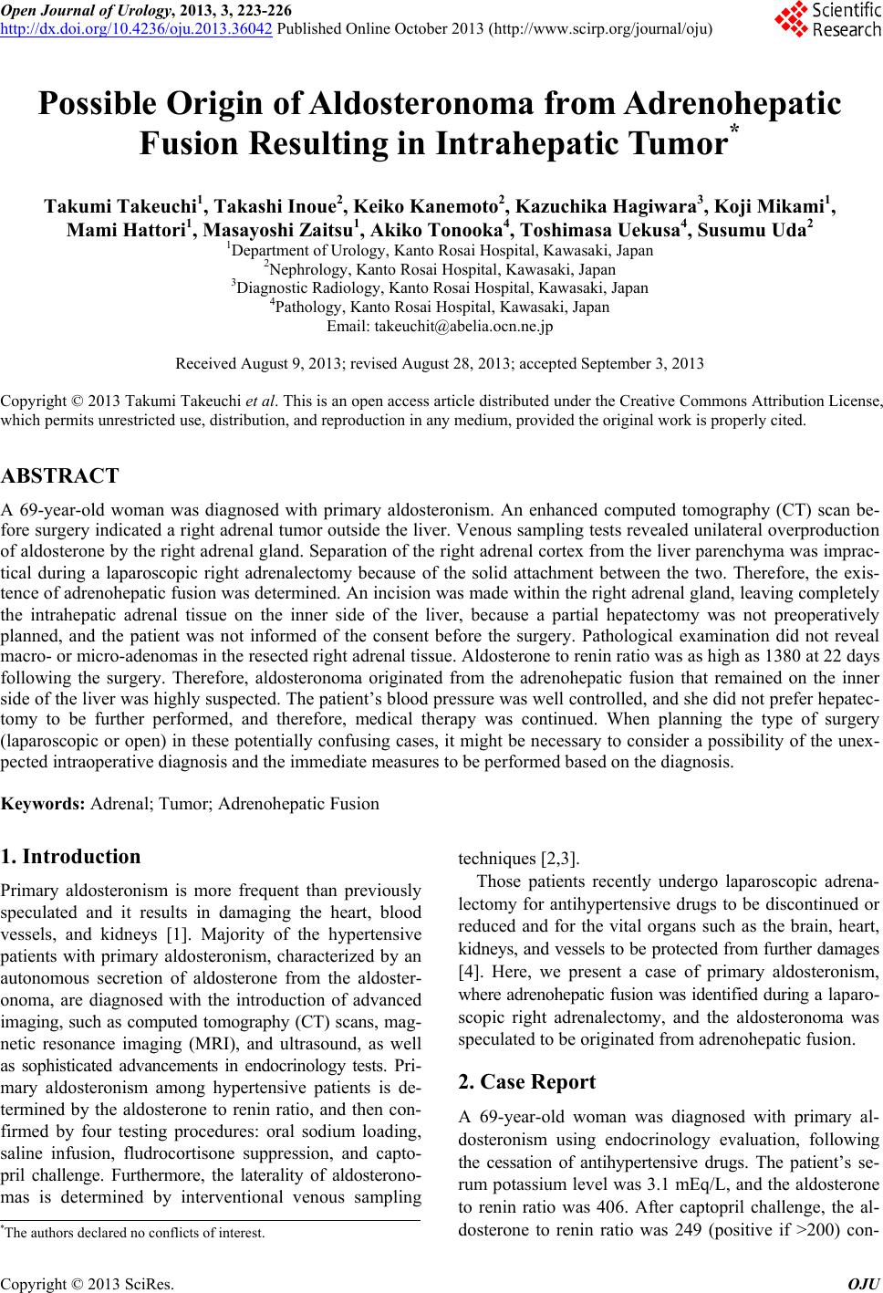
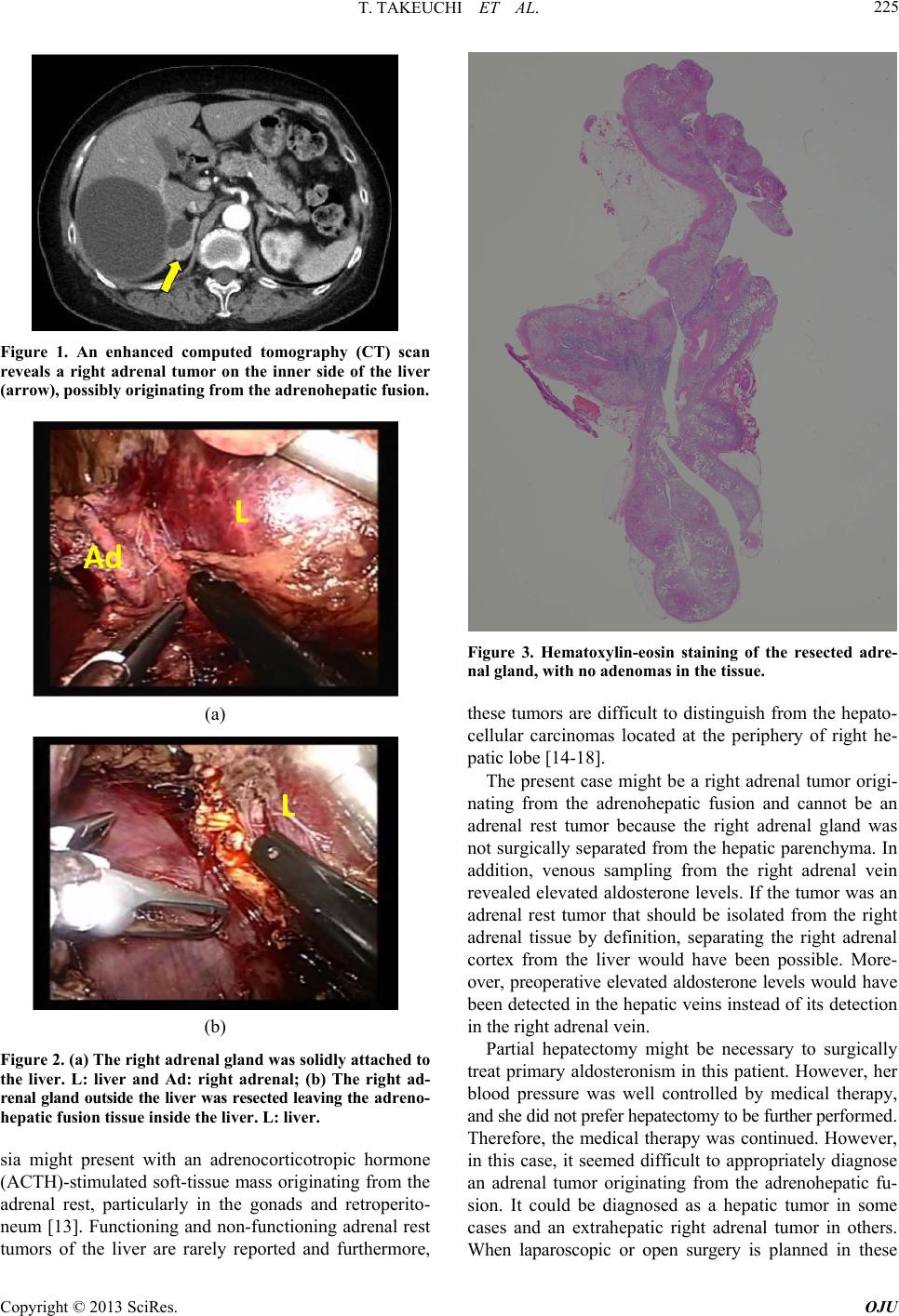
potentially confusing cases, it might be necessary to con-
sider a possibility of the unexpected intraoperative diag-
nosis and the immediate measures to be performed based
on the diagnosis. According to the medical databases,
there are no previous cases of patients where intrahepatic
adrenal tumors originating from the adrenohepatic fusion
were identified during the laparoscopic or open surgery.
REFERENCES
[1] G. P. Rossi, “A Comprehensive Review of the Clinical
Aspects of Primary Aldosteronism,” Nature Reviews En-
docrinology, Vol. 7, No. 8, 2011, pp. 485-495.
http://dx.doi.org/10.1038/nrendo.2011.76
[2] M. Salvà, M. V. Cicala and F. Mantero, “Primary Aldos-
teronism: The Role of Confirmatory Tests,” Hormone
and Metabolic Research, Vol. 44, No. 3, 2012, pp. 177-
180. http://dx.doi.org/10.1055/s-0032-1304661
[3] P. Mulatero, S. Monticone, C. Bertello, G. Mengozzi, D.
Tizzani, A. Iannaccone and F. Veglio, “Confirmatory
Tests in the Diagnosis of Primary Aldosteronism,” Hor-
mone and Metabolic Research, Vol. 42, No. 6, 2010,pp.
406-410. http://dx.doi.org/10.1055/s-0029-1246186
[4] O. Steichen, F. Zinzindohoué, P. F. Plouin and L. Amar,
“Outcomes of Adrenalectomy in Patients with Unilateral
Primary Aldosteronism: A Review,” Hormone and Meta-
bolic Research, Vol. 44, No. 3, 2012, pp. 221-227.
http://dx.doi.org/10.1055/s-0031-1299681
[5] L. H. Honoré and K. E. O’Hara, “Combined Adrenorenal
Fusion and Adrenohepatic Adhesion: A Case Report with
Review of the Literature and Discussion of Pathogene-
sis,” Journal of Urology, Vol. 115, No. 3, 1976, pp. 323-
325.
[6] R. Chamanza, H. A. Marxfeld, A. I. Blanco, S. W. Naylor
and A. E. Bradley, “Incidences and Range of Spontane-
ous Findings in Control Cynomolgus Monkeys (Macaca
fascicularis) Used in Toxicity Studies,” Journal of Toxi-
cologic Pathology, Vol. 38, No. 4, 2010, pp. 642-657.
http://dx.doi.org/10.1177/0192623310368981
[7] O. Quesada-Canales, A. Suárez-Bonnet, G. A. Ramírez,
M. Aguirre-Sanceledonio, M. Andrada, M. Rivero and A.
Espinosa de Los Monteros, “Adrenohepatic Fusion in Do-
mestic Ferrets (Mustela putorius furo),”Journal of Com-
parative Pathology, Vol. 149, No. 2-3, 2013, pp. 314-317.
http://dx.doi.org/10.1016/j.jcpa.2013.02.003
[8] S. Iwamoto, K. Okuda, N. Takeda, K. Sonoda and H.
Sanefuji, “Case Report: Right-Sided Periadrenal Metasta-
sis Supplied by the Hepatic Artery. Clue to the Genesis of
Pedunculated Hepatocellular Carcinoma,” Journal of Ga-
stroenterology and Hepatology, Vol. 12, No. 5, 1997, pp.
392-397.
http://dx.doi.org/10.1111/j.1440-1746.1997.tb00449.x
[9] K. Okuda, M. Arakawa, Y. Kubo, K. Sakata, M. Kage, S.
Iwamoto, S. Takeda, K. Sonoda and H. Sanefuji, “Right-
Sided Pedunculated Hepatocellular Carcinoma: A Form
of Adrenal Metastasis,” Hepatology, Vol. 27, No. 1, 1998,
pp. 81-85. http://dx.doi.org/10.1002/hep.510270114
[10] K. Okano, H. Usuki and H. Maeta, “Adrenal Metastasis
from Hepatocellular Carcinoma through an ADRENO-
HEPATIC Fusion,” Journal of Clinical Gastroenterology,
Vol. 38, No. 10, 2004, p. 912.
http://dx.doi.org/10.1097/00004836-200411000-00019
[11] H. S. Woo, K. H. Lee, S. Y. Park, H. S. Han, C. J. Yoon
and Y. H. Kim, “Adrenal Cortical Adenoma in Adreno-
hepatic Fusion Tissue: A Mimic of Malignant Hepatic
Tumor at CT,” American Journal of Roentgenology, Vol.
188, No. 3, 2007, pp. W246-W248.
http://dx.doi.org/10.2214/AJR.05.0498
[12] B. K. Park, C. K. Kim, B. C. Jung and Y. L. Suh, “Corti-
cal Adenoma in Adrenohepatic Fusion Tissue: Clue to
Making a Correct Diagnosis at Preoperative Computed
Tomography Examination,” European Urology, Vol. 56,
No. 6, 2009, pp. 1082-1085.
http://dx.doi.org/10.1016/j.eururo.2009.05.007
[13] T. D. Barwick, A. Malhotra, J. A. Webb, M. O. Savage
and R. H. Reznek, “Embryology of the Adrenal Glands
and Its Relevance to Diagnostic Imaging,” Clinical Radi-
ology, Vol. 60, No. 9, 2005, pp. 953-959.
http://dx.doi.org/10.1016/j.crad.2005.04.006
[14] Y. M. Shin, “Hepatic Adrenal Rest Tumor Mimicking
Hepatocellular Carcinoma,” Korean Journal of Hepatol-
ogy, Vol. 16, No. 3, 2010, pp. 338-341.
http://dx.doi.org/10.3350/kjhep.2010.16.3.338
[15] Y. Baba, T. Beppu, K. Imai, T. Masuda, K. Iyama, H.
Sasano and H. Baba, “A Case of Adrenal Rest Tumor of
the Liver: Radiological Imaging and Immunohistochemi-
cal Study of Steroidogenic Enzymes,” Hepatology Re-
search, Vol. 38, No. 11, 2008, pp. 1154-1158.
http://dx.doi.org/10.1111/j.1872-034X.2008.00360.x
[16] K. Arai, H. Muro, M. Suzuki, N. Oba, K. Ito and H. Sa-
sano, “Adrenal Rest Tumor of the Liver: A Case Report
with Immunohistochemical Investigation of Steroido-
genesis,” Pathology International, Vol. 50, No. 3, 2000,
pp. 244-248.
http://dx.doi.org/10.1046/j.1440-1827.2000.01029.x
[17] P. Contreras, E. Altieri, C. Liberman, A. Gac, A. Rojas, A.
Ibarra, M. Ravanal, M. Serón-Ferré, “Adrenal Rest Tu-
mor of the Liver Causing Cushing’s Syndrome: Therapy
with Ketoconazole Preceding an Apparent Surgical Cure,”
The Journal of Clinical Endocrinology & Metabolism,
Vol. 60, No. 1, 1985, pp. 21-28.
http://dx.doi.org/10.1210/jcem-60-1-21
[18] E. Z. Wallace, J. R. Leonidas, A. E. Stanek and A. Avra-
mides, “Endocrine Studies in a Patient with Functioning
Adrenal Rest Tumor of the Liver,” American Journal of
Medicine, Vol. 70, No. 5, 1981, pp. 1122-1125.
http://dx.doi.org/10.1016/0002-9343(81)90886-X
Copyright © 2013 SciRes. OJU