International Journal of Otolaryngology and Head & Neck Surgery, 2013, 2, 195-200
http://dx.doi.org/10.4236/ijohns.2013.25041 Published Online September 2013 (http://www.scirp.org/journal/ijohns)
Preliminary Hearing Results of Tympanomastoidectomies
Using Titanium Pros theses: Scenario in a Developing
Country*
Ankush Sayal, Virangna Taneja, Achal Gulati#
Department of Otolaryngology and Head & Neck Surgery, MAM College and Assoc., LN Hospital, New Delhi, India
Email: #achalgulati@rediff.com, ankushsayal@yahoo.com, virangnataneja@ymail.com
Received June 23, 2013; revised July 20, 2013; accepted August 8, 2013
Copyright © 2013 Ankush Sayal et al. This is an open access article distributed under the Creative Commons Attribution License,
which permits unrestricted use, distribution, and reproduction in any medium, provided the original work is properly cited.
ABSTRACT
The study was done to review postoperative hearing gain of patients with different Middle Ear Risk Index (MERI) un-
dergoing Tympanomastoidectomies with titanium prostheses reconstruction. A Retrospective chart review was per-
formed from September 2009 to December 2011. Of the 17 cases, 9 had moderate MERI while 8 had severe MERI. 9
patients underwent Tympanomastoidectomy with Total Ossicular Reconstruction Prostheses (TORP) and 8 w ith Partial
Ossicular Reconstruction Prostheses (PORP). Hearing gain of 25.31 dB was achieved in cases with moderate MERI
compared to 29.37 dB in severe MERI. Less than 20 dB average air-bone gap was achieved in 75% of PORP and
77.77% of TORP reconstructions. In developing countries with limited resources, decision regarding ossicular recon-
struction should be made taking into account MERI, intra operative findings and type of surgery. Best results are
achieved in cases of CWD with TORP and ICW with PORP.
Keywords: Midddle Ear Risk Index; Titanium; Total Ossicular Replacement Prosthesis; Partial Ossicular Replacement
Prosthesis
1. Introduction
The foremost objective of surgery done for chronic otitis
media is complete exteriorisation of disease. Although
the concept of disease removal was resolved much earlier,
the problem of reconstruction and giving better listening
to the patient have remained a challenge for the otologist.
In early 1960, John Sheehy described th e use of prosthe-
sis for reconstruction of the ossicular chain and since
then various prostheses have been used in the form of hy-
droxylapatite, Plastipore, Polyethylene and most recently
titanium prosthesis. The titanium implant is compatible,
readily available, light weight (4 mg) and technically
easy to use [1-2]. These titanium implants have over-
come the problems associated with the use of autologus
implants (usually necrosed due to underlying disease)
and homograft’s (risk of transmission of underlying dis-
ease), but their use is limited due to high costs.
In the p ast studies by Tos [3], Hirsch [4], Vartianer [5],
they concluded that disease exteriorisation as well as
ossicular reconstruction should be done in the same set-
ting. Contrary to this, studies by Sheehy [6] and Shelton
[7] advocated the role of staged surgery to provide a bet-
ter hearing outcome. They concluded that the 2nd stage
surgery should be performed after a period of 6 - 24
months to relook for any recurrence and for the purpose
of ossicular reconstruction. But the concept of staging
has since then remained a matter of controversy and the
decision is best left to the operating surgeon who may
decide depending on the intra operative findings and the
chances of the patient returning for regular follow-up, a
situation often encountered in the developing countries.
In order to grade the severity of middle ear disease. Kar-
tush developed a practical reporting protocol to stratify
patient groups to allow for meaningful study compari-
sons. This numerical indicator has been referred to as
“Middle Ear Risk Index”.
To determine Middle Ear Risk Index (MERI) of a pa-
tient, a specific value is assigned for each risk factor, and
then the value which includes Belluci criteria to assess
the degree of otorrhoea, Austin/Kartush criteria for ossi-
cular status, perforation, Cholesteatoma middle ear granu-
lations/effusion and history of previous surgery [8] was
*Financial Disclosure: The study was conducted at MAM College and
Assoc. LN Hospital, and the author and co-authors are employed at the
same institution. There are no financial interests involved.
#Corresponding author.
C
opyright © 2013 SciRes. IJOHNS
A. GULATI ET AL.
196
added. The suggested risk categories can be derived from
MERI as follows: MERI 0 = Normal; MERI 1 - 3 = Mild
disease; MERI 4 - 6 = Moderate disease; MERI 7 - 12 =
Severe disease.
Being in a developing country with limited economic
and human resources, an important objective of ear sur-
gery is not only to provide good listening to the individ-
ual, but also provide the same at affordable costs. Paucity
of resources and the fact that most patients in our country
present with severe MERI, the use of such costly tita-
nium prostheses becomes limited only to patients where
maximum benefit is expected. Therefore in such a sce-
nario planning, -ossicular, -reconstructions in any patient
undergoing Tympanomastoidectomy requires precise ju-
dgement and population-based studies for the effective
use of limited resources. But even after an extensive re-
view of literature, we found that similar studies are lack-
ing. This study compares the audiological improvement
in patients with moderate/severe MERI undergoing Tym-
panomastoidectomies (Canal Wall Up or Canal Wall
Down) and reconstruction using titan ium TORP or PORP.
We have used Spiggle and Theis titanium TORP and
PORP in this study for the ossicular reconstruction. Avai-
lable length for TORP varies from 7 mm to 3.5 mm, com-
pared to 3.5 mm to 0.5 mm for PORP.
2. Materials and Methods
A retrospective analysis was done for all the patients un-
dergoing tympanomastoidectomy with ossicular chain re-
construction from September 2009 to December 2011
(28 months). Patients of chronic otitis media with Cho-
lesteatoma or granulations with a purely conductive hear-
ing loss were included in the study. Those with history of
complications or have undergone previous ear surgeries
were excluded from the study. Of the total, complete fol-
low up with post operative audiological analysis was
available for 17 cases.
Data was analysed in terms of middle ear risk index,
surgical procedure being done (ICW or CWD), method
of reconstruction (TORP or PORP), single/2nd stage re-
construction, and compared with the audiological im-
provement. Pre operative as well as post operative Air-
Bone gap was calculated at 0.5, 1, 2 and 3 kHz in accor-
dance with the guidelines of American Academy of Oto-
laryngology—Head and Neck Surgery committee on
hearing and equilibrium for hearing evaluation [9].
All the surgeries were performed under General An-
aesthesia via Post aural approach by the same surgeon
(A.S). The MERI index, disease exten t and the condition
of middle ear mucosa were used as criteria by the oper-
ating surgeon intra operatively for deciding whether to
go for canal wall up or canal wall down surgery, and also
for deciding whether the reconstruction would be done as
a primary reconstruction or to subject the patient to a sec-
ond stage reconstruction. Depending on the ossicular sta-
tus, TORP or PORP were placed over the stapes foot-
plate or supra structure respectively. The size of the pros-
thesis was calculated using a measuring gauge. For
TORP, the length of the prostheses required was calcu-
lated from footplate of stapes while for PORP, the stapes
head was used as a reference. The upper limit was taken
till the facial ridge in case of CWD and till the an nulus in
case of ICW surgery for both the prostheses. A small
piece of conchal cartilage (0.3 mm thickness) was har-
vested in all the cases and placed over the prostheses.
3. Results
Of the 17 cases that fulfilled the inclusion criteria, 9 had
moderate MERI and 8 had severe MERI. 7 patients un-
derwent mastoidectomies with canal wall down, while 10
had intact canal wall surgery. 8 underwent reconstru ction
using PORP, whereas 9 cases underwent reconstruction
using TORP. 13 cases were taken up for disease removal
and reconstruction in single setting, whereas 4 cases were
taken up fo r 2 n d stage reconst ru ct ion (Table 1).
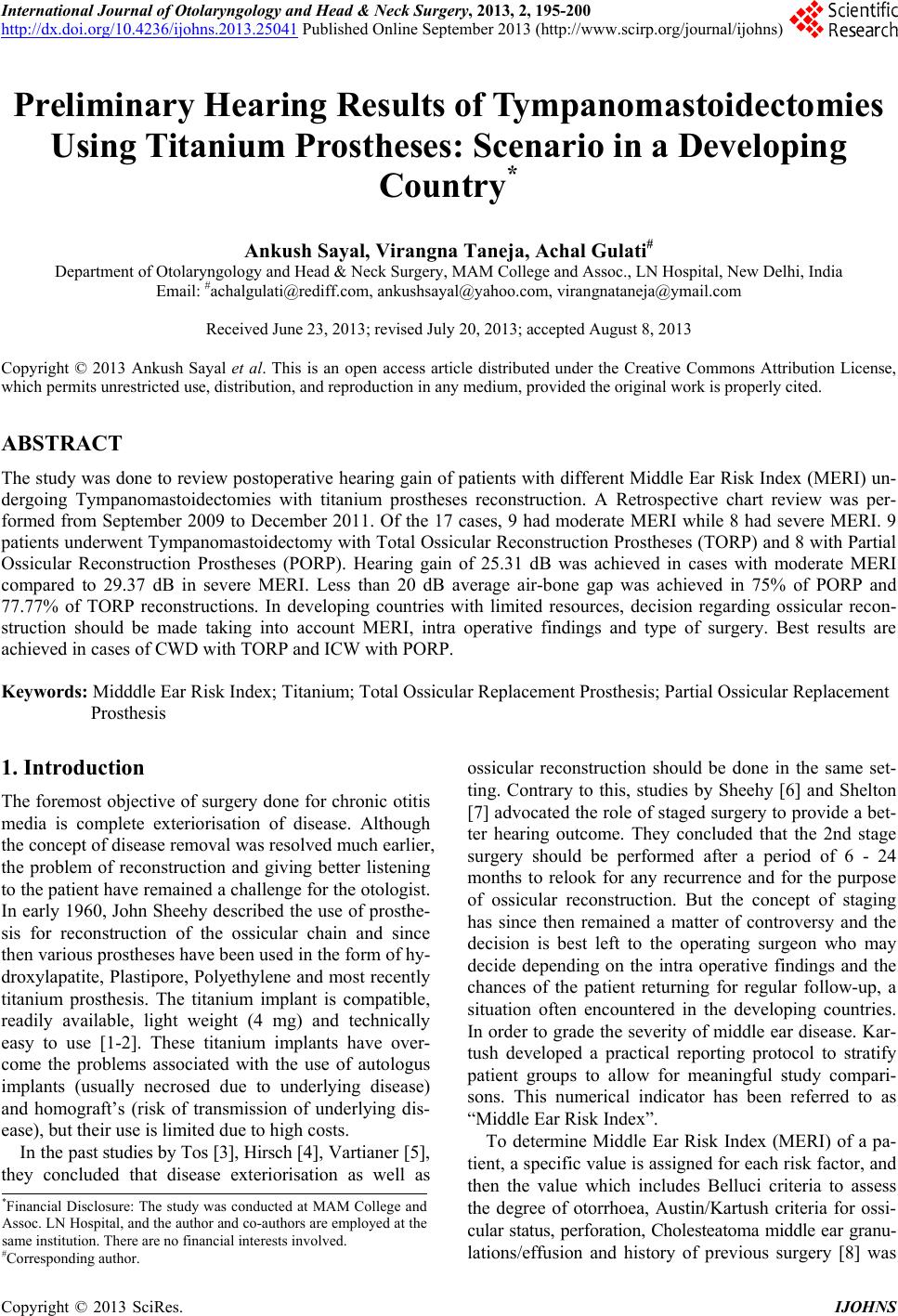
6 cases underwent ICW with PORP reconstruction and
4 cases underwent ICW with TORP reconstruction. In 5
Canal wall down with TORP reconstruction was at-
tempted and 2 cases underwent CWD with PORP recon-
struction (Figure 1). Average hearing gain was found to
be 28.75 dB in 10 cases undergoing ICW, compared to 7
cases undergoing CWD who had average postoperative
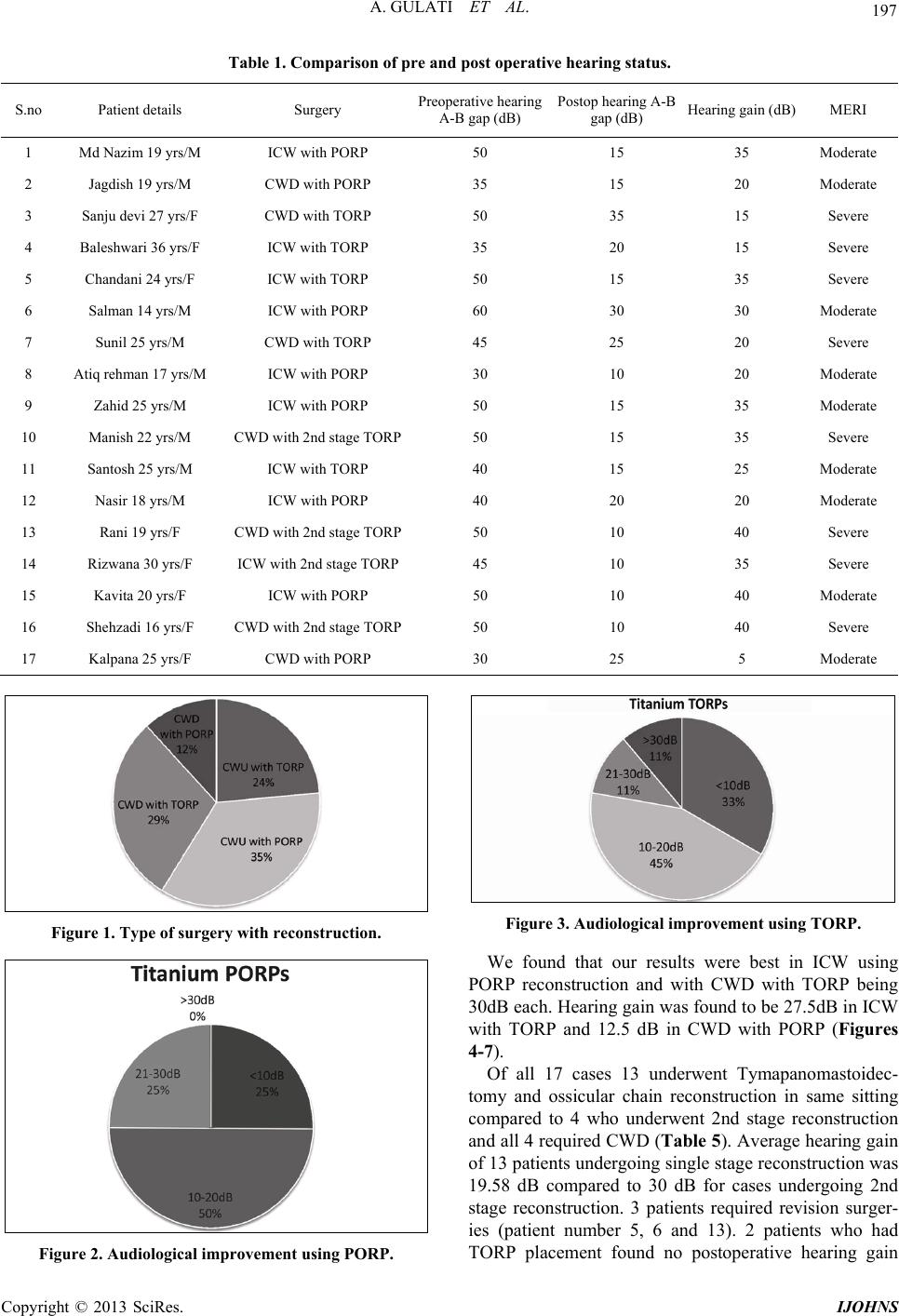
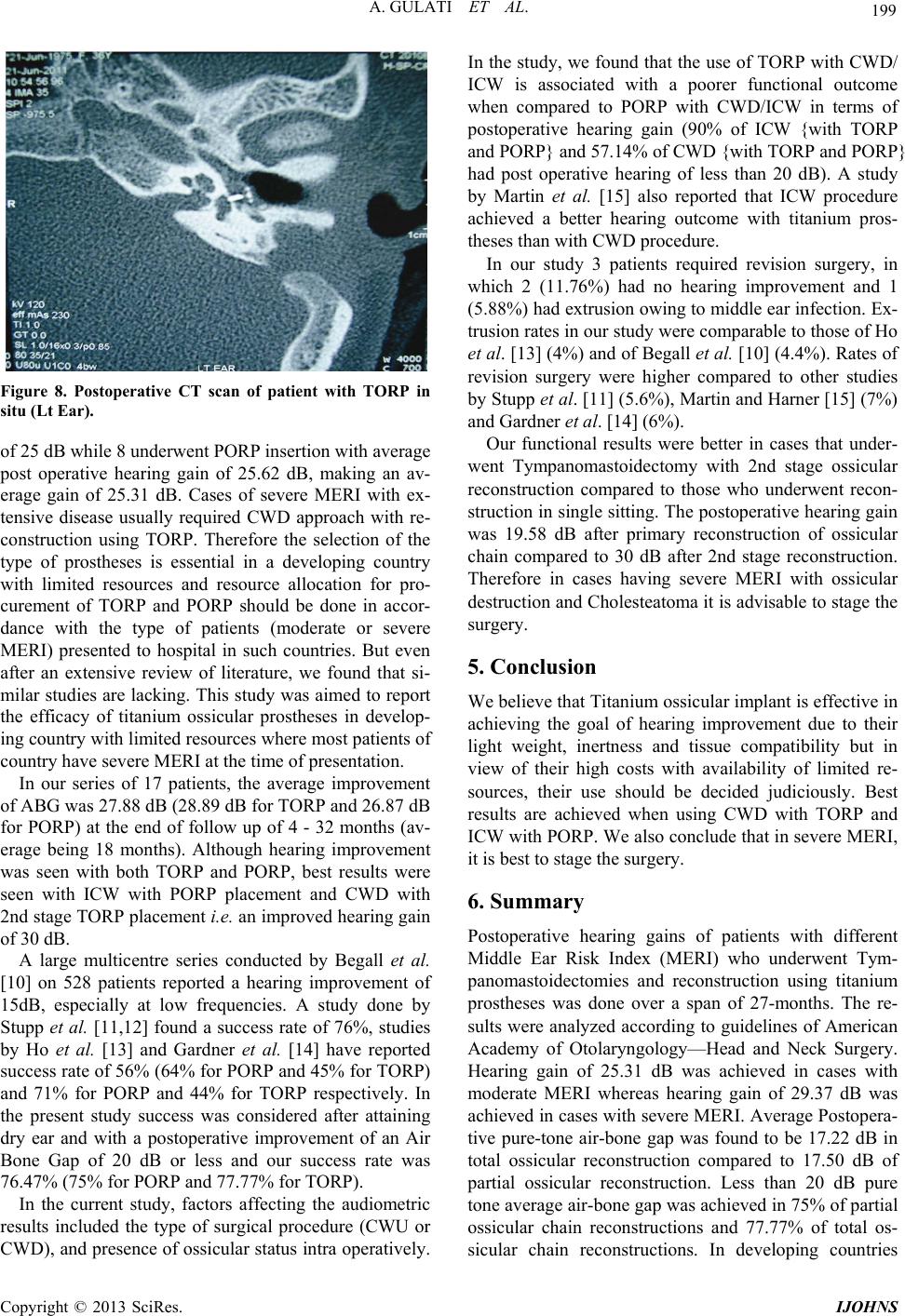
hearing of 21.25 dB. Po stoperatively, the mean Air-Bone
Gap was 17.5 dB for cases which underwent reconstruc-
tion using PORP (Figure 2) compared to 17.22 dB in
which TORP (Figure 3) were used. Mean hearing gain in
the PORP group was 26.87 dB, whereas for those under-
going reconstruction using TORP was 28.89 dB (Table
2). 50% of patients undergoing reconstruction with PORP
(4 out of 8) and 45% with TORP (4 out of 9) had postop-
erative hea r ing between 1 0 - 2 0 dB.
Of the 8 cases having severe MERI, 5 (62.5%) re-
quired CWD surgery whereas 3 (37.5%) required ICW.
On the other hand, of the 9 cases having moderate MERI,
only 2 (22.2%) required CWD compared to 7 (77.8%)
who required ICW. All the 8 cases having severe MERI
underwent reconstruction using TORP. On the other
hand 9 cases had moderate MERI of which 8 underwent
PORP insertion and 1 had TORP insertion. All 9 patients
having moderate MERI underwent single stage recon-
struction, whereas of the 8 cases having severe MERI, 4
underwent single stage and 4 underwent 2nd stage recon-
struction. 8 cases having severe MERI had average post
operative hearing gain of 29.37 dB. On the other hand 9
cases having moderate MERI had average post operative
hearing gain of 25.62 dB with PORP and hearing gain of
25 dB with TORP making an average gain of 25.31 dB
(Tables 3 and 4).
Copyright © 2013 SciRes. IJOHNS
A. GULATI ET AL.
Copyright © 2013 SciRes. IJOHNS
197
Table 1. Comparison of pre and post operative hearing status.
S.no Patient details Surgery Preoperative hear ing
A-B gap (dB) Postop hearing A-B
gap (dB) Hearing gain (dB) MERI
1 Md Nazim 19 yrs/M ICW with PORP 50 15 35 Moderate
2 Jagdish 19 yrs/M CWD with PORP 35 15 20 Moderate
3 Sanju devi 27 yrs/F CWD with TORP 50 35 15 Severe
4 Baleshwari 36 yrs/F ICW with TORP 35 20 15 Severe
5 Chandani 24 yrs/F ICW with TORP 5 0 15 35 Severe
6 Salman 14 yrs/M ICW with PORP 60 30 30 Moderate
7 Sunil 25 yrs/M CWD with TORP 45 25 20 Severe
8 Atiq rehman 17 yrs/M ICW with PORP 30 10 20 Moderate
9 Zahid 25 yrs/M ICW with PORP 50 15 35 Moderate
10 Manish 22 yrs/M CWD with 2nd stage TORP50 15 35 Severe
11 Santosh 25 yrs/M ICW with TORP 40 15 25 Moderate
12 Nasir 18 yrs/M ICW with PORP 40 20 20 Moderate
13 Rani 19 yrs/F CWD with 2nd stage TOR P50 10 40 Severe
14 Rizwana 30 yrs/F ICW with 2nd stage TORP45 10 35 Severe
15 Kavita 20 yrs/F ICW with PO R P 50 10 40 Moderate
16 Shehzadi 16 yrs/F CWD with 2nd stage TORP50 10 40 Severe
17 Kalpana 25 yrs / F CWD with PORP 30 25 5 Moderate
Figure 3. Audiological improvement using TORP.
Figure 1. Type of surgery with reconstruction.
We found that our results were best in ICW using
PORP reconstruction and with CWD with TORP being
30dB each. Hearing gain was found to be 27.5dB in ICW
with TORP and 12.5 dB in CWD with PORP (Figures
4-7).
Of all 17 cases 13 underwent Tymapanomastoidec-
tomy and ossicular chain reconstruction in same sitting
compared to 4 who underwent 2nd stage reconstruction
and all 4 required CWD (Table 5). Average hear ing gain
of 13 patients undergoing single stage reconstruction was
19.58 dB compared to 30 dB for cases undergoing 2nd
stage reconstruction. 3 patients required revision surger-
ies (patient number 5, 6 and 13). 2 patients who had
TORP placement found no postoperative hearing gain
Figure 2. Audiological improvement using PORP.
A. GULATI ET AL.
198
Table 2. Average audiological hearing gain post Tympano-
mastidectomy with ossicular reconstruction.
Average
Preoperative
hearing (dB)
Average
Postoperative
hearing (dB)
Average
Hearing
gain (dB)
ICW with TORP 42.5 20 27. 5
ICW with PORP 46.66 16.66 30
CWD with TORP 49 19 30
CWD with PORP 32.5 20 12.5
Table 3. Audiological hearing gain in terms of Middle Ear
Risk Index.
MERI Hearing gain in
patients with TORP Hearing gain in
patients with PORP
Moderate 25 25.625
Severe 29.37 no case
Table 4. Average audiological hearing gain in terms of
MERI and type of reconstruc tion.
MERI ICW with
PORP ICW with
TORP CWD with
PORP CWD with
TORP
Moderate 30 dB
(6 patients) 25 dB
(1 patient)12.5 dB
(2 patients) No patient
Severe No patient 28.33 dB
(3 patients) No patient 30dB
(5 patients)
Table 5. Patient distribution in terms of staged reconstruc-
tion.
MERI Single stage reconstruction 2nd stage reconstruction
Moderate 8 1
Severe 5 3
Figure 4. Hearing results of ICW with PORP.
and therefore exploration was done, whereas 1 patient
with PORP insertion (intra operative-limited cholestea-
toma confined to attic with necros es of incu s and malleus
Figure 5. Hearing results of ICW with TORP.
Figure 6. Hearing results of CWD with PORP.
Figure 7. Hearing results of CWD with TORP.
but intact stapes supra structure) who initially had hear-
ing gain later developed infection and had PORP extru-
sion, is still awaiting revision surgery ( Figure 8). Follow
up of cases was ranging from 4 month to 32 months (av-
erage 18 months).
4. Discussion
Of 8 cases having severe MERI, 5 (62.5%) required
CWD surgery and 3 (37.5%) required ICW. All under-
went reconstruction using TORP with an average post
operative hearing gain of 29.37 dB. On the other hand, of
the 9 cases having moderate MERI, only 2 (22.2%) re-
quired CWD while 7 (77.78%) underwent ICW. 1 patient
with CWD underwent TORP insertion with hearing gain
Copyright © 2013 SciRes. IJOHNS
A. GULATI ET AL. 199
Figure 8. Postoperative CT scan of patient with TORP in
situ (Lt Ear).
of 25 dB while 8 underwen t PORP insertion with average
post operative hearing gain of 25.62 dB, making an av-
erage gain of 25.31 dB. Cases of severe MERI with ex-
tensive disease usually required CWD approach with re-
construction using TORP. Therefore the selection of the
type of prostheses is essential in a developing country
with limited resources and resource allocation for pro-
curement of TORP and PORP should be done in accor-
dance with the type of patients (moderate or severe
MERI) presented to hospital in such countries. But even
after an extensive review of literature, we found that si-
milar studies are lacking. This study was aimed to report
the efficacy of titanium ossicular prostheses in develop-
ing country with limited resources where most patients of
country have severe MERI at the time of presentation.
In our series of 17 patients, the average improvement
of ABG was 27.88 dB (28.89 dB for TORP and 26.87 dB
for PORP) at the end of follow up of 4 - 32 months (av-
erage being 18 months). Although hearing improvement
was seen with both TORP and PORP, best results were
seen with ICW with PORP placement and CWD with
2nd stage TORP placement i.e. an improved hearing gain
of 30 dB.
A large multicentre series conducted by Begall et al.
[10] on 528 patients reported a hearing improvement of
15dB, especially at low frequencies. A study done by
Stupp et al. [11,12] found a success rate of 76%, studies
by Ho et al. [13] and Gardner et al. [14] have reported
success rate of 56% (64% for PORP and 45% for TORP)
and 71% for PORP and 44% for TORP respectively. In
the present study success was considered after attaining
dry ear and with a postoperative improvement of an Air
Bone Gap of 20 dB or less and our success rate was
76.47% (75% for PORP and 77.77% for TORP).
In the current study, factors affecting the audiometric
results included the type of surgical procedure (CWU or
CWD), and presence of ossicular status intra operatively.
In the study, we found that the use of TORP with CWD/
ICW is associated with a poorer functional outcome
when compared to PORP with CWD/ICW in terms of
postoperative hearing gain (90% of ICW {with TORP
and PORP} and 57.14% of CWD {with TORP and PORP}
had post operative hearing of less than 20 dB). A study
by Martin et al. [15] also reported that ICW procedure
achieved a better hearing outcome with titanium pros-
theses than with CWD procedure.
In our study 3 patients required revision surgery, in
which 2 (11.76%) had no hearing improvement and 1
(5.88%) had extrusion owing to middle ear infection. Ex-
trusion rates in our study were comparable to those of Ho
et al. [13] (4%) and of Begall et al. [10] (4.4%). Rates of
revision surgery were higher compared to other studies
by Stupp et al. [11] (5.6%), Martin and Harner [15] (7%)
and Gardner et al. [14] (6%).
Our functional results were better in cases that under-
went Tympanomastoidectomy with 2nd stage ossicular
reconstruction compared to those who underwent recon-
struction in single sitting. The postoperative hearing gain
was 19.58 dB after primary reconstruction of ossicular
chain compared to 30 dB after 2nd stage reconstruction.
Therefore in cases having severe MERI with ossicular
destruction and Cholesteatoma it is advisable to stage the
surgery.
5. Conclusion
We believe that Titanium ossicular implant is effective in
achieving the goal of hearing improvement due to their
light weight, inertness and tissue compatibility but in
view of their high costs with availability of limited re-
sources, their use should be decided judiciously. Best
results are achieved when using CWD with TORP and
ICW with PORP. We also conclude that in severe MERI,
it is best to stage the surg ery.
6. Summary
Postoperative hearing gains of patients with different
Middle Ear Risk Index (MERI) who underwent Tym-
panomastoidectomies and reconstruction using titanium
prostheses was done over a span of 27-months. The re-
sults were analyzed according to guidelines of American
Academy of Otolaryngology—Head and Neck Surgery.
Hearing gain of 25.31 dB was achieved in cases with
moderate MERI whereas hearing gain of 29.37 dB was
achieved in cases with severe MERI. Average Postopera-
tive pure-tone air-bone gap was found to be 17.22 dB in
total ossicular reconstruction compared to 17.50 dB of
partial ossicular reconstruction. Less than 20 dB pure
tone average air-bone gap was achieved in 75% of partial
ossicular chain reconstructions and 77.77% of total os-
sicular chain reconstructions. In developing countries
Copyright © 2013 SciRes. IJOHNS
A. GULATI ET AL.
Copyright © 2013 SciRes. IJOHNS
200
with limited resources, decision regarding ossicular re-
construction should be made taking into account MERI,
intra operative findings and type of surgery to achieve
better post operative hearing results. Best results are achi-
eved in cases of CWD with TORP and ICW with PORP.
REFERENCES
[1] L. M. Menendez-Colino, M. Bernal-Sprekelsen, I. Alobid
and J. Traserra-Coderch, “Preliminary Functional Results
of Tympanoplasty with Titanium Prostheses,” Otolaryn-
gology—Head and Neck Surgery, Vol. 131, No. 5, 2004,
pp. 747-749. doi:10.1016/j.otohns.2004.04.017
[2] C. V. Dalchow, D. Grun and H. F. Stupp, “Reconstruc-
tion of the Ossicular Chain with Titanium Implants,” Oto-
laryngology—Head and Neck Surgery, Vol. 125, No. 6,
2001, pp. 628-630. doi:10.1067/mhn.2001.120397
[3] M. Tos, “Late Results in Tympanoplasty,” Acta Oto-La-
ryngologica, Vol. 82, No. 1, 1976, pp. 282-285.
doi:10.3109/00016487609120906
[4] B. E. Hirsch, D. B. Kamerer and S. Doshi, “Single Stage
Management of Cholesteatoma,” Otolaryngology—Head
and Neck Surgery, Vol. 106, 1992, pp. 351-354.
[5] E. Vartianen and J. Nuutinen, “Long Term Hearing Re-
sults of One Stage Tympanoplasty for Chronic Otitis Me-
dia,” European Archives of Oto-Rhino-Laryngology, Vol.
249, No. 6, 1992, pp. 329-331.
[6] J. L. Sheehy and J. A. Crabtree, “Tympanoplasty: Staging
the Operation,” Laryngoscope, Vol. 83, No. 10, 1973, pp.
1594-1621. doi:10.1288/00005537-197310000-00003
[7] C. Shelton and J. L. Sheehy, “Tympanoplasty: Review of
400 Staged Cases,” Laryngoscope, Vol. 100, No. 7, 1990,
pp. 679-681.
[8] J. M. Kartush, “Ossicular Chain Reconstruction. Capitu-
lum to Malleus,” Otolaryngologic Clinics of North Amer-
ica, Vol. 27, No. 4, 1994, pp. 689-715.
[9] American Academy of Otolaryngology-Head and Neck
Surgery Foundation, Inc., “Committee on Hearing and
Equilibrium Guidelines for the Evaluation of Results of
Treatment of Conductive Hearing Loss,” Otolaryngology
—Head and Neck Surgery, Vol. 113, No. 3, 1995, pp.
186-187. doi:10.1016/S0194-5998(95)70103-6
[10] K. Begall and H. Zimmermann, “Reconstruction of the
Ossicular Chain with Titanium Implants. Results of a
Multicenter Study,” Laryngorhinootologie, Vol. 79, No. 3,
2000, pp. 139-145. doi:10.1055/s-2000-298
[11] C. H. Stupp, C. Dalchow, D. Grun, H. F. Stupp and J.
Wustrow, “Three Years of Experience with Titanium Im-
plants in the Middle Ear,” Laryngorhinootologie, Vol. 78,
No. 6, 1999, pp. 299-303. doi:10.1055/s-2007-996875
[12] C. H. Stupp, H. F. Stupp and D. Grun, “Replacement of
Ear Ossicles with Titanium Prostheses,” Laryngorhino-
otologie, Vol. 75, No. 6, 1996, pp. 335-337.
doi:10.1055/s-2007-997590
[13] S. Y. Ho, R. A. Battista and R. J. Wiet, “Early Results
with Titanium Ossicular Implants,” Otology & Neurotol-
ogy, Vol. 24, No. 2, 2003, pp. 149-152.
doi:10.1097/00129492-200303000-00005
[14] E. K. Gardner, C. G. Jackson and D. M. Kaylie, “Results
with Titanium Ossicular Reconstruction Prostheses,” La-
ryngoscope, Vol. 114, No. 1, 2004, pp. 65-70.
doi:10.1097/00005537-200401000-00011
[15] A. D. Martin and S. G. Harner, “Ossicular Reconstruction
with Titanium Prostheses,” Laryngoscope, Vol. 114, No.
1, 2004, pp. 61-64.
doi:10.1097/00005537-200401000-00010