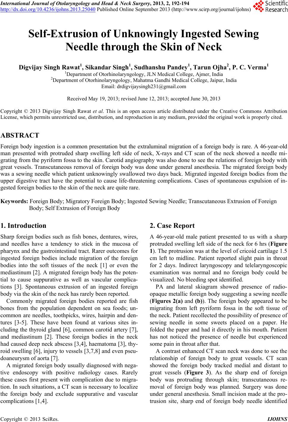
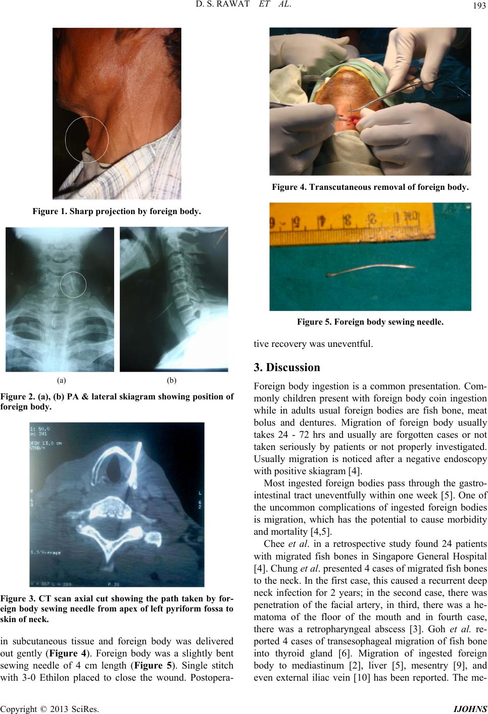
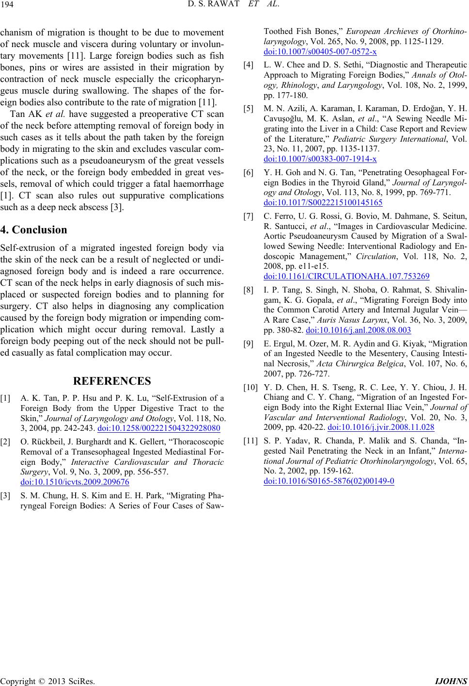
International Journal of Otolaryngology and Head & Neck Surgery, 2013, 2, 192-194
http://dx.doi.org/10.4236/ijohns.2013.25040 Published Online September 2013 (http://www.scirp.org/journal/ijohns)
Self-Extrusion of Unknowingly Ingested Sewing
Needle through the Skin of Neck
Digvijay Singh Rawat1, Sikandar Singh1, Sudhanshu Pandey1, Tarun Ojh a2, P. C. Verma1
1Department of Otorhinolaryngology, JLN Medical College, Ajmer, India
2Department of Otorhinolaryngology, Mahatma Gandhi Medical College, Jaipur, India
Email: drdigvijaysingh231@gmail.com
Received May 19, 2013; revised June 12, 2013; accepted June 30, 2013
Copyright © 2013 Digvijay Singh Rawat et al. This is an open access article distributed under the Creative Commons Attribution
License, which permits unrestricted use, distribution, and reproduction in any medium, provided the original work is properly cited.
ABSTRACT
Foreign body ingestion is a common presentation but the extraluminal migration of a foreign body is rare. A 46-year-old
man presented with protruded sharp swelling left side of neck, X-rays and CT scan of the neck showed a needle mi-
grating from the pyriform fossa to the skin. Carotid angiography was also done to see the relations of foreign body with
great vessels. Transcutaneous removal of foreign body was done under general anesthesia. The migrated foreign body
was a sewing needle which patient unknowingly swallowed two days back. Migrated ingested foreign bodies from the
upper digestive tract have the potential to cause life-threatening complications. Cases of spontaneous expulsion of in-
gested foreign bodies to the skin of the neck are quite rare.
Keywords: Foreign Body; Migratory Foreign Body; Ingested Sewing Needle; Transcutaneous Extrusion of Foreign
Body; Self Extrusion of Foreign Body
1. Introduction
Sharp foreign bodies such as fish bones, dentures, wires,
and needles have a tendency to stick in the mucosa of
pharynx and the gastrointestinal tract. Rarer outcomes for
ingested foreign bodies include migration of the foreign
bodies into the soft tissues of the neck [1] or even the
mediastinum [2]. A migrated foreign body has the poten-
tial to cause suppurative as well as vascular complica-
tions [3]. Spontaneous extrusion of an ingested foreign
body via the skin of the neck has rarely been reported.
Commonly migrated foreign bodies reported are fish
bones from the population dependent on sea foods; un-
common are needles, toothpicks, wires, hairpin and den-
tures [3-5]. These have been found at various sites in-
cluding the thyroid gland [6], common carotid artery [7],
and mediastinum [2]. These foreign bodies in the neck
had caused deep neck abscess [3,4], haematoma [3], thy-
roid swelling [6], injury to vessels [3,7,8] and even pseu-
doaneurysm of aorta [7].
A migrated foreign body usually diagnosed with nega-
tive endoscopy with positive radiology cases. Rarely
these cases first present with complication due to migra-
tion. In such situations, a CT scan is necessary to localize
the foreign body and exclude suppurative and vascular
complications [1,4].
2. Case Report
A 46-year-old male patient presented to us with a sharp
protruded swelling left side of the neck for 6 hrs (Figure
1). The protrusion was at the level of cricoid cartilage 1.5
cm left to midline. Patient reported slight pain in throat
for 2 days. Indirect laryngoscopy and telelaryngoscopic
examination was normal and no foreign body could be
visualized. No bleeding spot identified.
PA and lateral skiagram showed presence of radio-
opaque metallic foreign body suggesting a sewing needle
(Figures 2(a) and (b)). The foreign body appeared to be
migrating from left pyriform fossa in the soft tissue of
the neck. Patient recollected the possibility of presence of
sewing needle in some sweets placed on a paper. He
folded the paper and had it directly in his mouth. Patient
has not noticed the presence of needle but experienced
some pain in throat after that.
A contrast enhanced CT scan neck was done to see the
relationship of foreign body to great vessels. CT scan
showed the foreign body tracked medial and distant to
great vessels (Figure 3). As the sharp end of foreign
body was protruding through skin; transcutaneous re-
moval of foreign body was planned. Surgery was done
under general anesthesia. Small incision made at the pro-
trusion site, sharp end of foreign body needle identified
C
opyright © 2013 SciRes. IJOHNS
D. S. RAWAT ET AL. 193
Figure 1. Sharp projection by fore i gn body.
(a) (b)
Figure 2. (a), (b) PA & lateral skiagram showing position of
foreign body.
Figure 3. CT scan axial cut showing the path taken by for-
eign body sewing needle from apex of left pyriform fossa to
skin of neck.
in subcutaneous tissue and foreign body was delivered
out gently (Figure 4). Foreign body was a slightly bent
sewing needle of 4 cm length (Figure 5). Single stitch
with 3-0 Ethilon placed to close the wound. Postopera-
Figure 4. Transcutaneous removal of foreign body.
Figure 5. Foreign body sewing needle.
tive recovery was uneventful.
3. Discussion
Foreign body ingestion is a common presentation. Com-
monly children present with foreign body coin ingestion
while in adults usual foreign bodies are fish bone, meat
bolus and dentures. Migration of foreign body usually
takes 24 - 72 hrs and usually are forgotten cases or not
taken seriously by patients or not properly investigated.
Usually migration is noticed after a negative endoscopy
with positive skiagram [4].
Most ingested foreign bodies pass through the gastro-
intestinal tract uneventfully within one week [5]. One of
the uncommon complications of ingested foreign bodies
is migration, which has the potential to cause morbidity
and mortality [4,5].
Chee et al. in a retrospective study found 24 patients
with migrated fish bones in Singapore General Hospital
[4]. Chung et al. presented 4 cases of migrated fish bones
to the neck. In the first case, this caused a recurrent deep
neck infection for 2 years; in the second case, there was
penetration of the facial artery, in third, there was a he-
matoma of the floor of the mouth and in fourth case,
there was a retropharyngeal abscess [3]. Goh et al. re-
ported 4 cases of transesophageal migration of fish bone
into thyroid gland [6]. Migration of ingested foreign
body to mediastinum [2], liver [5], mesentry [9], and
even external iliac vein [10] has been reported. The me-
Copyright © 2013 SciRes. IJOHNS
D. S. RAWAT ET AL.
Copyright © 2013 SciRes. IJOHNS
194
chanism of migration is thought to be due to movement
of neck muscle and viscera during voluntary or involun-
tary movements [11]. Large foreign bodies such as fish
bones, pins or wires are assisted in their migration by
contraction of neck muscle especially the cricopharyn-
geus muscle during swallowing. The shapes of the for-
eign bodies also contribute to the rate of migration [11].
Tan AK et al. have suggested a preoperative CT scan
of the neck before attempting removal of foreign body in
such cases as it tells about the path taken by the foreign
body in migrating to the skin and excludes vascular com-
plications such as a pseudoaneurysm of the great vessels
of the neck, or the foreign body embedded in great ves-
sels, removal of which could trigger a fatal haemorrhage
[1]. CT scan also rules out suppurative complications
such as a deep neck abscess [3].
4. Conclusion
Self-extrusion of a migrated ingested foreign body via
the skin of the neck can be a result of neglected or undi-
agnosed foreign body and is indeed a rare occurrence.
CT scan of the neck helps in early diagnosis of such mis-
placed or suspected foreign bodies and to planning for
surgery. CT also helps in diagnosing any complication
caused by the foreign body migration or impending com-
plication which might occur during removal. Lastly a
foreign body peeping out of the neck should not be pull-
ed casually as fatal complication may occur.
REFERENCES
[1] A. K. Tan, P. P. Hsu and P. K. Lu, “Self-Extrusion of a
Foreign Body from the Upper Digestive Tract to the
Skin,” Journal of Laryngology and Otology, Vol. 118, No.
3, 2004, pp. 242-243. doi:10.1258/002221504322928080
[2] O. Rückbeil, J. Burghardt and K. Gellert, “Thoracoscopic
Removal of a Transesophageal Ingested Mediastinal For-
eign Body,” Interactive Cardiovascular and Thoracic
Surgery, Vol. 9, No. 3, 2009, pp. 556-557.
doi:10.1510/icvts.2009.209676
[3] S. M. Chung, H. S. Kim and E. H. Park, “Migrating Pha-
ryngeal Foreign Bodies: A Series of Four Cases of Saw-
Toothed Fish Bones,” European Archieves of Otorhino-
laryngology, Vol. 265, No. 9, 2008, pp. 1125-1129.
doi:10.1007/s00405-007-0572-x
[4] L. W. Chee and D. S. Sethi, “Diagnostic and Therapeutic
Approach to Migrating Foreign Bodies,” Annals of Otol-
ogy, Rhinology, and Laryngology, Vol. 108, No. 2, 1999,
pp. 177-180.
[5] M. N. Azili, A. Karaman, I. Karaman, D. Erdoğan, Y. H.
Cavuşoğlu, M. K. Aslan, et al., “A Sewing Needle Mi-
grating into the Liver in a Child: Case Report and Review
of the Literature,” Pediatric Surgery International, Vol.
23, No. 11, 2007, pp. 1135-1137.
doi:10.1007/s00383-007-1914-x
[6] Y. H. Goh and N. G. Tan, “Penetrating Oesophageal For-
eign Bodies in the Thyroid Gland,” Journal of Laryngol-
ogy and Otology, Vol. 113, No. 8, 1999, pp. 769-771.
doi:10.1017/S0022215100145165
[7] C. Ferro, U. G. Rossi, G. Bovio, M. Dahmane, S. Seitun,
R. Santucci, et al., “Images in Cardiovascular Medicine.
Aortic Pseudoaneurysm Caused by Migration of a Swal-
lowed Sewing Needle: Interventional Radiology and En-
doscopic Management,” Circulation, Vol. 118, No. 2,
2008, pp. e11-e15.
doi:10.1161/CIRCULATIONAHA.107.753269
[8] I. P. Tang, S. Singh, N. Shoba, O. Rahmat, S. Shivalin-
gam, K. G. Gopala, et al., “Migrating Foreign Body into
the Common Carotid Artery and Internal Jugular Vein—
A Rare Case,” Auris Nasus Larynx, Vol. 36, No. 3, 2009,
pp. 380-82. doi:10.1016/j.anl.2008.08.003
[9] E. Ergul, M. Ozer, M. R. Aydin and G. Kiyak, “Migration
of an Ingested Needle to the Mesentery, Causing Intesti-
nal Necrosis,” Acta Chirurgica Belgica, Vol. 107, No. 6,
2007, pp. 726-727.
[10] Y. D. Chen, H. S. Tseng, R. C. Lee, Y. Y. Chiou, J. H.
Chiang and C. Y. Chang, “Migration of an Ingested For-
eign Body into the Right External Iliac Vein,” Journal of
Vascular and Interventional Radiology, Vol. 20, No. 3,
2009, pp. 420-22. doi:10.1016/j.jvir.2008.11.028
[11] S. P. Yadav, R. Chanda, P. Malik and S. Chanda, “In-
gested Nail Penetrating the Neck in an Infant,” Interna-
tional Journal of Pediatric Otorhinolaryngology, Vol. 65,
No. 2, 2002, pp. 159-162.
doi:10.1016/S0165-5876(02)00149-0