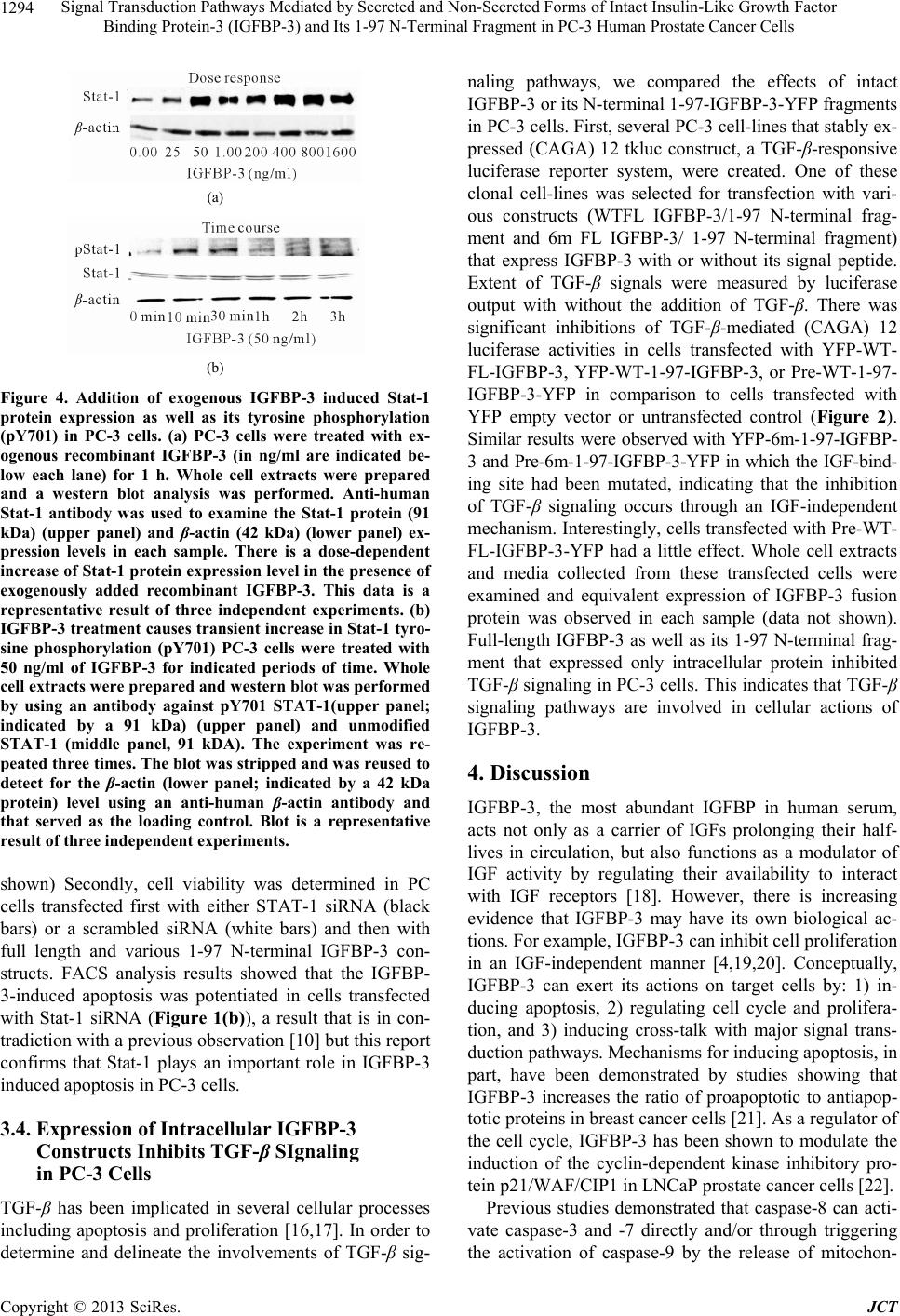
Signal Transduction Pathways Mediated by Secreted and Non-Secreted Forms of Intact Insulin-Like Growth Factor
Binding Protein-3 (IGFBP-3) and Its 1-97 N-Terminal Fragment in PC-3 Human Prostate Cancer Cells
1296
Chemistry, Vol. 281, No. 34, 2006, pp. 24588-24601.
http://dx.doi.org/10.1074/jbc.M509463200
[6] R. Rajah, B. Valentinis and P. Cohen, “Insulin-Like Growth
Factor (IGF)-Binding Protein-3 Induces Apoptosis and
Mediates the Effects of Transforming Growth Factor-Be-
ta1 on Programmed Cell Death through a p53- and IGF-
Independent Mechanism,” Journal of Biological Chemis-
try, Vol. 272, No. 18, 1997, pp. 12181-12188.
http://dx.doi.org/10.1074/jbc.272.18.12181
[7] A. J. Butt, K. A. Fraley, S. M. Firth and R. C. Baxter,
“IGF-Binding Protein-3-Induced Growth Inhibition and
Apoptosis Do Not Require Cell Surface Binding and Nu-
clear Translocation IN Human Breast Cancer Cells,” Endo-
crinology, Vol. 143, No. 7, 2002, pp. 2693-2699.
http://dx.doi.org/10.1210/en.143.7.2693
[8] K.W.Lee, L. Ma, X. Yan, et al., “Rapid Apoptosis Induc-
tion by IGFBP-3 Involves an Insulin-Like Growth Factor-
Independent Nucleomitochondrial Translocation of RXR-
alpha/Nur77,” Journal of Biological Chemistry, Vol. 280,
No. 17, 2005, pp. 16942-16948.
http://dx.doi.org/10.1074/jbc.M412757200
[9] L. J. Schedlich, T. F. Young, S. M. Firth and R. C. Baxter,
“Insulin-Like Growth Factor-Binding Protein (IGFBP)-3
and IGFBP-5 Share a Common Nuclear Transport Path-
way in T47D Human Breast Carcinoma Cells,” Journal of
Biological Chemistry, Vol. 273, No. 29, 1998, pp. 18347-
18352. http://dx.doi.org/10.1074/jbc.273.29.18347
[10] A. Spagnoli, M. Torello, S. R. Nagalla, et al., “Identifica-
tion of STAT-1 as a Molecular Target of IGFBP-3 in the
Process of Chondrogenesis,” Journal of Biological Chemis-
try, Vol. 277, No. 21, 2002, pp. 18860-18867.
http://dx.doi.org/10.1074/jbc.M200218200
[11] G. Zappalà and M. M. Rechler, “IGFBP-3, Hypoxia and
TNF-Alpha Inhibit Adiponectin Transcription,” Bioche-
mical and Biophysical Research Communications, Vol.
382, No. 4, 2009, pp. 785-789.
http://dx.doi.org/10.1016/j.bbrc.2009.03.112
[12] S. M. Leal, S. S. Huang and J. S. Huang, “Interactions of
High Affinity Insulin-Like Growth Factor-Binding Pro-
teins with the Type V Transforming Growth Factor-Be-
tareceptor in Mink Lung Epithelial Cells,” Journal of
Biological Chemistry, Vol. 274, No. 10, 1999, pp. 6711-
6717. http://dx.doi.org/10.1074/jbc.274.10.6711
[13] L. C. Giudice, E. M. Farrell, H. Pham, et al., “Insulin-
Like Growth Factor Binding Proteins in Maternal Serum
throughout Gestation and in the Puerperium: Effects of a
Pregnancy-Associated Serum Protease Activity,” The
Journal of Clinical Endocrinology & Metabolism, Vol. 71,
No. 4, 1990, pp. 806-816.
http://dx.doi.org/10.1210/jcem-71-4-806
[14] P. Angelloz-Nicoud, C. Lalou and M. Binoux, “Prostate
Carcinoma (PC-3) Cell Proliferation Is Stimulated by the
22 - 25-kDa Proteolytic Fragment (1-160) Andinhibited by
the 16-kDa Fragment (1 - 95) of Recombinant Human Insu-
lin-Like Growth Factor Binding Protein-3,” Growth Hor-
mone & IGF Research, Vol. 8, No. 1, 1998, pp. 71-75.
[15] H. Shahjee, N. Bhattacharyya and G. Zappala, “An N-Ter-
minal Fragment of Insulin-Like Growth Factor Binding
Protein-3 (IGFBP-3) Induces Apoptosis in Human Pros-
tate Cancer Cells in an IGF-Independent Manner,” Growth
Hormone & IGF Research, Vol. 18, No. 3, 2008, pp. 188-
197.
[16] Y. Oh, H. L. Muller, L. Ng and R. G. Rosenfeld, “Trans-
forming Growth Factor-Beta-Induced Cell Growth Inhi-
bition in Human Breast Cancer Cells Is Mediated through
Insulin-Like Growth Factor-Binding Protein-3 Action,”
Journal of Biological Chemistry, Vol. 270, No. 23, 1995,
pp. 13589-13592.
http://dx.doi.org/10.1074/jbc.270.23.13589
[17] Z. S. Gucev, Y. Oh, K. M. Kelley and R. G. Rosenfeld,
“Insulin-Like Growth Factor Binding Protein 3 Mediates
Retinoic Acid- and Transforming Growth Factor Beta 2-
Induced Growth Inhibition in Human Breast Cancer
Cells,” Cancer Research, Vol. 56, 1996, pp. 1545-1550.
[18] G. R. Devi, D. L. Graham, Y. Oh and R. G. Rosenfeld,
“Effect of IGFBP-3 on IGF and IGF-Analogue-Induced
Insulin-Like Growth Factor-I Receptor (IGFIR) Signal-
ling,” Growth Hormone & IGF Research, Vol. 11, No. 4,
2001, pp. 231-239.
http://dx.doi.org/10.1054/ghir.2001.0231
[19] C. Lalou, C. Lassarre and M. Binoux, “A Proteolytic
Fragment of Insulin-Like Growth Factor (IGF) Binding
Protein-3 That Fails to Bind IGFs Inhibits the Mitogenic
Effects of IGF-I and Insulin,” Endocrinology, Vol. 137,
No. 8, 1996, pp. 3206-3212.
http://dx.doi.org/10.1210/en.137.8.3206
[20] K. W. Colston, C. M. Perks, S. P. Xie and J. M. Holly,
“Growth Inhibition of Both MCF-7 and Hs578T Human
Breast Cancer Cell Lines by Vitamin D Analogues Is As-
sociated with Increased Expression of Insulin-Like Growth
Factor Binding Protein-3,” Journal of Molecular Endo-
crinology, Vol. 20, 1998, pp. 157-162.
[21] A. J. Butt, S. M. Firth, M. A. King and R. C. Baxter, “In-
sulin-Like Growth Factor-Binding Protein-3 Modulates
Expression of Bax and Bcl-2 and Potentiates p53-Inde-
pendent Radiation-Induced Apoptosis in Human Breast
Cancer Cells,” Journal of Biological Chemistry, Vol. 275,
No. 50, 2000, pp. 39174-39181.
http://dx.doi.org/10.1074/jbc.M908888199
[22] B. J. Boyle, X. Y. Zhao, P. Cohen and D. Feldman, “Insu-
lin-Like Growth Factor Binding Protein-3 Mediates 1 Al-
pha, 25-Dihydroxyvitamin d(3) Growth Inhibition in the
LNCaP Prostate Cancer Cell Line through p21/ WAF1,”
Journal of Urology, Vol. 165, No. 4, 2001, pp. 1319-1324.
http://dx.doi.org/10.1016/S0022-5347(01)69892-6
[23] D. R. Green, “Apoptotic Pathways: The Roads to Ruin,”
Cell, Vol. 94, No. 6, 1998, pp. 695-698.
http://dx.doi.org/10.1016/S0092-8674(00)81728-6
[24] S. Fanayan, S. M. Firth, A. J. Butt and R. C. Baxter,
“Growth Inhibition by Insulin-Like Growth Factor-Bind-
ing Protein-3 in T47d Breast Cancer Cells Requires Tran-
sforming Growth Factor-Beta (TGF- Beta) and the Type
II TGF-Beta Receptor,” Journal of Biological Chemistry,
Vol. 275, No. 50, 2000, pp. 39146-39151.
http://dx.doi.org/10.1074/jbc.M006964200
[25] S. Fanayan, S. M. Firth and R. C. Baxter, “Signaling
Copyright © 2013 SciRes. JCT