Pharmacology & Pharmacy, 2013, 4, 496-501
http://dx.doi.org/10.4236/pp.2013.46072 Published Online September 2013 (http://www.scirp.org/journal/pp)
The Pharmacodynamic Study of Qin Bing Eye Drop on
Photokeratoconjunctivitis*
Ruifang Xie1, Qiuhua Zhao2, Zhicheng Li3, Xin Zhou1#
1Department of Pharmacy, Longhua Hospital, Shanghai, China; 2National Medical Department, Youjiang Medical University for
Nationalities, Baishe, China; 3Department of Surgery, Pudong Hospital, Shanghai, China.
Email: #2479707904@qq.com
Received May 29th, 2013; revised July 12th, 2013; accepted July 26th, 2013
Copyright © 2013 Ruifang Xie et al. This is an open access article distributed under the Creative Commons Attribution License,
which permits unrestricted use, distribution, and reproduction in any medium, provided the original work is properly cited.
ABSTRACT
Background: Qin Bing eye drop is prepared with cortex fraxini and borneol. It has been commonly used to relieve the
symptoms of photokeratoconjunctivitis (PKC), chronic conjunctivitis and keratitis for more than forty years. However,
the origins of cortex fraxini are various, which may result in the unstable quality. Methods: The animal model of PKC
was established using rabbit’s eyes exposure to Ultraviolet (UV); levofloxaxin drop, normal saline, Qin Bing eye drops
made of herbs from Hebei province and Shanxi province were respectively instilled to both eyes of rabbits; the symp-
toms of rabbits’ eyes were observed at different time points and scores based on weights were recorded; the recovery
rates of different groups were evaluated. Results: The scores of both eyes for all rabbits were above 9 points, indicating
animal models for PKC were successful; the average recovery rates of therapeutic groups, including Qin Bing eye drop
groups, were higher than untreated group at different time points; the slopes of recovery rate in Qin Bing eye drop (He-
bei) were above Shanxi. Conclusion: PKD animal model can be established successfully; Qin Bing eye drop groups
promoted the symptoms of PKD recovering quicker than untreated group. Furthermore, Qin Bing eye drop made of
herbs from province Hebei had better effects than Shanxi, which was consistent with chemical results. This indicated
that chemical constituents of herbs were able to reflect the effects to a certain extent.
Keywords: Qin Bing Eye Drop; Photokeratoconjunctivitis (PKC); Cortex Fraxini
1. Introduction
Cortex fraxini is the dried bark of fraxinus rhynchophylla
Hance, fraxinus chinensis Roxb., fraxinus chinensis Roxb.
var. acuminate Lingelsh. and fraxinus stylosa Lingelsh.,
and called “Qin pi” in Chinese. It is a commonly used
Chinese herb belonging to the “heat-clearing” category
according to the classification of traditional Chinese
medicine (TCM) and has the efficacy of clearing heat
and eliminating dampness, improving acuity of vision,
etc [1]. Pharmacological researches demonstrated that it
possessed anticancer, anticoagulant, antioxidant, antial-
lergic, antibacterial, diuretic and central nervous system
protecting activities [2]. The origins of cortex fraxini are
various, including province Shanxi, Sichuan, Hebei,
Liaoning, et al. [3], which may lead to the unstable qual-
ity.
Qin Bing eye drop is prepared with cortex fraxini and
borneol. This eye drop can get rid of heat and dampness,
improve eyesight, relieve pain. It has been commonly
used to relieve the symptoms of photokeratoconjunctivi-
tis (PKC), chronic conjunctivitis and keratitis in our hos-
pital for more than forty years and exhibits good thera-
peutic function [4].
In our previous paper, HPLC chromatographic finger-
prints of cortex fraxini were established and estimated by
synthetic weighing method. Herbs from different origins
were compared. The results showed the synthetic scores
of province Heibei were higher than Shanxi [5]. Never-
theless, whether chemical fingerprints reflect effects need
to be further confirmed.
In current paper, the animal model of PKC was es-
tablished by making rabbit’s eyes exposure to Ultra-
violet (UV). The effects of Qin Bing eye drops made
of herbs from province Hebei and Shanxi were com-
pared. The relationship between origin and effects was
discussed.
*Declaration of Interest: The authors declare no conflict of interest and
are solely responsible for the content of this paper.
#Corresponding author.
Copyright © 2013 SciRes. PP
The Pharmacodynamic Study of Qin Bing Eye Drop on Photokeratoconjunctivitis 497
2. Materials and Methods
2.1. Materials and Reagents
The cortex fraxini of different origins and batches were
purchased from Wan Shi Cheng Limited Company
(Shanghai, China). Levofloxacin eye drop (Lot 00706213)
and normal saline (Lot 10082406) were respectively ob-
tained from Zhuhai United Pharmaceutical Limited Com-
pany (Zhongshan, China) and Shanghai Changzheng
Fumin Jinshan Pharmaceutical Limited Company (Shang-
hai, China). Pentobarbital (Lot 20100312, Fruck, Ger-
many) were also used in this experiment.
Four batches of Qin Bing eye drops (Lot 100713,
100715, 100811 and 100819) were prepared at Long hua
hospital (Shanghai, China). According to the content of
main ingredients and synthetic scores of HPLC finger-
prints [5] two batches respectively from province Heibei
(Lot100715) and Shanxi (Lot 100819) were selected for
animal experiments (Table 1).
2.2. Animals
16 of New Zealand White Rabbits (1.5 - 2.5 kg), male
and female half, were provided from the Yingen Rabbit
Room [Shanghai, China, Licensed No. SCXK (hu) 2005-
0003] and were housed in Animal Laboratory of Fu Dan
University [Shanghai, China, Licensed No. SYXK (hu)
2009-0082]. The room temperature maintained at 20˚C
- 25˚C under a 12-h light-12-h dark cycle (07:00 -
19:00) and humidity were between 40% and 70%. All
rabbits were raised in stainless steel cages with 2 rab-
bits per cage. Food and water were given ad libitum.
The raising conditions were in accordance with the
environmental and facilities standards of experimental
animals for Health Department of People’s Republic of
China.
2.3. Introduction of PKC Model
Solution of pentobarbital (30 mg/ml dissolved in normal
saline) was administrated intravenously at 30 mg/kg in
order to anaesthetize a rabbit and keep its eyes open.
Next, one side of this rabbit head was placed in the light
box of SJ-1031A UV Detector (Hongo Corporation, To-
kyo) and the eye was exposed to ultraviolet rays for 30
min, twenty centimeters away from light emission source
at 90 degrees (Figure 1), then switched to the other side.
Both eyes of each rabbit were radiated twice daily. The
incubation period is usually two to ten hours after expo-
sure. Next morning both eyes of rabbits were monitored
at nine o’clock by two independent observers. Typical
extra-ocular symptoms of rabbits were evaluated: pain,
photophobia tears, discharge, conjunctiva hyperemia, and
so on. The cornea was observed using YZ5F1 slit lamp
microscope (ShuZhou LiuLiu Vision technological Ltd.
Table 1. The contents and synthetic scores of HPLC finger-
prints in Qin Bing eye drops.
Batch Origin Esculin
(μg/mg)
Esculetin
(μg/mg) Synthetic scores
100713 Hebei 2.009 0.105 49.13
100715 Hebei 2.022 0.113 98.33
100811 Shanxi0.586 0.111 61.16
100819 Shanxi0.591 0.113 69.18
Figure 1. The electric opthalmia rabbit model by ultraviolet
irradiation.
Company) with sodium fluorescein test paper (Lot
20101706, Tianjin JinMing New Technology Limited
Company). Each symptom was scored as follows: 0, no
change; 1, slight; 2, moderate; 3, severe by a well-trained
observer to minimize the variation in score. These data
were recorded as vehicle.
2.4. Administration and Assessment of
Symptoms
The rabbits were randomly divided into four group (n =
4): one group receiving levofloxacin eye drop as positive
control, one untreated group as negative control group,
and two treatment groups receiving Qin Bing eye drops
(origins of cortex fraxini were respectively from Heibei
and Shanxi in China). Two drops of drug solution were
instilled into the right eye three times daily while the
same drops of normal saline into the left eye as control
until symptoms recovered completely. After first admini-
stration, symptoms of rabbits’ eyes were respectively
observed based on weight (Tab le 2) at 3 h, 6 h, 24 h, 27
h, 30 h, 48 h, 51 h, 54 h, 72 h, 75 h and 78 h.
2.5. Statistical Analysis
The scores of each group were represented as mean ± SD
(Standard Deviation); Recovery rate was calculated by
he following equation: t
Copyright © 2013 SciRes. PP
The Pharmacodynamic Study of Qin Bing Eye Drop on Photokeratoconjunctivitis
Copyright © 2013 SciRes. PP
498
Scores after exposure to UVScores after administration
Recovery rate100%
Scores after exposure to UV

Linear regression was performed between recovery
rate and time, and there was good linear relationship
when the correlation coefficient was above 0.9. Data
from therapeutic group and control group were evaluated
by Student-test, P < 0.05 was considered statistically
significant. Above analysis was carried out using Excel
and SPSS 17.0 software.
3. Results
3.1. Evaluation of Animal Model
After exposure to UV for two to ten hours, eyes of rab-
bits exhibited the symptoms of photophobia tears, secre-
tions and so on (Figure 2(a)). The scores of both eyes for
all rabbits were above 9 points (Figure 3), indicating
animal models for PKC were successful. There were no
significant differences between right and left eye of rab-
bits (P < 0.05), showing these animals were appropriate
for following experiment.
3.2. Improvement of PKC Symptoms
After instillation of eye drops, the symptoms such as
photophobia tears and discharge were improved (Figures
2(b) and (c)). Thirty hours after administration, the re-
covery rate of each group could reach 59% except un-
treated group (Table 3). The slopes of each group before
and after 30 h showed that the symptoms recover quicker
in the early stage than later period (Table 4). Further-
more, a good linear regression was observed between time
and recovery rate for each group with correlation coeffi-
cient above 0.90 (Table 4), which implied that the longer
period passed the better the rabbits recovered (Figure 4).
After three days, the recovery rates of groups including
Table 2. Criteria for scoring ocular symptoms.
Ocular Parameters Scores
0 1 2 3
Pain Absent Mild Moderate Severe
Photophobia tears Absent Mild Moderate Severe
Symptoms of conjunctiva Absent Mild Moderate Severe
Discharge Absent Slight Moderate Profuse
Corneal fluorescence staining Negative <1/3 1/3 - 2/3 >2/3
Symptoms were scored by two independent observers using the scoring system described in Supplementary. The five parameters were assessed. Each parameter
was scored 0 (no disease) to 3 (maximum disease), and the sum of all five parameters represented the degree of symptoms after UV radiation and treatment.
Thus, a normal eye would have a score of 0 and an eye with most severe disease would have a score of 15; Pain was assessed by mild pain or sense of foreign
objects in eye (1), moderate pain (2), to severe pain of both eyes; Photophobia was graded as follows: 1) tears pooling at lower lid margin; 2) tears flowing over
lower lid and staining eyelid; 3) tears staining upper and lower lids with extension below the eye socker (pesudomembranes); Symptoms of conjunctiva was
assigned as follows: 0, conjunctiva without congestion; 1, conjunctiva mild congestion; 2, eyelid and facial skin flushing, conjunctiva congestion; 3, ble-
phaospasm, blurring of vision and rainbow, significant conjunctiva congestion and edema, contracted pupil and slow reaction to light; Discharge was graded as
follows: 1) discharged located to medial canthus; 2) discharge collecting along the lid margin; 3) profuse discharge matting fur around the eyes (and pseu-
domembranes); Corneal was observed using slit lamp microscope with sodium fluorescein test paper. The area of fluorescence staining was graded as 1, <1/3
whereas 3 was >1/3.
(a) (b) (c)
Figure 2. The alteration of symptoms for rabbit eyes: after UV radiation (a), partly relief after administration (b) and recov-
ery completely after administration (c).
The Pharmacodynamic Study of Qin Bing Eye Drop on Photokeratoconjunctivitis 499
Figure 3. Scores of both eyes for animal models before administration (n = 4).
Table 3. Recovery rate of every group at different time points.
Time (h) Levofloxacin
group (%) (n = 4)
Qingbin eye drop
(Hebei, Lot. 100715)
group (%) (n = 4)
Qingbin eye drop
(Shanxi, Lot 100819)
group (%) (n = 4)
Normal saline#
(%) (n = 16)
Untreated group
(%) (n = 4)
0 14.77 ± 7.01 16.74 ± 11.11 9.03 ± 6.94 11.35 ± 10.32 12.50 ± 10.75
3 30.78 ± 5.70 24.10 ± 12.43 22.22 ± 3.93 24.05 ± 10.72 16.67 ± 13.61
6 33.90 ± 11.29 40.07 ± 15.9 32.29 ± 5.24 32.90 ± 12.35 25.00 ± 11.79
24 51.33 ± 9.10 61.18 ± 4.65** 45.14 ± 13.68 49.13 ± 12.72 41.67 ± 6.80
27 56.53 ± 7.56 69.58 ± 3.94* 54.17 ± 15.96 57.66 ± 12.23 52.08 ± 10.49
30 64.87 ± 16.10 69.58 ± 3.94* 59.38 ± 11.97 60.96 ± 12.58 54.17 ± 10.76
48 65.06 ± 11.42 77.29 ± 8.75 79.86 ± 4.17* 74.04 ± 9.74 58.33 ± 16.67
51 79.17 ± 14.43 87.36 ± 10.40 88.89 ± 10.39 83.47 ± 10.96 68.75 ± 15.77
54 87.50 ± 10.21 92.36 ± 10.49 88.89 ± 10.39 87.24 ± 10.43 70.83 ± 15.96
72 93.75 ± 12.50 90.28 ± 11.45 96.88 ± 6.25* 91.75 ± 11.02 72.92 ± 18.48
75 97.92 ± 4.17 95.14 ± 5.73 96.88 ± 6.25 94.53 ± 6.70 79.17 ± 15.96
78 97.92 ± 4.17 97.22 ± 5.56 100.00 ± 0.00* 95.83 ± 6.57 79.17 ± 15.96
;XSD **Compared with untreated group P < 0.01; *Compared with untreated group P < 0.05; #The data of Normal saline group were from the data of left
eyes of all the rabbits, which were instilled by normal saline.
Table 4. Regression analysis between recovery rate and time.
Before 30 h After 30 h
Group Regression Correlation coefficient Regression Correlation coefficient
Levofloxacin group y = 0.0986x + 0.0751 0.9851 y = 0.064x + 0.642 0.9423
Qingbin eye drop (Hebei) y = 0.1205x + 0.0469 0.9842 y = 0.034x + 0.778 0.9088
Qingbin eye drop (Shanxi) y = 0.1030x + 0.01 0.9961 y = 0.037x + 0.786 0.9503
Normal saline y = 0.1043x + 0.0283 0.9931 y = 0.041x + 0.731 0.9592
Untreated group y = 0.0946 + 0.0056 0.9790 y = 0.039x + 0.577 0.9471
untreated group were above 80% (Figure 4). This indi-
cated the symptoms of PKC could be self-healing.
3.3. Effects Comparison of Different Groups
For the tendency of recovery, the average recovery rates
of therapeutic groups at different time points were higher
than untreated group, which indicated that instillation of
eye drops including normal saline promoted the symp-
toms of PKC (Table 3). Especially for Qing Bin eye drop
groups, there were statistical differences between Qing
Bin eye drop (Hebei) group and un-treatment group at
Copyright © 2013 SciRes. PP
The Pharmacodynamic Study of Qin Bing Eye Drop on Photokeratoconjunctivitis
500
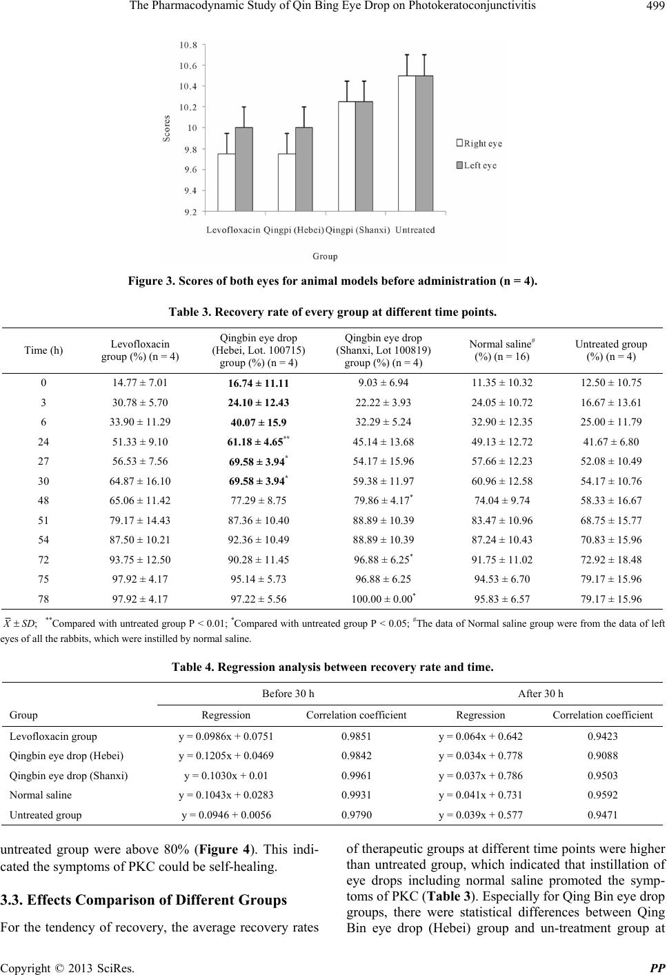
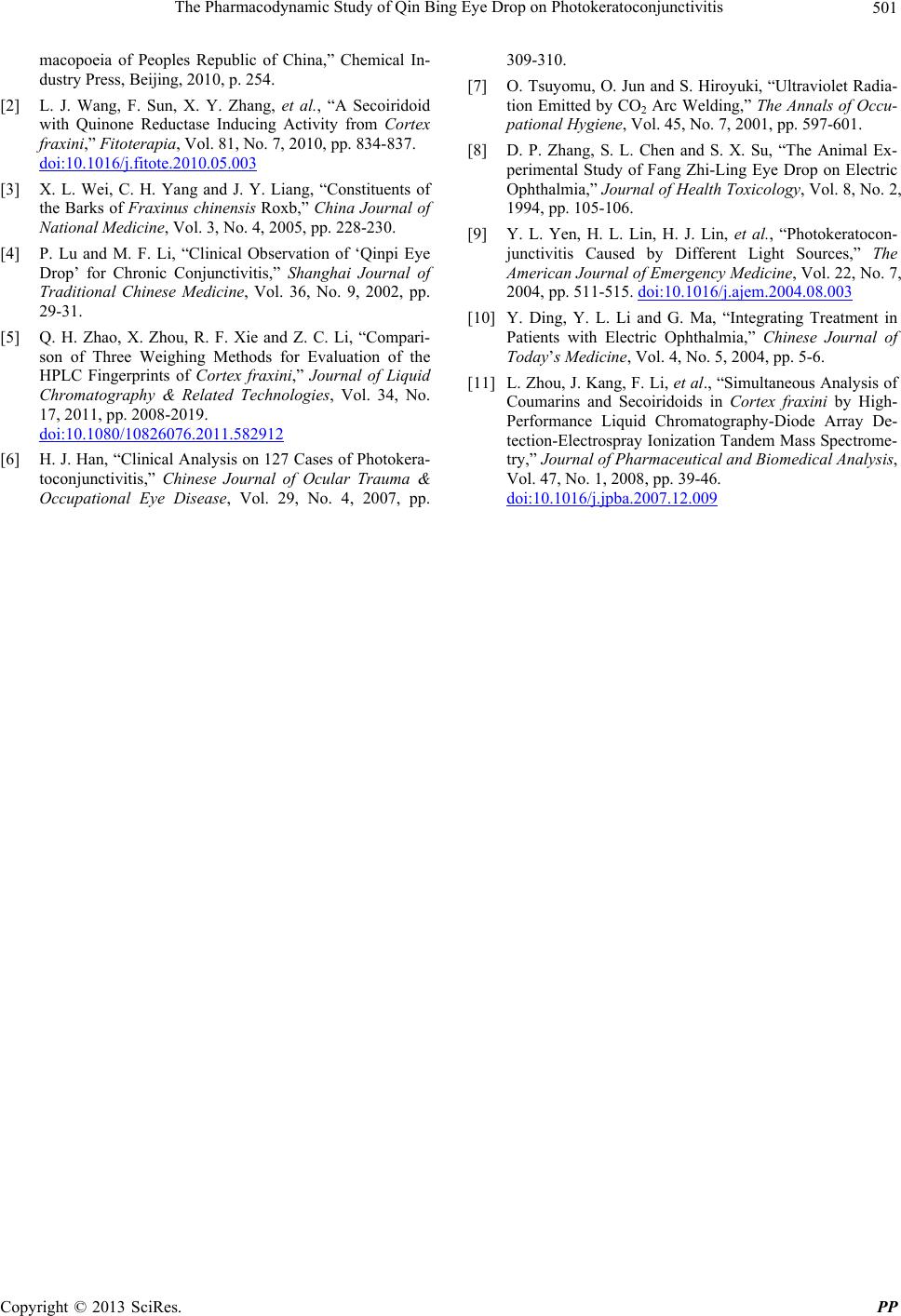
Figure 4. Effects of different drugs on recovery rates (n =
4).
24 h, 27 h and 30 h as well as Qing Bin eye drop (Shanxi)
group and un-treatment group at 48 h, 72 h and 78 h (Ta-
ble 3). This demonstrated Qing Bin eye drops could be
effective for PKC. The recovery rates of Qing Bin eye
drop (Hebei) in 30 h were above Qing Bin eye drop
(Shanxi).
For the speed of recovery (Table 4), the slopes of
therapeutic groups were more than untreated group in 30
h, which implied all the eye drops including normal sa-
line could relieve the symptoms of PKC quicker than
untreated group, especially in the early stage. Among all
the eye drop groups, the slopes of Qing Bin eye drop
groups in 30 hours, including Hebei and Shanxi, were
higher than levofloxacin group, which suggested Qing
Bin eye drops could be better than control groups. Fur-
thermore, Qing Bin eye drop group (Hebei) was the
highest (0.1205). This indicated that Qingbin eye drop
(Hebei) could be superior to Qing Bin eye drop (Shanxi).
4. Discussions
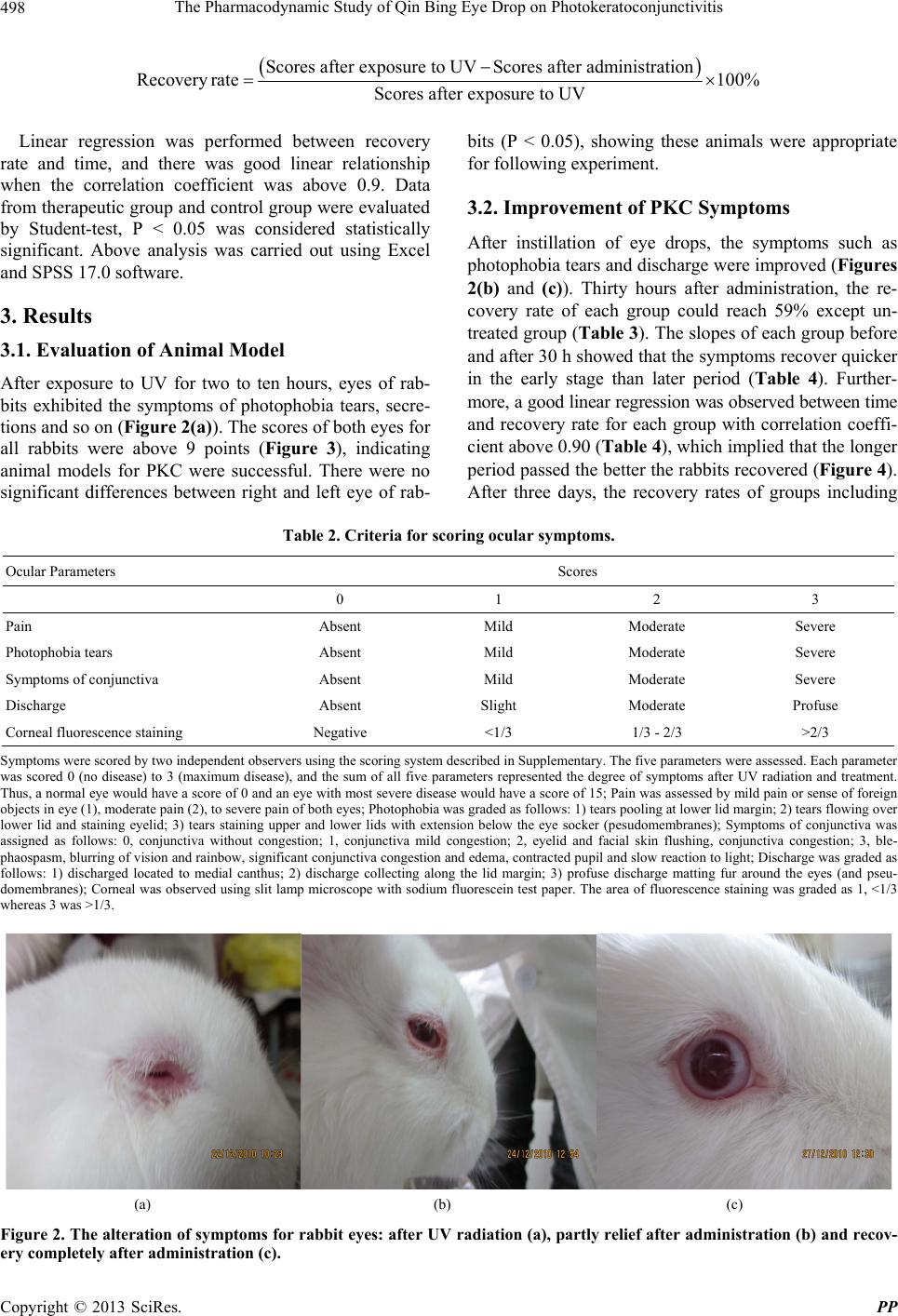
Photokeratoconjunctivitis (PKD) is caused by excessive
exposure to UV radiation. Excessive UV hydrolyzes the
water molecular in superficial layers of the eye to pro-
duce a large number of oxygen free radicals, which at-
tack cells through various pathways and make cells ne-
crosis and dropping off [6]. As a result, lots of inflam-
matory stimulation factors are released to induce symp-
toms such as ocular pain, tearing, a sense of sand in the
eye and photophbia [7].
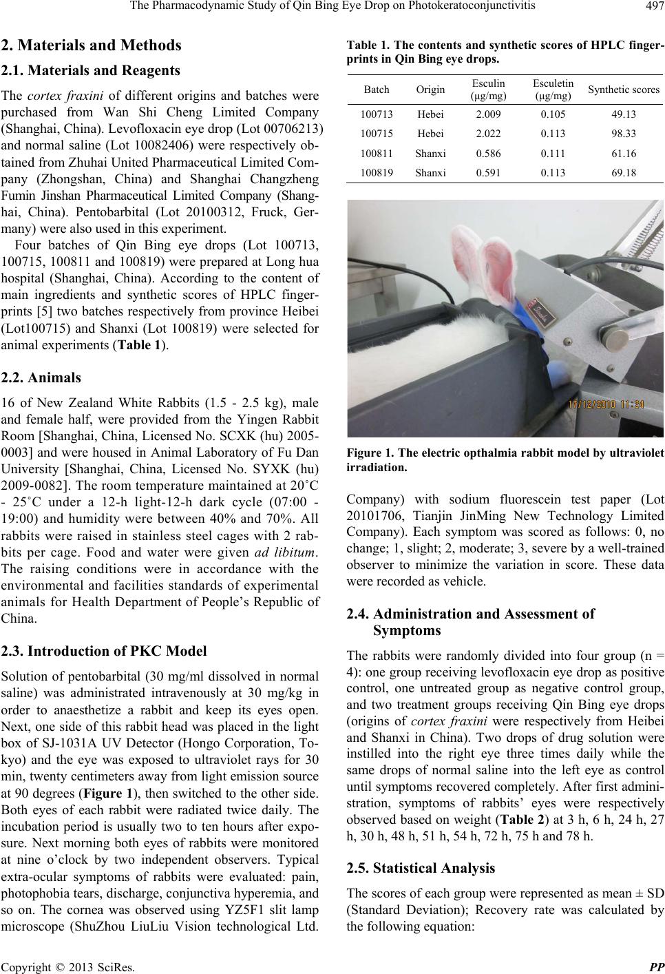
Few studies on the animal’s model of electric oph-
thalmia have been reported. In this paper, animal model
was established according to above mechanism of PKD
[8]. As we known, rabbits closed eyes protectively when
exposure to UV. However, they naturally opened eyes
under the statue of anaesthetization. So in present ex-
periment, intravenously anaesthetization was performed
to keep animals cooperative. The injury degree depended
on the intensity, distance and period of UV irradiation [9].
If the period was too long and UV intensity was too large,
eyes of rabbits would be irreversibly damaged and in that
case the effects of eye drops would be inconvenient to be
observed. In contrast, if the degree of electric ophthamia
was too mild, the rabbits would recover quickly and it
was not suitable for observation. After several trials, the
final parameters were optimized: the rabbits’ eyes were
twice exposure to both 254 nm and 365 nm of UV de-
tector for 30 min, twenty centimeters away from light
emission source at 90 degrees. After 2 - 8 hours, the
symptoms of PKD were apparent and the scores were
above 9, which demonstrated the models were success-
ful.
In most clinical cases of PKD, if no inflammation,
symptoms will be self-relief in 6 - 8 hours and gone
within a week or so. This situation explains that the PKD
symptoms of animal model are self-curative. Clinically,
symptomatic treatments are commonly adopted: in early
stage anaesthetic eye drops are instilled to relive pain and
antibiotic eye drops or eye ointments are applied for
prophylaxis of inflammation [10]. According to this rou-
tine therapeutic method, our experiment chose levoflox-
acin eye drop and normal saline as control medicine.
The results showed Qing Bin eye drop groups could
promote the symptoms of PKD recovering quicker than
untreated group. Furthermore, Qing Bin eye drop (Hebei)
had better effects than Shanxi. According to our previous
results (Table 1), the amount of esculin in Qing Bin eye
drop (Hebei, lot. 100715) is more than that of Shanxi (lot.
100819) as well as the synthetic scores. Coumarins are
proven to be the active constituents, which were reported
to have activities such as anti-inflammation, antivirus,
antiarthritis and anticancer [11]. Our results of effects
were consistent with chemical results. This indicated that
chemical constituents of herbs could reflect the effects in
part.
To sum up, in this paper, the animal model of PKC
was successfully established using rabbits’ eyes exposure
to Ultraviolet (UV). The Qin Bing eye drops made of
herbs from Hebei and Shanxi were compared. Results
showed that the effects of Qin Bing Eye drop (Hebei)
were better than Shanxi, which was in consistence with
the chemical results. This demonstrated that main con-
stituents in cortex fraxini as marks of pharmacological
effects were reasonable.
5. Acknowledgements
This research was supported by Longhua Medcial Project
(LYTD-14) and Chinese Medicine Shanghai Health Bu-
reau scientific research fund (No. 2008X002A).
REFERENCES
[1] National Commission of Chinese Pharmacopoeia, “Phar-
Copyright © 2013 SciRes. PP
The Pharmacodynamic Study of Qin Bing Eye Drop on Photokeratoconjunctivitis
Copyright © 2013 SciRes. PP
501
macopoeia of Peoples Republic of China,” Chemical In-
dustry Press, Beijing, 2010, p. 254.
[2] L. J. Wang, F. Sun, X. Y. Zhang, et al., “A Secoiridoid
with Quinone Reductase Inducing Activity from Cortex
fraxini,” Fitoterapia, Vol. 81, No. 7, 2010, pp. 834-837.
doi:10.1016/j.fitote.2010.05.003
[3] X. L. Wei, C. H. Yang and J. Y. Liang, “Constituents of
the Barks of Fraxinus chinensis Roxb,” China Journal of
National Medicine, Vol. 3, No. 4, 2005, pp. 228-230.
[4] P. Lu and M. F. Li, “Clinical Observation of ‘Qinpi Eye
Drop’ for Chronic Conjunctivitis,” Shanghai Journal of
Traditional Chinese Medicine, Vol. 36, No. 9, 2002, pp.
29-31.
[5] Q. H. Zhao, X. Zhou, R. F. Xie and Z. C. Li, “Compari-
son of Three Weighing Methods for Evaluation of the
HPLC Fingerprints of Cortex fraxini,” Journal of Liquid
Chromatography & Related Technologies, Vol. 34, No.
17, 2011, pp. 2008-2019.
doi:10.1080/10826076.2011.582912
[6] H. J. Han, “Clinical Analysis on 127 Cases of Photokera-
toconjunctivitis,” Chinese Journal of Ocular Trauma &
Occupational Eye Disease, Vol. 29, No. 4, 2007, pp.
309-310.
[7] O. Tsuyomu, O. Jun and S. Hiroyuki, “Ultraviolet Radia-
tion Emitted by CO2 Arc Welding,” The Annals of Occu-
pational Hygiene, Vol. 45, No. 7, 2001, pp. 597-601.
[8] D. P. Zhang, S. L. Chen and S. X. Su, “The Animal Ex-
perimental Study of Fang Zhi-Ling Eye Drop on Electric
Ophthalmia,” Journal of Health Toxicology, Vol. 8, No. 2,
1994, pp. 105-106.
[9] Y. L. Yen, H. L. Lin, H. J. Lin, et al., “Photokeratocon-
junctivitis Caused by Different Light Sources,” The
American Journal of Emergency Medicine, Vol. 22, No. 7,
2004, pp. 511-515. doi:10.1016/j.ajem.2004.08.003
[10] Y. Ding, Y. L. Li and G. Ma, “Integrating Treatment in
Patients with Electric Ophthalmia,” Chinese Journal of
Todays Medicine, Vol. 4, No. 5, 2004, pp. 5-6.
[11] L. Zhou, J. Kang, F. Li, et al., “Simultaneous Analysis of
Coumarins and Secoiridoids in Cortex fraxini by High-
Performance Liquid Chromatography-Diode Array De-
tection-Electrospray Ionization Tandem Mass Spectrome-
try,” Journal of Pharmaceutical and Biomedical Analysis,
Vol. 47, No. 1, 2008, pp. 39-46.
doi:10.1016/j.jpba.2007.12.009