V. VERMA ET AL.
Copyright © 2013 SciRes. SS
409
teoarthritis. In displaced fractures of the talar body, the
risk of avascular necrosis has been reported to be about
27% [10]. The published results of fractures after fixation
reveal high incidence of both early and late complica-
tions, with osteonecrosis occuring in ten out of 26 cases
and full talar body collapse in five out of those ten cases
[11].
A major difficulty in management of these patients
remains the diagnosis. Forty-seven percent of patients
with chronic instability of ankle show evidence of previ-
ous talar osteochondral fracture on magnetic resonance
imaging, which had not been previously diagnosed [12].
Therefore it is likely that a significant number of talar
injuries may go undiagnosed. The clinical signs are vari-
able but can include localised tenderness at the site of
fracture, which can be associated with a decreased range
of motion, and in more severe cases effusion and crepitus
[2]. However it is important to note that there may be
little or even no signs. If there is any dou bt at the time of
presentation as to wh ether there is a talar fracture further
imaging is recommended. In case of late recognition of
talus fractures, catastrophic results may occur in the
hind-foot [13,14]. Hence it is prime that these fractures
be diagnosed well in time to avert such complications.
Following such intra-articular injuries of ankle joint,
good anatomical reduction with osteosynthesis when
appropriate are mandatory even in an immature skeleton,
in order to achieve a good result. Preservation of blood
supply is paramount. Removal of metal work is recom-
mended after there is evidence of healing [1]. Patient
should be mobilised early without weight bearing and
should remain non-weight bearing until there is evidence
of sound radiological union. It is now worthwhile to
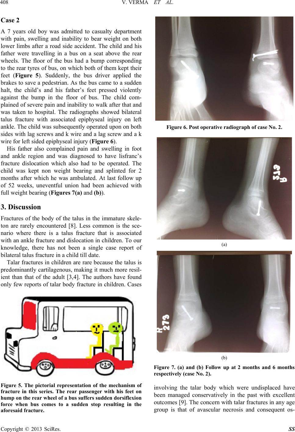
suggest that looking at the mode of injury in this series
our public transport buses should be modified accord-
ingly or manufacturers should refrain from keeping the
seat near rear wheels just after the bump. It is suggested
that it should either be just below the seat or the area
should be free of seating to avoid similar injuries.
4. Conclusion
Talar body injuries are uncommon, particularly in chil-
dren. There exist significant differences in outcomes and
complications in adult and children. These injuries can be
difficult to be diagnosed and a CT scan or magnetic reso-
nance imaging may be required. If already diagnosed, a
CT scan is advised to clarify the nature of injury. A
minimal or undisplaced fracture of talus is less likely to
undergo avascular necrosis than a displaced fracture but
even with optimal treatment, avascular necrosis may still
occur. Therefore an appropriate length of follow-up is
advisable. The authors advocate use of non-operative
management in cases where the fracture is minimally
displaced or undisplaced and in cases where there is a
displacement, patient should undergo anatomical reduc-
tion and internal fix a tion.
REFERENCES
[1] W. E. Linhart and M. Hollwarth, “Fractures of the Talus
in Children,” Unfallchirurg, Vol. 88, No. 4, 1985, pp.
168-174.
[2] D. B. Thordarson, “Talar Body Fractures,” Foot Ankle
Trauma, Vol. 32, 2001, pp. 65-77.
[3] R. M. Letts and D. Gibeault, “Fractures of the Neck of the
Talus in Children,” Foot Ankle Trauma, Vol. 1, No. 12,
1980, pp. 74-77.
[4] I. Spark, “Fractures of the Talus in Children,” Acta Chi-
rurgica Scandinavica, Vol. 107, No. 6, 1954, pp. 553-
566.
[5] J. T. Smith, T. A. Curtis, S. Spencer, J. R. Kasser, S. T.
Mahan, “Complications of Talus Fractures in Children,”
Journal of Pediatric Orthopaedics, Vol. 30, No. 8, 2010,
pp. 779-784. doi:10.1097/BPO.0b013e3181f73e6e
[6] R. Eberl, G. Singer, J. Schalamon, P. Hausbrandt and M.
E. Hoellwarth, “Fractures of the Talus—Differences be-
tween Children and Adolescents,” Journal of Trauma,
Vol. 68, No. 1, 2010, pp. 126-130.
doi:10.1097/TA.0b013e3181a74667
[7] H. Thermann, T. Hu¨fner, M. Richter, E. Schratt and H.
Tscherne, “Paediatricfoot Fractures,” Journal of Foot and
Ankle Surgery, Vol. 7, No. 2, 2001, pp. 61-76.
doi:10.1046/j.1460-9584.2001.00253.x
[8] O. Sneppen, S. Bach Christensen, O. Krogsoe and J. Lor-
entzen “Fracture of the Body of the Talus,” Acta Ortho-
paedica Scandinavica, Vol. 48, No. 3, 1977, pp. 317-324.
doi:10.3109/17453677708988775
[9] I. Jensen, J. U. Wester and F. Rasmussen, “Prognosis of
Fracture of the Talus in Children,” Acta Orthopaedica
Scandinavica, Vol. 65, No. 4, 1994 pp.398-400.
doi:10.3109/17453679408995478
[10] W. E. Linhart and M. Hollwarth, “Fractures of the Talus
in Children,” Unfallchirurg, Vol. 88, No. 4, 1985, pp.
168-174.
[11] H. A. Vallier, S. E. Nork, S. K. Benirschke and B. J.
Sangeorzan, “Surgical Treatment of talar Body Factures,”
The Journal of Bone & Joint Surgery, Vol. 85-A, Suppl. 1,
2004, pp. 180-192.
[12] I. F. Anderson, K. J. Crichton, T. Grattan-Smith, et al.,
“Osteochondral Fractures of the Dome of the Talus,” The
Journal of Bone & Joint Surgery, Vol. 71-A, No. 8, 1989,
pp. 1143-1152.
[13] N. Nenopoulos, V. A. Papavasiliou and A. V. Papavasi-
liou, “Talus Fracture Associated with a Fracture Disloca-
tion of the Distal Tibia in an Immature Skeleton,” Acta
Orthopædica Belgica, Vol. 69, No. 5, 2003, pp. 473-475.
[14] J. Gehr and W. Friedl, “Fracture of the Neck of the Talus
in a Child,” Unfallchirurg, Vol. 109 No. 10, 2006, pp.
910-913. doi:10.1007/s00113-006-1128-z