 Open Journal of Nephrology, 2013, 3, 152-160 http://dx.doi.org/10.4236/ojneph.2013.33028 Published Online September 2013 (http://www.scirp.org/journal/ojneph) Surface Treated Catheters for Vascular Access—Useful? Rolf Bambauer1*, Ralf Schiel2, Carolin Bambauer3, Reinhard Latza4 1Institute for Blood Purification, Homburg, Germany 2Inselklinik Heringsdorf GmbH, Seeheilbad Heringsdorf, Germany 3Main Hospital Darmstadt, Darmstadt, Germany 4Laboratorium of Medicine, St. Ingbert, Germany Email: *rolf.bambauer@t-online.de Received July 1, 2013; revised July 29, 2013; accepted August 11, 2013 Copyright © 2013 Rolf Bambauer et al. This is an open access article distributed under the Creative Commons Attribution License, which permits unrestricted use, distribution, and reproduction in any medium, provided the original work is properly cited. ABSTRACT Background: Catheter-related infections (CRI), thrombosis, and stenosis are among the most frequent complications associated with catheters which are inserted in vessels as vascular access. These problems are usually related to the handling of the staff, the catheter materials, and the surface properties of the catheter. To mitigate such complications surface treatment process of the outer surface, such as ion beam assisted deposition is investigated in a retrospective study from 1992 to 2007, to prove if the surface treatment of the catheters is a sufficient solution. Methods: This study (1992-2007) evaluated silver coated and non-coated implanted large-bore catheters used for extracorporeal detoxifica- tion. In 159 patients, 54 patients received a silver coated catheter (Spi-Argent, Spire Corporation, Bedford, MA, USA) and 105 patients, an untreated catheter served as controls. The catheters were inserted into the internal jugular or sub- clavian veins. After removal, the catheters were cultured for bacterial colonization using standard microbiologic assays. They were also examined using a scanning electron microscope (SEM). Results: The silver coated catheters showed a tendency towards longer in situ time. The microbiologic examinations of the catheter tips were in both catheter types high positive, but not significant. Conclusion: The silver coated catheters showed no significant reduction in infection rate by evaluation of all collected data in this retrospective study. There was no association between both catheters in significantly reducing patient discomfort. Other surface treatments which include the outer and inner surface are neces- sary. New developed catheter materials such as the microdomain structured inner and outer surface, as an example, are considered more biocompatible because they mimic the structure of natural biological surface. Keywords: Surface Treated Large Bore Catheters; Hemodialysis; Apheresis; Ion Been Assisted Deposition (IBAD); Microdomain-Structered Surface (PUR-SMA Coated Catheter) 1. Introduction Since the first introduction of large-bore catheters for acute hemodialysis in 1961 by Shaldon et al. [1], many problems with handling, materials, and contamination of these catheters have arisen. Nevertheless, vascular cathe- ters have become essential tools for management of hos- pitalized or chronically ill patients requiring intensive medical treatments [2]. The increased insertion of such devices has been accompanied by a corresponding in- crease in complications, such as bloodstream infection and thrombosis [3]. Infections are particular concern, be- cause they can appear at any time, even years after im- plantation, and are not material dependent. They are usu- ally attributed to microbial colonization of the skin or handling of the catheters by attending staff. Complication rates due to venous catheter-relates infection are reported to range from 34 to 40 percent [4,5]. Despite recent tech- nical innovations in hemodialysis, problems related to temporary or permanent vascular access have found no satisfactory solutions. Temporary vascular access is par- ticularly problematic. Catheterization of the femoral vessels produces more complications than the catheterization of the superior vena cava (SVC). Cannulation of the SVC versus the subclavian vein is difficult to implement and involves a high complication rate [6]. Using the infraclavicular catheterization technique is often difficult to push the large-bore catheter under the clavicle. Because of the anatomical position of the subclavian vein, perforation is more likely with a rigid, large-bore catheter, apart from the danger of causing a pneumothorax or a hemathorax [7-10]. *Corresponding author. C opyright © 2013 SciRes. OJNeph  R. BAMBAUER ET AL. 153 Therefore, Bambauer et al. have introduced the inser- tion of large-bore catheters in the superior vena cava rather than in the internal jugular or the subclavian veins [11,12]. Dialysis catheters are used for vascular access in 65% of incident hemodialysis (HD) patients, and in 25% of the prevalent HD populations [13]. Today, the first choice of vascular access is the vena cava superior over the internal jugular vein. Catheter-related bacteremia is a major cause of mor- bidity among hemodialysis patients. Treatment with sys- temic antibiotics alone without removal of the catheter fails to definitely eradicate the infection in most patients [14]. Catheter-related bacteremia must be managed by either catheter removal with delayed placement of a new catheter or management of the infected catheter with a new catheter over a guide-wire and additional systemic antibiotic therapy. These catheter-related complications are contributing factors to increasing cost of medical care. They are responsible for patient readmissions and longer hospital stays as well as patient discomfort, morbidity, and occasional mortality. The source of catheter-related bacteremia is in most patients a bacterial biofilm that forms in the catheter lu- men or on the outer surface. This biofilm, most consist- ing of staphylococcus aureus, cannot be destroyed or eliminated by a systemic antibiotic therapy because of antimicrobial resistance [15]. Bacteriae could most of the time colonize rough surfaces [16]. The combination of rough surfaces and protein deposits should be an ideal situation for the colonization of bacteria. The bacteria could produce and become covered with a slime layer, in which case antibiotic drugs have no influence on the bacteria. The bacteria under the slime layer use the or- ganic substances of the catheter material for their me- tabolism. The toxins of the bacteria can penetrate the slime layer and enter the patient blood provoking a catheter infection [16]. Biofilm is a microbial derived sessile community characterized by cells that are irre- versibly attached to a substratum or interface to each other, embedded in a matrix of extracellular polymeric substances that have produced [17]. Such a biofilm can be the origin of fibrin sheath formations leading to catheter dysfunction due to blood reducing and to blood disturbances. The therapy must be to remove the catheter immediately, or exchange it over a guide-wire with a new catheter and additional systemic antibiotic therapy. Biocompatibility of synthetic materials is another ma- jor problem. The interaction of blood with a synthetic surface causes coagulation and activation of the com- plement system. This can lead to the adsorption of vari- ous proteins and the formation of a layer of protein on the synthetic surface. Thrombocytes, other cells and bac- teria adhere to this layer of protein so that thrombi may form which can lead to blood flow disturbances and catheter dysfunction [18]. To influence catheter-related bacteremia different new developments are available today, such as new catheter materials, coating of the catheter surface with antibiotic- heparin, or silver and silicone, cuffs on the outer surface, catheter for tunnelling, installation of an antibiotic-anti- coagulant lock into the catheter lumen after the HD, etc. [14,19,20]. The first results with available catheters which coated on the outer surface with silver or silicone were encouraged [18,21]. In a retrospective study from 1992 to 2007, outer sur- face treated catheters with silver versus untreated cathe- ter in 159 patients, who needed a large-bore catheter, were investigated. The results of a preliminary study from 2001, which showed 75% decline in the infection rate with the surface treated catheters cannot be confirmed with the present study. One reason may be that on the surface treated catheters only the outer surface was coated with silver and the possibility of contamination by the handling during the extracorporeal treatments. Therefore, new materials and surface treatment tech- nologies are needed to save health care costs for hemodi- alysis catheters, to reduce infection rates and thrombus formations and to help improve the patients’ outcome. The handling of the catheter by the attending staff must be improved and done after the guidelines of different medical communities [22]. In this study, the authors try to give an overview of the surface treated catheters and show a new technique of microdomain-structured surface catheters (PUR-SMA coated catheters, (Gambro, Ger- many) as an example if these technologies are useful in reducing catheter-related infections and thrombogenic- ities. 2. Catheter and Material All available single-, double-, or triple-lumen catheters have some deficiencies depending on the material. Not all catheters are radiopaque. No problem is experienced with the polyurethane catheters after the incorporation of contrast media; however, the latter material may affect catheter durability when using Teflon. This problem was overcome by making a thicker catheter wall, but this caused endothelial irritation and early thrombus forma- tion [23]. Catheters providing radio contrast are not ab- solutely necessary however, because their position can be controlled more simply and gently with an intra-atrial electrocardiogram (ia-ECG) lead [24]. The three most important criteria of any catheter material are a good tolerance, a low thrombogenicity, and a low infection rate. Rarely do the material properties perfectly match every requirement in a given application and biomaterials are no exception. Therefore, it often becomes necessary to strike a compromise so that a material has acceptable Copyright © 2013 SciRes. OJNeph  R. BAMBAUER ET AL. 154 properties. For example, in a product such a hemodialy- sis catheter, which demands both good flexibility and low surface friction, the best candidate may be a slippery, less flexible material rather than a more supple one with unacceptable high friction [25]. The importance of surface-engineered biomaterials has been recognized by major medical device companies, because surface modification processes can reduces the rate of infection, thrombogenicity, and other cathe- ter-related complications without adversely affecting the basic design function of catheters. Although the field of surface-engineered biomaterials is still essentially in its infancy, the range of services currently offered by surface treated vendors is varied and continually expanding. Surface modification processes can reduces the rate of infection, thrombogenicity, and other catheter-related complications without adversely affecting the basic design function of catheters. Exam- ples include conventional coating process such as de- pending and spraying: vacuum-deposition techniques (e.g., sputtering), and surface modification approaches such as diffusion (e.g., nitriding, carburizing), laser and plasma processes, chemical plating, grafting or bonding, and bombardment with energetic particles (as in plasma immersion or ion implantation). Of the available tech- niques, those based on ionised particle bombardment have been particularly successful in biomaterial surface modification, primarily because they combine versartility and low-temperatures processing with superior control, reliability, and reproducibility [25,26]. The ion beam-based technology used for the treatment of catheters covered herein is ion beam-assisted deposi- tion (IBAD: Spi-Argent®, Spire corporation, Bedford, MA, USA) [21,25,27]. The process is typically per- formed at low temperature under high vacuum. The af- fected layer in the typical films deposited by the IBAD process, is in the order of 1 μm or less vacuum- compati- ble catheter materials may, therefore, be treated without adversely affecting bulk mechanical properties. The IBAD is line-of-directly: however, parts with compli- cated geometries may be manipulated for uniform cov- erage of all surfaces [26]. Silver has been indicated as a good prospect for an in- fection-resistant coating material for catheters. The pro- blem previously preventing the use of silver on catheters has been the inability to deposit adherent films of silver on flexible polymeric substances. The IBAD process permits the formation of silver coatings at a relatively low temperature with extremely good adhesion that pre- vents delamination of the film during extended exposure to bodily fluids. The IBAD silver-deposited film has a low coefficient of friction, is highly uniform, and has a cytotoxicity test and the USP Systemic Injection Test. Excellent results were obtained in both tests [25,26, 28,29]. Another possibility shows the new developed catheter material, the microdomain structured surface (PUR-SMA coated catheter, Gambro Germany) [23]. Microdomain surfaces are considered the most biocompatible because the mimic the structure of natural biological surfaces. Microdomain structures are used to match the multiple requirements for improved catheter surfaces, that is re- duced thrombogenicity and improved antimicrobial properties. An SMA-modified polyurethane coating con- sists of hydrophobic and hydrophilic microdomain in range below 50 nmm. Up to 50 percent of the molecule is presented to the surface and creates microdomain struc- tured surfaces. If the domains are below a critical dimen- sion of approximately 100 nmm, theoretical considera- tions indicate that interaction with proteins, blood cells, or even bacteria will be unstable and therefore not occur as frequently as on non-microdomain structured surfaces. 3. Patients In the retrospective study of a single center from 1992 to 2007, all catheter data of all included patients were col- lected from the patients´s charts. The inclusion criteria were patients >18 years of age who required a large-bore catheter (in-/outpatient), were free of bacteremia and provided informed consent. The exclusion criteria were a pregnant or lactating female, a hypersensitivity of silver and a bacteremia at the time of catheter insertion. An IRB approval was in 1992 not necessary [30]. After the patients had given their consent to this study, the physi- cian chose the catheter which he inserted after a ran- domization of one surface treated catheter, and than two untreated catheters, and so on. In the study a total of 159 patients (age 66.5 ± 13.2 years, female n = 94 (59%)) are involved. Large-bore, single-lumen catheters were inserted percutaneously in the internal jugular or subclavian veins. The percutane- ously catheterization was necessary in renal failure be- cause of acute kidney injury (AKI) for hemodialysis due to cardio-vascular disease, postoperative AKI etc., and in end-stage renal disease (ESRD) because of clotting fis- tula, septicaemia, abscess and catheter thrombosis and faults in the catheter material (n = 138 (86.8%)). Further indications of catheterization were access problems in patients with familial hypercholesterolemia (n = 12 (7.5%)) under LDL-apheresis treatment, different indica- tions for plasmapheresis (n = 7 (4.4%)) and in 2 patients with carcinoma (n =2) (1.3%)). In 54 patients (34%) a catheter with silver coating on the outer surface (Spi-Argent®, Spire, Bedford, MA, USA) was inserted and 105 patients (66%) received un- treated catheters after a randomization of one treated and two untreated catheters. Patients with untreated catheters were younger (62.2 ± 16.2 versus 68.8 ± 10.7, p = 0.003) Copyright © 2013 SciRes. OJNeph  R. BAMBAUER ET AL. 155 but there were no differences between the groups re- garding gender distribution, diagnosis, or extracorporeal detoxification methods. Catheterization must always be done under aseptic conditions (wearing sterile gown each time, sterile gloves, mask etc). The patient should be correctly positioned, according to the vascular access point to be used and should be given adequate local an- esthetic. The catheters were placed by nephrologists after the Seldinger technique and/or under fluoroscopic guidance. Before percutaneous insertion each patient skin was dis- infected using a consistent method, and a sterile skin smear was taken for microbiologic examination, and than the catheter was inserted. Before fixing the catheter with a suture, its position (particularly the catheter tip) should be checked with a normal radiological control and/or with an ia ECG [24]. Before and after the extracorporeal detoxification procedures, the staff worked under sterile conditions with disinfection and sterile gloves etc. In long-term catheters, a blood smear was taken every 4 weeks or earlier if an inflammation was seen on the in- sertion side of the catheter to screen for bacteria. Cathe- ters were removed either when other vascular access routes became available or when serious infections de- veloped, or if the catheter was not longer necessary. Before catheter removal, a skin smear was taken. The catheters were then removed under sterile conditions, and the tip was examined bacteriologically. The remainder of the catheter was rinsed in physiological saline solution and fixed in a solution of phosphate buffer containing glutaraldehyde and formaldehyde for histological inves- tigation. 4. Statistical Analysis Statistical analysis was performed using the Statistical Package for Social Sciences (SPSS 13.0). All continuous data are presented as mean ± standard deviation (SD) or if the data showed no normal distribution, as median and range. Dichtomous data were presented as a number (n) or in percent (%). Univariante, unadjusted analysis were performed with the independent samples t-test, chi- square test, Fisher´s exact test for frequencies at or below 5 and the Wilcoxon´s rank sum test. Pearson’s correla- tion coefficient was calculated and multivaria’s analysis was used to evaluate the presence of associated variables. Significance was defined at the 0.05 level. 5. Results The median in situ period untreated and silver coated catheters were 138.9 (range, 1 - 1,845) and 115.0 (range, 4 - 1,348) days respectively (p = 0.653). Calculating the in situ times after classification for different age groups, it will be overt, that in patients older than 45 years in situ times were significantly longer (p < 0.01). Comparing the in situ times of untreated catheters after classification for in situ times, there was a tendency towards longer in situ times for the silver coated catheters. In the median catheters were used for 44 (range, 1 - 670) treatment ses- sions. Untreated catheters were used for 51 (range, 1 - 625) treatments, silver coated catheters for 39 (range, 1 - 670, p = 0.849) treatment sessions [30]. Performing microbiologic examinations of the catheter tips some differences were overt. Of the untreated cathe- ter tips 55 % cultured positive for bacteria. Of the cul- tures in patients with surface treated catheters 52 % were positive, not significantly lower. Although untreated catheters showed a lower infection rate with Staphylo- coccus aureus, in treated catheters the infection rate with Staphylococcus epidermidis, pseudomonas, and others such as saphrophytes was not significantly lower (Table 1). A catheter thrombosis rate was not proved in the re- moved catheters. Performing multivariante analysis there was a strong association between catheters’ in situ period (R-square = 0.96), the number of treatment sessions (ß = 0.97, p < 0.001) and patients’ age (ß = 0.095, p = 0.002). There was no association between the in situ time and silver coated/untreated catheters, results of the bacteriological examination, and patients diagnosis outcome. Catheter malfunction or fibrin sheath formation as an outcome of both groups was not investigated. The decrease of the infection rate in surface treated catheter in the preliminary study from 2001 cannot be seen in this presented study from 1992 to 2007. An ex- planation could be that all and more available data are now evaluated. The untreated catheters showed a higher positive culture for bacteria of 55% versus 52% to the surface treated catheters, but without significance. The procedure for both studies was the same. The PUR-SMA coating prevents contact of blood components with barium sulphate, possibly leading to leaching as particles or dissolved in the surrounding me- dia. The advantage of the PUR-SMA surface treatment is the coating of the inner and the outer surface in contrast to the ion beam-based surface treatment technologies in which can be treated only the outer surface of the cathe- ters. The preliminary results with these PUR-SMA coated catheters showed a good biocompatibility without any blood deposits and a low thrombogenicity and co- agulation activity. The microbiological results were low and of those from the Spi-Argent® catheters [30]. 6. Discussion Catheter-related infections are the most dangerous com- plications of large-bore catheter aside from accidential puncture of an artery. In addition to colonization, bio- compatibility of a catheter material is an important con- Copyright © 2013 SciRes. OJNeph 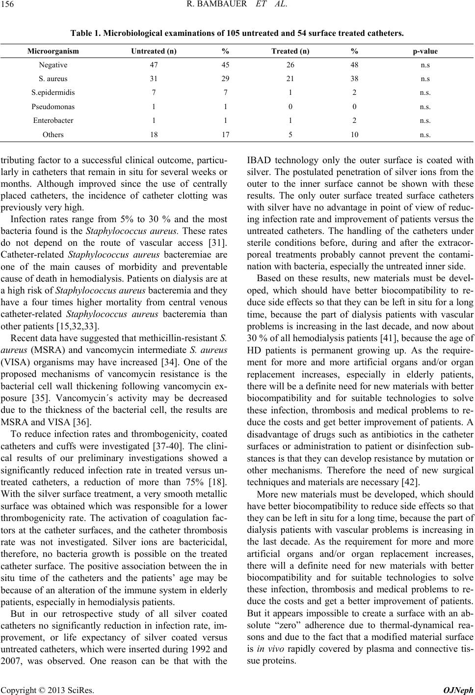 R. BAMBAUER ET AL. Copyright © 2013 SciRes. OJNeph 156 Table 1. Microbiological examinations of 105 untreated and 54 surface treated catheters. Microorganism Untreated (n) % Treated (n) % p-value Negative 47 45 26 48 n.s S. aureus 31 29 21 38 n.s S.epidermidis 7 7 1 2 n.s. Pseudomonas 1 1 0 0 n.s. Enterobacter 1 1 1 2 n.s. Others 18 17 5 10 n.s. tributing factor to a successful clinical outcome, particu- larly in catheters that remain in situ for several weeks or months. Although improved since the use of centrally placed catheters, the incidence of catheter clotting was previously very high. Infection rates range from 5% to 30 % and the most bacteria found is the Staphylococcus aureus. These rates do not depend on the route of vascular access [31]. Catheter-related Staphylococcus aureus bacteremiae are one of the main causes of morbidity and preventable cause of death in hemodialysis. Patients on dialysis are at a high risk of Staphylococcus aureus bacteremia and they have a four times higher mortality from central venous catheter-related Staphylococcus aureus bacteremia than other patients [15,32,33]. Recent data have suggested that methicillin-resistant S. aureus (MSRA) and vancomycin intermediate S. aureus (VISA) organisms may have increased [34]. One of the proposed mechanisms of vancomycin resistance is the bacterial cell wall thickening following vancomycin ex- posure [35]. Vancomycin´s activity may be decreased due to the thickness of the bacterial cell, the results are MSRA and VISA [36]. To reduce infection rates and thrombogenicity, coated catheters and cuffs were investigated [37-40]. The clini- cal results of our preliminary investigations showed a significantly reduced infection rate in treated versus un- treated catheters, a reduction of more than 75% [18]. With the silver surface treatment, a very smooth metallic surface was obtained which was responsible for a lower thrombogenicity rate. The activation of coagulation fac- tors at the catheter surfaces, and the catheter thrombosis rate was not investigated. Silver ions are bactericidal, therefore, no bacteria growth is possible on the treated catheter surface. The positive association between the in situ time of the catheters and the patients’ age may be because of an alteration of the immune system in elderly patients, especially in hemodialysis patients. But in our retrospective study of all silver coated catheters no significantly reduction in infection rate, im- provement, or life expectancy of silver coated versus untreated catheters, which were inserted during 1992 and 2007, was observed. One reason can be that with the IBAD technology only the outer surface is coated with silver. The postulated penetration of silver ions from the outer to the inner surface cannot be shown with these results. The only outer surface treated surface catheters with silver have no advantage in point of view of reduc- ing infection rate and improvement of patients versus the untreated catheters. The handling of the catheters under sterile conditions before, during and after the extracor- poreal treatments probably cannot prevent the contami- nation with bacteria, especially the untreated inner side. Based on these results, new materials must be devel- oped, which should have better biocompatibility to re- duce side effects so that they can be left in situ for a long time, because the part of dialysis patients with vascular problems is increasing in the last decade, and now about 30 % of all hemodialysis patients [41], because the age of HD patients is permanent growing up. As the require- ment for more and more artificial organs and/or organ replacement increases, especially in elderly patients, there will be a definite need for new materials with better biocompatibility and for suitable technologies to solve these infection, thrombosis and medical problems to re- duce the costs and get better improvement of patients. A disadvantage of drugs such as antibiotics in the catheter surfaces or administration to patient or disinfection sub- stances is that they can develop resistance by mutation or other mechanisms. Therefore the need of new surgical techniques and materials are necessary [42]. More new materials must be developed, which should have better biocompatibility to reduce side effects so that they can be left in situ for a long time, because the part of dialysis patients with vascular problems is increasing in the last decade. As the requirement for more and more artificial organs and/or organ replacement increases, there will a definite need for new materials with better biocompatibility and for suitable technologies to solve these infection, thrombosis and medical problems to re- duce the costs and get a better improvement of patients. But it appears impossible to create a surface with an ab- solute “zero” adherence due to thermal-dynamical rea- sons and due to the fact that a modified material surface is in vivo rapidly covered by plasma and connective tis- sue proteins.  R. BAMBAUER ET AL. 157 Therefore other concepts of the prevention of implant- associated infections must involve the impregnation of the devices the inner and outer surface with antibiotics, antimicrobial substances and/or metals [43,44]. Another point is to understand the processes leading to the devel- opment of catheter-related bacteremia in order to can offer effective preventive and therapeutic possibilities [45] such as new polymer-antibiotic systems in inhibiting bacterial biofilm formation and in reducing neutrophil activation after surface contact on different biomaterials, thus reducing the risk for biomaterial-mediated inflam- matory reactions [46-48], or the development of new biofilm to serve in a communication system termed quo- rum sensing [49], or molecules that inhibit quorum sens- ing signal generation among organism could block mi- crobial biofilm formation [50]. These catheters related complications are contributing factors to the increasing cost of medical care. They are responsible for patient readmissions and longer hospital stays as well as patients discomfort, morbidity, and occa- sional mortality. Feldman et al. calculated in 1996 the costs of the morbidity due to catheter infections will soon exceed $ 1 billion per year [51]. Therefore he demanded to reduce vascular access-related morbidity, that strate- gies must be developed not only to prevent and detect appropriately early synthetic vascular access dysfunction, but to better identify the patients in a whom radial arte- rio-venous fistula is a viable clinical option. The repre- sentative health care cost savings for hemodialysis cathe- ters, given specific infection rates and potential infection rate reductions achieved by treated catheters [23]. The cost analysis was calculated using the literature and the available costs of different companies which dis- tribute these catheters [52]. Potential health care cost reductions that could be achieved through the use of sur- face treated-catheters by an annual usage of 125,971 he- modialysis catheter devices and an infection rate of 5% - 20%, savings per year of $ 17.7 million, reduction about 40% [52]. Besides a high number of patients who die to CRI, the costs of these infections are increasingly steady. After Schwebel et al. the costs are $ 2118/intensive care unit day, and after Pronovost et al. $ 45,000 per each infection [53,54]. Tacconelli et al. estimated in 2009 the costs associated with CRI in four European countries (France, Germany, Italy, and UK) between € 35.9 and € 163.9 million per year [55]. Due to these tremendous high costs it must be possible of scientists, physicians, bioengineers and others to de- velop new techniques and new materials to reduce these high costs and to increase the improvement of patients. But besides the high costs due catheter-related infec- tions, the patients´ longer hospital stays, and patients discomfort, mortality, and occasionally mortality are the most important problems which must be resolved. To reduce these complications it is necessary that the han- dling of the catheters must be done first after the numer- ous recommendation and guidelines available in the lit- erature [22,56,57]. Surface treatment of catheter is necessary, but of the inner and the outer surface. Therefore new technology must be developed for the surface treatment with antibi- totics, antimicrobial substances and/or metal. New mate- rial and new-polymer-antibiotic systems are demanded. The developed of new biofilm to serve in a communica- tion system termed quorum sensing [49], or molecules which inhibit quorum sensing signal generation among organism could block microbial biofilm formation [50]. This reqirement shows perhaps the new developed cathe- ter material, the microdomain-structured surface [PUR- SMA-coated catheters, Gambro, Germany) [18]. Micro- domain surfaces are considered the most biocompatible because they mimic the structure of natural biological surfaces. Microdomain structures are used to match the multiple requirements for improved catheter surfaces, that is reduced thromgenecity and improved antimicro- bial properties. First results with these catheters are very encouraged. Most important is the improvement of the handling of the catheters by the attending staff which is recommend- ed in numerous available guidelines to reduce the tre- mendous high costs to treat the CRI and the discomfort and morbidity of the patients. 7. Conclusion The results showed that catheters which were surface treated only on the outside had no advantages versus un- treated catheters. In a retrospective study from 1992 to 2007, outer surface treated catheters with silver versus untreated catheters in 159 patients, who needed a large bore catheter, were investigated. There was no associa- tion between the in situ time and silver coated/uncoated catheters, resulting in the bacteriological examination, and patients’ diagnosis or outcome. Reasons may be that on the surface treated catheters only the outer surface was coated with silver and the possibility of contamination by the handling during the extracorporeal treatments. There- fore, new materials and surface treatment on both sur- faces, the inner and outer surface, are needed to save the tremendous high health costs for hemodialysis catheters, to reduce infection rates, and thrombus formations and to improve the patients’ outcome. REFERENCES [1] L. Beloqui, S, Shaldon, L, Chiandussi and B. Higgs, “Ha- emodialysis by Percutaneous Catheterization of the Fe- moral Artery and Vein with Reginal Heparinisation,” Lancet, Vol. 11, 1961, p. 857. Copyright © 2013 SciRes. OJNeph  R. BAMBAUER ET AL. 158 [2] D. G. Maki, “Infection Due to Infusion Therapy,” In: I. V. Bennett and P. S. Brachman, Eds., Hospitals Infections, Little Brown and Co., Boston, 1992, pp. 849-859. [3] J. S. Groegor, A. B. Lucas and H. A. T. Thaler, “Infec- tious Morbidity Associated with Long-Term Use of Ve- nous Access Devices in Patients with Cancer,” Annals of Internal Medicine, Vol. 119, No. 12, 1993, pp. 1168-1174. doi:10.7326/0003-4819-119-12-199312150-00003 [4] R. Bambauer, P. Mestres and K. J. Pirrung, “Frequency, Therapy, and Prevention of Infections Associated with Large-Bore Catheter,” ASAIO Journal, Vol. 38, No. 2, 1992, pp. 96-101. [5] R. Bambauer, R. Inninger and K. J. Pirrung, “Complica- tions and Side Effects Associated with Large-Bore Cathe- ters in the Suclavian and Internal Veins,” Artificial Or- gans, Vol. 18, No. 4, 1994, pp. 318-321. doi:10.1111/j.1525-1594.1994.tb02204.x [6] C. M. Kjellstrand, G. G. Merino, S. M. Mauer, et al., “Complications of Percutaneous Femoral Vein Catheteri- zation for Hemodialysis,” Clinical Nephrology, Vol. 4, No. 1, 1975, pp. 37-40. [7] A. Schwarzbeck, W. D. Brittinger, G. E. von Henning, et al., “Cannulation of Subclavian Vein for Hemodialysis Using Seldinger Technique,” Transactions of American Society for Artificial Internal Organs, Vol. 27, No. 1, 1978, pp. 24-31. [8] A. De Cubber, C. De Wolf, N. Lameire, et al., “Single Needle Hemodialysis with Double Headpump via the Subclavian Vein,” Dialysis & Transplantation, Vol. 7, No. 11, 1978, pp. 1261-1263. [9] P. R. Uldall, P. R. Dyck and F. Woods, “A Subclavian Cannula for Temporary Vascular Access for Hemodialy- sis or Plasmapheresis,” Dialysis & Transplantation, Vol. 8, No. 9, 1979, pp. 963-968. [10] C. T. Flynn and R. McGowan, “Subclavian Vein Catheter and Clockwork Pump,” Dialysis & Transplantation, Vol. 9, No. 5, 1980, pp. 556-616. [11] R. Bambauer and G. A. Jutzler, “Jugularis-Interna Punk- tion zur Shaldon-Katheterisierung. Ein neuer Zugang für Akute Hämodialysen,” Nieren-Hochdruckh, Vol. 9, No. 3, 1980, pp. 109-116 [12] R. Bambauer and G. A. Jutzler, “Transcutaneous Inser- tion of the Shaldon Catheter through the Internal Jugular Vein as Access for Acute Hemodialysis,” Dialysis & Transplantation, Vol. 11, No. 9, 1982, pp. 766-771. [13] D. Philibert, M. Agharazii, E. Audy, et al., “Clinical Ex- perience with a Chronic Hemodialysis Catheter with Sym- metrical Tip Configuration (Palindrone TM),” JASN, Vol. 16, 2005. [14] M. Allon, “Dialysis Catheter-Related Bacteremia Treat- ment and Prophylaxis,” American Journal of Kidney Dis- eases, Vol. 44, No. 5, 2004, pp. 779-791. [15] J. Nielsen, H. J. J. Kolosk and F. Espersen, “Staphylo- coccus aureus Bacteremia among Patients Undergoing Dialysis: Focus Dialysis Catheter-Related Cases,” Ne- phrology Dialysis Transplantation, Vol. 13, No. 1, 1988, pp. 139-145. doi:10.1093/ndt/13.1.139 [16] R. Locci, G. Peters and G. Pulverer, “Microbiological Colonization of Prosthetic Devices. Microtopological Cha- racteristics of Intravenous Catheters as Detected by Scan- ning Electron Microscopy,” Zentralblatt für Bakteriologie, Mikrobiologie und Hygiene, Vol. 173, No. 2, 1981, pp. 285-292. [17] J. W. Costerton, L. Montanaro and C. R. Arciola, “Bio- film in Implant Infections: Its Production and Regulation,” International Journal of Artificial Organs, Vol. 28, No. 9, 2005, pp. 1065-1068. [18] R. Bambauer, R. Schiel, S. Bambauer and P. Sioshansi, “Large Bore Catheters with Surface Treatments versus Untreated Catheters for Blood Access,” The Journal of Vasclar Access, Vol. 2, No. 3, 2001, pp. 97-105. [19] J. D. Maya, D. Carlton, E. Estrada, et al., “Treatment of Dialysis Catheter-Related Staphylococcus aureus Bacte- remia with an Antibiotic Lock: A Quality Improvement Report,” American Journal of Kidney Diseases, Vol. 50, No. 2, 2007, pp. 289-295. doi:10.1053/j.ajkd.2007.04.014 [20] G. K. Dogra, H. Herson, B. Hutchison, et al., “Prevention of Tunnelled Hemodialysis Catheter-Related Infections Using Catheter Restricted Filling with Gentamycin and Citrate: A Randomized Controlled Study,” Journal of the American Society of Nephrology, Vol. 13, No. 8, 2002, pp. 2133-2139. doi:10.1097/01.ASN.0000022890.29656.22 [21] R. Bambauer, R. Schiel, P. Mestres and P. Sioshansi, “Scanning Electron Microscopic Investigation of Cathe- ters for Blood Access,” International Journal of Artificial Organs, Vol. 18, No. 5, 1995, pp. 326-331. [22] R. Hollenbeck, V. Mickley, J. Brunkwall, et al., “Gefäß- zugang zur Hämodialyse” Der Nephrologe, Vol. 4, No. 2, 2009, pp. 158-176. doi:10.1007/s11560-009-0281-0 [23] R. Bambauer, R. Schiel, S. Bambauer and P. Sioshansi, “Long-Term Catheters for Apheresis and Dialysis with Surface Treatment with Infection Resistance and Low Thrombogenicity,” Therapeutic Apheresis and Dialysis, Vol. 7, No. 2, 2003, pp. 225-231. doi:10.1046/j.1526-0968.2003.00042.x [24] R. Bambauer and G. A. Jutzler, “Lagekontrolle Großlu- miger Zentraler Venenkatheter Mittels Intrakardialer Ele- ktrographie,” Intensivmed, Vol. 17, No. 6, 1980, pp. 12- 17. [25] P. Sioshansi and E. J. Tobin, “Surface Treatment of Bio- materials by Ion-Beam Processes,” Medical Plastics and Biomaterials, Vol. 2, 1995, pp. 50-59. [26] P. Sioshansi, “New Process for Surface Treatment of Ca- theters,” Artificial Organs, Vol. 18, No. 4, 1994, pp. 226- 271. doi:10.1111/j.1525-1594.1994.tb02193.x [27] R. Bambauer, P. Mestres, R. Schiel, et al., “New Surface Treatment Technologies for Catheters Used for Extracor- poreal Detoxication Methods,” Dialysis & Transplanta- tion, Vol. 24, No. 5, 1995, pp. 228-237. [28] P. Sioshansi, “Ion Implantation of Cobalt Chronicum Prosthetic Components to Reduce Polyethylene Wear,” Orthopedics Today, Vol. 11, No. 1, 1991, pp. 24-25. [29] D. Schierholz, A. Rump and W. Pulverer, “Klinische und Präklinische Effizienz Antimikrobieller Katheter,” Ana- esthesiol Intensivmed Notfallmed Schmerzther, Vol. 32, No. 5, 1997, pp. 289-305. doi:10.1055/s-2007-995058 Copyright © 2013 SciRes. OJNeph  R. BAMBAUER ET AL. 159 [30] R. Bambauer, R. Schiel, C. Bambauer and R. Latza, “Sur- face-Treted versus Untreated Large-Bore Catheters as Vascular Access in Hemodialysis and Apheresis,” Inter- national Journal of Nephrology, Vol. 2012, 2012, Article ID: 956136, 8 pages. http://www.Hindawi.com/journals/ijn(2012/956136/ [31] R. Vanholder, N. Hoenich, S. Ringoir, et al., “Morbidity and Mortality of Central Venous Catheter Hemodialysis, a Review of 10 Years’ Experience,” Nephron, Vol. 47, No. 4, 1987, pp. 247-279. doi:10.1159/000184523 [32] G. Jean, B. Charra, C. Chazot, et al., “Risk Factor Analy- sis for Long-Term Tunnelled Dialysis Catheter-Related Bacteremias,” Nephron, Vol. 91, No. 2, 2002, pp. 399- 405. doi:10.1159/000064279 [33] S. H. Kim, K. I. Song, J. W. Chang, et al., “Prevention of Uncuffed Hemodialysis Catheter-Related Bacteremia Us- ing an Antibiotic Lock Technique: A Prospective, Ran- domised Clinical Trial,” Kidney International, Vol. 69, No. 1, 2006, pp. 161-164. doi:10.1038/sj.ki.5000012 [34] E. A. King, D. McCoy, S. Desai, et al., “Vancomycin Resistant Enterococcal Bacteraemia and Daptomycin: Are Higher Doses Necessary?” Journal of Antimicrobial Chemotherapy, Vol. 66, No. 9, 2011, pp. 2112-2118. doi:10.1093/jac/dkr255 [35] I. Cui, A. Iwamoto and I. O. Lian, “Novel Mechanism of Antibiotic Resistance Originating in Vancomycin-Inter- mediate Staphylococcus aureus,” Antimicrobial Agents and Chemotherapy, Vol. 50, No. 2, 2006, pp. 428-438. doi:10.1128/AAC.50.2.428-438.2006 [36] I. Cui, F. Tominage, H. M. Nenh, et al., “Correlation between Reduced Daptomycin Suscetibility and Van- comycin Resistance in Vancomycin-Intermediate Staphy- lococcus aureus,” Antimicrobial Agents and Chemothe- rapy, Vol. 50, No. 3, 2006, pp. 1079-1082. doi:10.1128/AAC.50.3.1079-1082.2006 [37] D. G. Maki, J. K. Garman, J. M. Shapiro, et al., “An At- tachable Silver-Impregnated Cuff for Prevention of Infec- tion with Central Venous Catheters: A Prospective Ran- domized Multicenter Trial,” The American Journal of Medicine, Vol. 85, No. 3, 1988, pp. 307-314. doi:10.1016/0002-9343(88)90579-7 [38] K. S. Tweden, J. D. Cameron, A. J. Razzouk, et al., “Sil- ver Modification of Polyethylene Terphthalate Textile for Antimicrobial,” ASAIO Journal, Vol. 43, No. 2, 1997, pp. M475-M481. doi:10.1097/00002480-199703000-00088 [39] A. Oloffs, C. Gosse-Siestrup, S. Bisson, et al., “Biocom- patibility of Silver Coated Polyurethane Catheters and Silver Coated Dacron Material,” Biomaterials, Vol. 15, No. 10, 1994, pp. 753-758. doi:10.1016/0142-9612(94)90028-0 [40] R. Bambauer, P. Mestres, R. Schiel, et al., “Surface Treated Large-Bore Catheters with Silver Based Coating Versus Untreated Catheters for Extracorporeal Detoxica- tion Methods,” ASAIO Journal, Vol. 44, No. 4, 1998, pp. 3003-3008. doi:10.1097/00002480-199807000-00013 [41] K. S. Rabindranath, T. Bansal, J. Adams, et al., “Syste- matic Review of Antimicrobials for Prevention of Hemo- dialysis Catheter-Related Infections,” Nephrology Dialy- sis Transplantation, Vol. 24, No. 12, 2009, pp. 3763-3774. doi:10.1093/ndt/gfp327 [42] A. A. Hampton and R. J. Sheretz, “Vascular Access In- fections in Hospitalised Patients,” The Surgical Clinics of North America, Vol. 68, No. 1, 1988, pp. 57-71. [43] C. Von Eif, W. Kohnen, K, Becker, et al., “Modern Strategies in the Prevention of Implant-Associated Infec- tions,” The International Journal of Artificial Organs, Vol. 28, No. 11, 2005, pp. 1146-1156. [44] J. F. Timsit, O. Mimoz, B. Mourviller, et al., “Rando- mized Controlled Trial of Chlorhexidine Dressing and Highly Adhesive Dressing for Preventing Catheter-Re- lated Infections in Critically Ill Adults,” American Jour- nal of Respiratory and Critical Care Medicine, Vol. 186, No. 12, 2012, pp. 1272-1278. doi:10.1164/rccm.201206-1038OC [45] L. Troidle and F. O. Finkelstein, “Catheter-Related Bac- teremia in Hemodialysis Patients: The Role of the Central Venous Catheter in Prevention and Therapy,” The In- ternational Journal of Artificial Organs, Vol. 31, No. 9, 2008, pp. 827-833. [46] S. Cicalini, F. Palmieri and N. Petrosillo, “Clinical Re- view: New Technologies for Prevention of Intravascular Catheter-Related Infections,” Critical Care, Vol. 8, 2004, pp. 157-162. doi:10.1186/cc2380 [47] G. Donelli, I. Francolini, A. Piozzi, et al., “New Polymer- Antibiotic Systems to Inhibit Bacterial Biofilm Formation: A Suitable Approach to Prevent Central Venous Catheter- Associated Infections,” Journal of Chemotherapy, Vol. 14, No. 5, 2002, pp. 501-507. [48] S. Schmitt, G. Haase, E. Csomor, et al., “Inhibitor of Complement, Compstatin, Prevents Polymer-Mediated Mac-1 Up-Regulation of Human Neutrophils Independent of Biomaterial Type Tested,” Journal of Biomedical Ma- terials Research, Vol. 66A, No. 3, 2003, pp. 491-499. doi:10.1002/jbm.a.10031 [49] M. R. Parsek, D. L. Val, B. L. Hanzelka, et al., “Acyl Homoserine-Lactone Quorum-Sensing Signal Genera- tion,” Proceedings of the National Academy of Sciences of the United States of America, Vol. 96, No. 8, 1999, pp. 4360-4365. doi:10.1073/pnas.96.8.4360 [50] D. G. Davies, M. R. Parsek, J. P. Pearson, et al., “The Involvement of Cell-to-Cell Signals in the Development of a Bacterial Biofilm,” Science, Vol. 280, No. 5361, 1998, pp. 295-298. doi:10.1126/science.280.5361.295 [51] H. J. Feldmann, S. Kobrina and A. Wasserstein, “Hemo- dialysis Vascular Morbidity,” Journal of the American Society of Nephrology, Vol. 7, No. 4, 1996, 523- 535. [52] S. Bambauer, “Cost Reduction Benefits of Applying an Antimicrobial Surface Treatment to Catheters,” Personnel Communication, 1996. [53] C. Schwebel, J. C. Lucet, A. Vesin, et al., “Economic Evaluation of Chlorhexidine-Impregnated Sponges for Preventing Catheter-Related Infection in Critically Ill Adults in the Dressing Study,” Critical Care Medicine, Vol. 40, No. 1, 2012, pp. 11-17. doi:10.1097/CCM.0b013e31822f0604 [54] P. J. Pronovost, C. A. Goeschel, E. Colantuoni, et al., “Sustaining Reductions in Catheter-Related Bloodstream Infections in Michigan Intensive Care Units: Observa- Copyright © 2013 SciRes. OJNeph  R. BAMBAUER ET AL. Copyright © 2013 SciRes. OJNeph 160 tional Study,” British Medical Journal, Vol. 340, 2010, p. c309. doi:10.1136/bmj.c309 [55] E. Toccanelli, G. Smith, A. Hiske, et al., “Epidemiology, Medical Outcomes and Costs of Catheter-Related Blood- stream Infections in Intensive Care Units of Four Euro- pean Countries: Literature- and Registry-Based Esti- mates,” Journal of Hospital Infection, Vol. 72, No. 2, 2009, pp. 97-103. doi:10.1016/j.jhin.2008.12.012 [56] N. P. O’Grady, M. Alexander, L. A. Burns, et al., “Gui- delines for the Preventions of Intravascular Catheter-Re- lated Infections,” Clinical Infectious Diseases, Vol. 52, No. 9, 2011, pp. e162-e193. [57] T. Nagai, S. Kohsaka, T. Anzai, et al., “Low Incidence of Catheter-Related Complications in Patients with Ad- vanced Pulmonary Arterial Hypertension Undergoing Con- tinuous Epoprostenol Infusions,” Chest, Vol. 141, No. 1, 2012, pp. 272-273.doi:10.1378/chest.11-1893
|