Open Journal of Nephrology, 2013, 3, 148-151
http://dx.doi.org/10.4236/ojneph.2013.33027 Published Online September 2013 (http://www.scirp.org/journal/ojneph)
Renal Allograft Thrombosis in the Early Post
Transp la nt P er io d
M. Budruddin1*, Issa Al Salmi2, R. Shilpa2
1Nephrology Department, Tawam Hospital, Al Ain, UAE
2Nephrology Department, Royal Hospital, Muscat, Oman
Email: *drbudruddin@gmail.com, isa.al.salmi@gmail.com, ramaiah_shilpa@yahoo.co.in
Received June 26, 2013; revised July 23, 2013; accepted August 10, 2013
Copyright © 2013 M. Budruddin et al. This is an open access article distributed under the Creative Commons Attribution License,
which permits unrestricted use, distribution, and reproduction in any medium, provided the original work is properly cited.
ABSTRACT
Renal allograft thrombosis invo lving either the renal artery or the veins is a well known entity in clinical p ractice. This
complication of the renal transplant surgery is more common in the early part of the post transplant period and it is usu-
ally associated with acute antibody mediated rejection. This more commonly occurs in the paediatric renal transplant
and also seems to have some relation to the duration of peritoneal dialysis pretransplant. However, the occurrence of
graft thrombosis in isolation without clinical or histological graft rejection is not rare. We encountered a patient in
whom the renal allograft thrombosis occurred after 6 weeks of commercial renal transplantation without any histologi-
cal evidence of rejection.
Keywords: Allograft Thrombosis; Immunosuppression; Cyclosporine; OKT3; Hemodialysis; Renogram; Rejection and
Pyelonephritis
1. Introduction
Renal allograft thrombosis is not an uncommon compli-
cation of renal transplantation. It usually occurs in the
early post renal transplant period. Several factors have
been implicated with this notorious complication. Throm-
bosis can even occur in the late part of the post transplant
period. Irrespective of the time of development of throm-
bosis the outcome is equ a lly bad.
2. Case Summary
A 58-year-old Omani male with background of long
standing type I diabetes along with its complications of
diabetic retinopathy, Nephropathy, Neuropathy, Ischemic
heart disease and paroxysmal atrial fibrillation. He was
initiated on haemodialysis in late 2009. He received
commercial renal transplantation in October 2010 from
Pakistan and was put on triple immunosuppression with
Cyclosporin, Mycophenolate and prednisolone. Details
of induction immunosuppression were not available in
the discharge notes.
On the 8th post operative day on his arrival in our hos-
pital he had mild leucocytosis, surgical wound infection,
uncontrolled blood sugars and the renal functions were
deranged with e-GFR 21 ml/min. He was started empiri-
cally on Tazocbactam after sending the samples for cul-
tures and blood sugars were controlled with insulin. At
this stage graft kidney biopsy was proposed but patient
and his relatives did not agree. An impression of delayed
graft function was made and he was treated for infection.
Ultrasound of the graft kidney showed good blood flow
in the graft kidney and the resistive index was 0.6 - 0.8
and there were no collection around the renal allograft.
The renal functions gradually improved with decrease in
the serum creatinine from 255 umol/l on presentation to
190 umol/l on discharge. DTPA renogram was planned
but we could not get it done as the family wanted to take
him to the peripheral Nephrology Unit close to their
home, for further care.
During his stay there in hospital after a week he de-
veloped tenderness over the graft and the Doppler ultra-
sound of the graft showed perinephric collection of 5.7 ×
2.9 × 1.4 cm with high suspicion of bleed (Figure 1). In
the mean time he developed Symptomatic atrial fibrilla-
tion. Apart from the rate control drugs he was also war-
farinised on the suggestion of cardiologist. Subsequent
three days were uneventfull with the serum creatinine
improving further to 119 umol/l and also with improve-
ment in the graft tenderness. He was hence discharged
with follow up appoin tment in the cardiology and Trans-
*Corresponding author.
C
opyright © 2013 SciRes. OJNeph
M. BUDRUDDIN ET AL. 149
Figure 1. Perigraft haematoma shown by colour Doppler
ultrasound.
plant clinic. There his medications on discharge included
Ciclosporin 150 mg bid, MMF 1gm bid, prednisolone 20
mg bid, warfarin 2 mg od, amlodepine 10mg od and
isosorbid 10 mg tid.
Five days later he again presen ted to the emergency of
that hospital with history of fever associated with chills,
decreased urine output and loss of consciousness. Ex-
amination revealed hypotension blood pressure 80/50
mmhg, congested chest, bluish discolouration associated
with tenderness over the right iliac fossa. Immediate re-
suscitation was done and the laboratory investigations
showed serum creatinine of 230 umol/l, Hb of 7.6 gm%
which was earlier 9.4 gm% on discharge, leucocytosis of
14000 cumm and INR of 3. Urgent ultrasound of the ab-
domen showed increase in the size of Haematoma to 11.7
× 7 × 9.1 cm (Figure 2). He was admitted under surgical
team and started on antibiotics, fresh frozen plasma along
with one dose of Vit K. Sequential ultrasound assesment
of the collection was planned and the second ultrasound
done the following day alarmingly showed no paren-
chymal blood flow and the renal functions worsened with
exponential rise in serum creatinine to >800 umol/l and
anuria. He was started on haemodialysis and CT scan of
the abdomen was done there showed large perinephric
collection with necrotic renal allograft and air bubbles
suggestive of emphysematous pyelonephritis. He was
shifted to our centre on 31st October for graft Nephrec-
tomy. On admission in our centre he was septic with ab-
dominal distention and severe tenderness over the Right
side of the abdomen. There was leucocytosis, thrombo-
cytopenia and the coagulation was deranged with fi-
brinogen degradation products (FDP) elevated. There
was no evidence of Deep vein thrombosis of the lower
limbs by Doppler ultrasound. He was urgently taken for
exploration which showed pale looking necrotic graft
Figure 2. Perigraft Haematoma shown by arrow on ultra-
sound.
Figure 3. Slide showing the blood vessels with thrombi.
(magnification ×400).
Figure 4. Tissue from the infracted kidney showing tubules
with bacterial colonies (magnification ×400).
Copyright © 2013 SciRes. OJNeph
M. BUDRUDDIN ET AL.
150
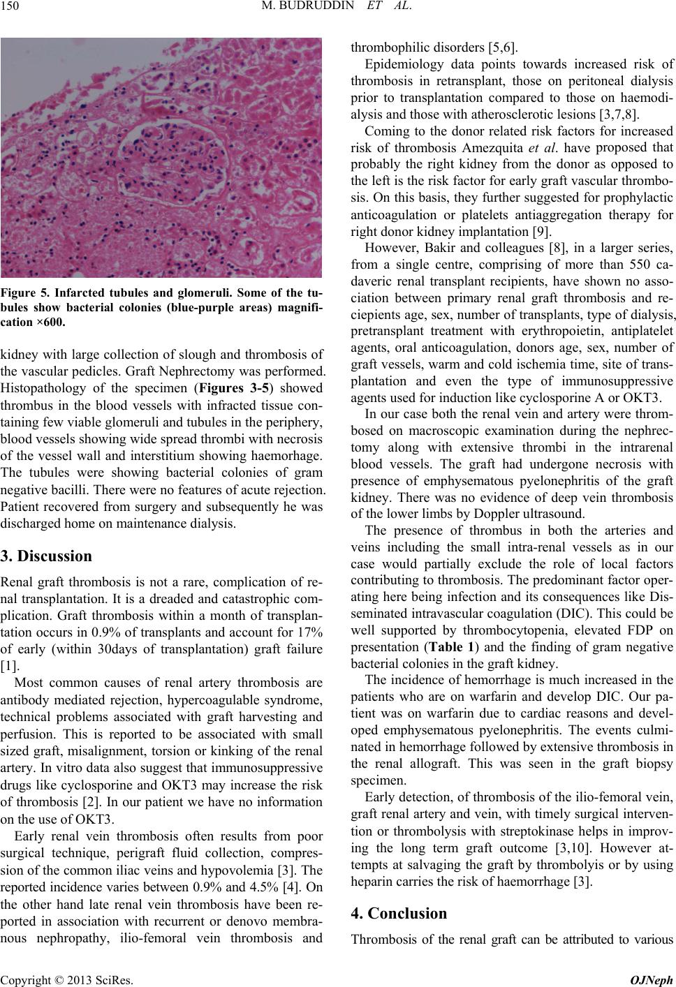
Figure 5. Infarcted tubules and glomeruli. Some of the tu-
bules show bacterial colonies (blue-purple areas) magnifi-
cation ×600.
kidney with large collection of slough and thrombosis of
the vascular pedicles. Graft Nephrectomy was performed.
Histopathology of the specimen (Figures 3-5) showed
thrombus in the blood vessels with infracted tissue con-
taining few viable glomeruli and tubules in th e periphery,
blood vessels sh owing wide sp read th ro mbi with ne cros is
of the vessel wall and interstitium showing haemorhage.
The tubules were showing bacterial colonies of gram
negative bacilli. Th ere were no features of acu te rejection .
Patient recovered from surgery and subsequently he was
discharged home on maintenance dialysis.
3. Discussion
Renal graft thrombosis is not a rare, complication of re-
nal transplantation. It is a dreaded and catastrophic com-
plication. Graft thrombosis within a month of transplan-
tation occurs in 0.9% of transplants and account for 17%
of early (within 30days of transplantation) graft failure
[1].
Most common causes of renal artery thrombosis are
antibody mediated rejection, hypercoagulable syndrome,
technical problems associated with graft harvesting and
perfusion. This is reported to be associated with small
sized graft, misalignment, torsion or kinking of the renal
artery. In vitro data also suggest that immunosuppressive
drugs like cyclosporine and OKT3 may increase the risk
of thrombosis [2]. In our patient we have no information
on the use of OKT3.
Early renal vein thrombosis often results from poor
surgical technique, perigraft fluid collection, compres-
sion of the common iliac veins and hypovolemia [3]. The
reported incidence varies between 0.9% and 4.5% [4]. On
the other hand late renal vein thrombosis have been re-
ported in association with recurrent or denovo membra-
nous nephropathy, ilio-femoral vein thrombosis and
thrombophilic disorders [5,6].
Epidemiology data points towards increased risk of
thrombosis in retransplant, those on peritoneal dialysis
prior to transplantation compared to those on haemodi-
alysis and those with atherosclerotic lesions [3,7,8].
Coming to the donor related risk factors for increased
risk of thrombosis Amezquita et al. have proposed that
probably the right kidney from the donor as opposed to
the left is the risk factor for early graft vascular thrombo-
sis. On this basis, they further sugge sted for prophylactic
anticoagulation or platelets antiaggregation therapy for
right donor kidney implantation [9].
However, Bakir and colleagues [8], in a larger series,
from a single centre, comprising of more than 550 ca-
daveric renal transplant recipients, have shown no asso-
ciation between primary renal graft thrombosis and re-
ciepients age, sex, number of transplants, type of dialysis,
pretransplant treatment with erythropoietin, antiplatelet
agents, oral anticoagulation, donors age, sex, number of
graft vessels, warm and cold ischemia time, site of trans-
plantation and even the type of immunosuppressive
agents used for in duction like cyclosporine A or OKT3 .
In our case both the renal vein and artery were throm-
bosed on macroscopic examination during the nephrec-
tomy along with extensive thrombi in the intrarenal
blood vessels. The graft had undergone necrosis with
presence of emphysematous pyelonephritis of the graft
kidney. There was no evidence of deep vein thrombosis
of the lower limbs by Doppler ultrasound.
The presence of thrombus in both the arteries and
veins including the small intra-renal vessels as in our
case would partially exclude the role of local factors
contributing to thrombo sis. The predominant factor oper-
ating here being infection and its consequences like Dis-
seminated intravascular coagu lation (DIC). This could be
well supported by thrombocytopenia, elevated FDP on
presentation (Table 1) and the finding of gram negative
bacterial colonies in the graft kidney.
The incidence of hemorrhage is much increased in the
patients who are on warfarin and develop DIC. Our pa-
tient was on warfarin due to cardiac reasons and devel-
oped emphysematous pyelonephritis. The events culmi-
nated in hemorrhage followed by extensive thrombosis in
the renal allograft. This was seen in the graft biopsy
specimen.
Early detection, of thrombos is of the ilio-femoral vein,
graft renal artery and vein, with timely surgical interven -
tion or thrombolysis with streptokinase helps in improv-
ing the long term graft outcome [3,10]. However at-
tempts at salvaging the graft by thrombolyis or by using
heparin carries the ri sk of haemorrhage [3].
4. Conclusion
Thrombosis of the renal graft can be attributed to various
Copyright © 2013 SciRes. OJNeph
M. BUDRUDDIN ET AL.
Copyright © 2013 SciRes. OJNeph
151
Table 1. Showing the levels of Hb, WBC, platelets, Prothrombin time PT, Firinogen degradation products and serum
creatinine over time.
Serum Creatinine
in mmol/l
Fibrinogen degradation
products in mg/lit
Prothrombin Time in secondsPlatelets × 10 × 9/litWBC × 10 × 9/lit Hb gm/dl Date
288 mmol/l 2.29 10.5 337 10.6 9.6 15 Oct
857 mmol/l 7.98 15.8 41 22.9 9.5 31 Oct
599 mmol/l 3.99 11.8 95 5.3 10.4 02 Nov
219 mmol/l 3.35 10.5 245 8.9 10.2 23 Nov
reasons. Once thrombosed, the loss of graft is inevitable.
However, prompt diagnosis and immediate attempt to
thrombolyse with streptokinase or surgical intervention
with endoluminal extraction of the thrombus do bear a
great promise in the immediate and long term survival of
the renal allograft.
REFERENCES
[1] J. A Bertolatus, “Etiology of Renal Artery Thrombosis in
Renal Transplantation,” Medscape Transplantation, Vol.
1, No. 1, 2000.
[2] E. Carlsen, “Enhancement of Procoagulant Activity in
Stimulated Mononuclear Blood Cells and Monocytes by
Cyclosporine,” Transplantation, Vol. 43, No. 4, 1987, pp.
543-548. doi:10.1097/00007890-198704000-00018
[3] S. Gang and M. Rajapurkar, “Vascular Complications
Following Renal Transplantation” JNRT, Vol. 2, No. 1,
2009, pp. 122-132.
[4] P. Giustacchini, F. Pisanti, F. Citterio, A. M. De Gaetano,
M. Castagneto and G. Nanni, “Renal Vein Thrombosis
after Renal Transplantation: An Important Cause of Graft
Loss,” Transplantation Proceedings, Vol. 34, No. 6, 2002,
pp. 2126-2127. doi:10.1016/S0041-1345(02)02876-2
[5] M. R. First, N. Mendoza, R. K. Maryniak and M. A.
Weiss, “Membranous Glomerulopathy Following Kidney
Transplantation. Association with Renal Vein Thrombo-
sis in Two of Nine Cases,” Transplantation, Vol. 38, No.
6, 1984, pp. 603-607.
doi:10.1097/00007890-198412000-00011
[6] P. J. Ramirez, R. Y. Gohh, A. Kestin, A. P. Monaco and
P. E. Morrissey, “Renal Allograft Loss Due to Proximal
Extension of the Ileofemoral Deep Venous Thrombosis,”
Clinical Transplantation, Vol. 16, No. 4, 2002, pp. 310-
313. doi:10.1034/j.1399-0012.2002.02006.x
[7] A. Sanni, C. H. Wilson, H. Wyrley-Birch, D. Vijayanand,
A. Navarro, S. Sohrabi, B. Jaques, D. Rix, N. Soomro, D.
Manas and D. Talbot, “Donor Risk Factor for Renal Graft
Thrombosis,” Transplantation Proceedings, Vol. 39, No.
1, 2007, pp. 138-139.
[8] N. Bakir, W. J. Sluiter, R. J. Ploeg, W. J. van Son and A.
M. Tegzess, “Primary Renal Graft Thrombosis,” Neph-
rology Dialysis Transplantation, Vol. 11, No. 1, 1996, pp.
140-147. doi:10.1093/oxfordjournals.ndt.a027030
[9] Y. Amezquita, C. Mendez, A. Fernandez, S. Caldes, J.
Pascual, A. Muriel, F. J. Burgos, R. Marcen and J. Ortuno,
“Risk Factors for Early Graft Thrombosis: A Case-Con-
trolled Study in Grafts from the Same Donor,” Trans-
plantation Proceedings, Vol. 40, No. 9, 2008, pp. 2891-
2893. doi:10.1016/j.transproceed.2008.09.014
[10] E. Renoult, L. Cormier, M. Claudon, T. Cao-Huu, L. Fri-
mat, O. Gaucher, J. Hubert and M. Kessler, “Successful
Surgical Thrombectomy of Renal Allograft Vein Throm-
bosis in the Early Postoperative Period,” American Jour-
nal of Kidney Diseases, Vol. 35, No. 5, 2000, p. E21.
doi:10.1016/S0272-6386(00)70286-1