Open Journal of Nephrology, 2013, 3, 117-119
http://dx.doi.org/10.4236/ojneph.2013.33021 Published Online September 2013 (http://www.scirp.org/journal/ojneph)
Xanthogranulomatous Pyelonephritis Associated with
Crohn’s Disease and Primary Hyperparathyroidism
Mekdess Abebe, Wing Mu, Heesuck Suh, Edward P. Nord,
Nand K. Wadhwa*
Division of Nephrology, Department of Medicine, School of Medicine, State University of New York at Stony Brook,
Stony Brook, USA
Email: *nand.wadhwa@stonybrookmedicine.edu
Received May 15, 2013; revised June 20, 2013; accepted July 3, 2013
Copyright © 2013 Mekdess Abebe et al. This is an open access article distributed under the Creative Commons Attribution License,
which permits unrestricted use, distribution, and reproduction in any medium, provided the original work is properly cited.
ABSTRACT
We report on a case of unilateral and diffuse xanthogranulomatous pyelonephritis (XGP) associated with Crohn’s dis-
ease and primary hyperparathyroidism (HPT). The patient had undiagnosed primary HPT for at least a year prior to
presentation. The co-existence of Crohn’s disease and primary HPT caused calcium oxalate stone likely by the known
mechanism of hypercalcemia and hyperoxaluria respectively. The patient developed XGP, a rare disease, as a result of
infected calcium oxalate stone which was treated with antibiotics and unilateral nephrectomy. To the best of our
knowledge, this is the first case of XGP associated with Crohn’s disease and primary HPT.
Keywords: Xanthogranulomatous Pyelonephritis; Hyperparath yro idism; Crohn’s; Hypercalcemia
1. Introduction
XGP is a rare, serious, chronic inflammatory disorder of
the kidney and constitutes less than 1% of chronic
pyelonephritis. It is characterized by destruction of renal
parenchyma and its replacement with granulomatous,
abscess and collection of lipid laden macrophage (xan-
thoma cells) [1]. The disease most frequently presents in
middle-aged groups with a predilection for female gender
[2]. XGP shares many characteristics with renal neo-
plasm in terms of its radio logic appearance and its ab ility
to invade adjacent structures. We report a case of uni-
lateral and diffuse XGP due to undiagnosed primary HPT
and Crohn’s disease leading to uni lateral nephrectomy.
2. Case Report
A 46-year-old white woman presented to emergency
department with a two week history of right flank pain
associated with generalized weakness, dizziness, decreas-
ed appetite and weight loss of 50 pounds in the last few
months. She also had shortness of breath at minimal
exertion. She denied any urinary complaint, fever, nausea
or vomiting. Her significant comorbidities included
Crohn’s disease diagnosed in 2009, small bowel obstruc-
tion due to Crohn’s disease related stricture resulting in
laparoscopic small bowel resection in 2010, chronic pan-
creatitis and bilateral kidney stones. Patient was not on
any medication prior to admission. She was an active
smoker but denied alcohol or illegal drug use.
At presentation, the patient was not in any distress.
Blood pressure was 97/53 mmHg, pulse 92 beats/minute
and temperature was 38.3˚C. She had dry oral mucosa.
Abdominal examination showed mildly distended abdo-
men with right costovertebral angle tenderness. No orga-
nomegally was appreciated. The remainder of the physi-
cal examination was within normal limits. Laboratory
evaluation showed white blood cell count of 11.8 × 10³
/mcL, hemoglobin 7.5 g/dL, hematocrit 22.4% and pla-
telet count of 397 × 10³ /mcL. Serum chemistry showed a
sodium of 134 mEq/L (134 mmol/L), potassium 3.8
mEq/L (3.8 mmol/L), chloride 111 mEq/L (111 mmol/L),
bicarbonate 13 mEq/L (13 mmol/L), blood urea nitrogen
60 mg/dL (21.4 mmol/L) , serum creatinine 3.6 mg/dL
(275 µmol/L) (her serum creatinine was 0.8 mg/dL (61
µmol/L) 6 months prior to this admission), ionized cal-
cium 5.4 mg/dL (1.35 mmol/L), total calcium 9.8 mg/dL
(2.45 mmol/L), corrected calcium for albumin 10.8 mg/
dL (2.7 mmol/L), phosphorous 5.2 mg/dL (1.68 mmol/L),
albumin 2.8 mg/dL and total protein 6.1 mg/dL. Liver
function tests were within normal limits. Anti nuclear
antibody was positive at 1:320. Serum protein electro-
p ho re si s (S PEP ), u ri ne protein electrophoresis (UPEP), C3,
*Corresponding author.
C
opyright © 2013 SciRes. OJNeph
M. ABEBE ET AL.
118
C4, antineutrophil cytoplasmic antibodies, Anti ds DNA
antibodies were negative. Urinalysis showed large leuko-
cyte esterase, WBC > 182 and protein 100 mg/dL. Urine
culture was negative.
Intravenous ceftriaxone and ciprofloxacin therapy was
started for presumed pyelonephritis. Patient also received
IV hydration with normal saline and 4 units of packed
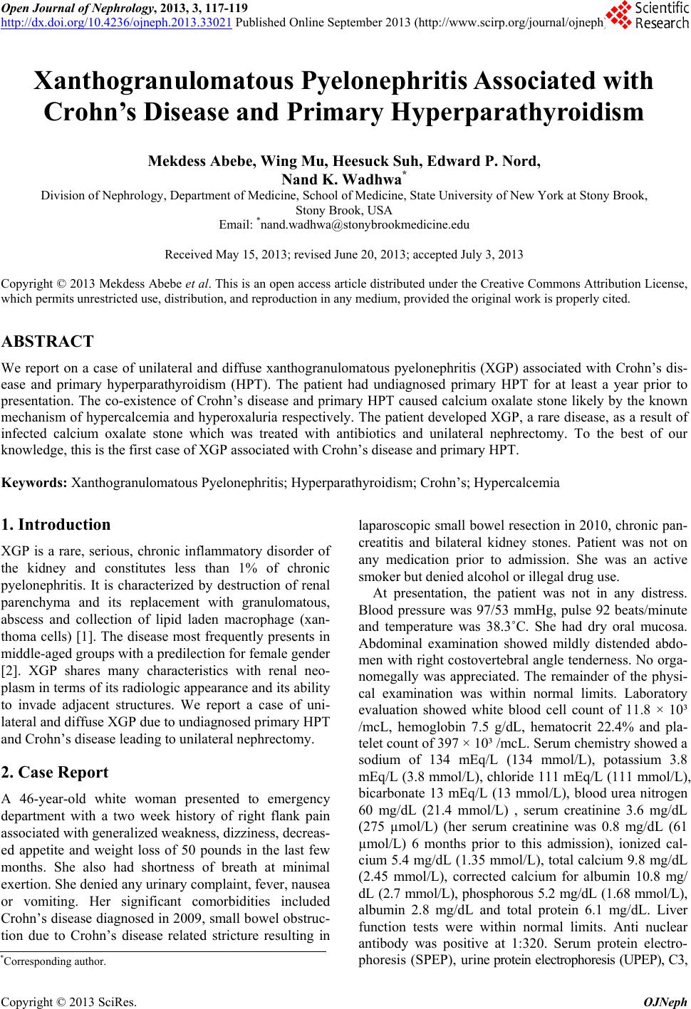
RBC. Renal ultrasound and non-co ntrast computed tomo-
graphy (CT) of the abdomen showed multiple hetero-
geneous and solid app earing lobular soft tissue masses at
the periphery of the mid and lower pole of the right
kidney that extends into the peri-renal space which was
suspicious for malignancy (Figure 1). Bilateral kidney
stones were recognized on CT. MRI of the abdomen
demonstrated enlarged right kidney with some increased
parenchymal signal intensity with poorly defined margin
with peri-nephric infection. However, Indium scan
revealed no evidence of pyelonephritis or renal abscess.
Medical therapy with IV hydration and antibiotics, ini-
tially ceftriaxone and ciprofloxacin, later aztreonam for a
total of 4 weeks were administered. In view of her hyper-
calcemia, she was investigated further and was found to
have elevated serum PTH of 390 pg/mL. A sestamebi
scan and an ultrasound of the neck revealed a right
parathyroid adenoma.
Two weeks after admission, because of persistence of
the right renal mass, CT guided percutaneous biopsy was
performed and revealed 43 cc of turbid fluid which was
negative for malignant cells and culture. Further testing
of the right kidney function using furosemide renal scan
demonstrated residual renal function of <10%. Sub-
sequently, due to suspicion of malignancy in the face of
non functioning and possibly infected right kidney,
patient underwent right nephrectomy 5 weeks after ad-
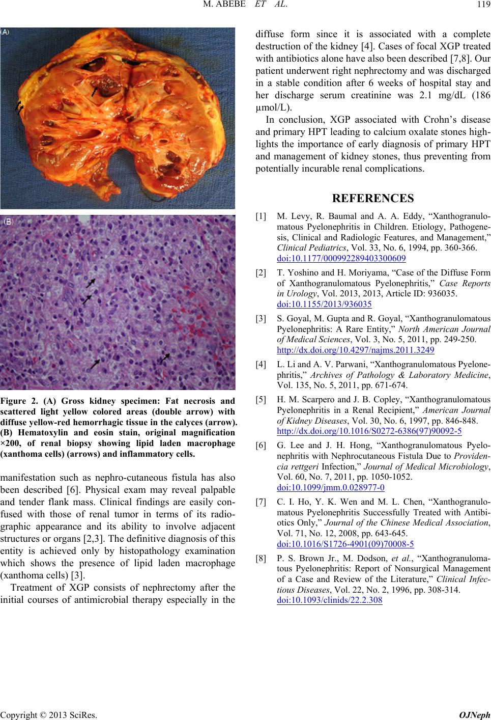
mission. Gross examination showed a peri-nephric fat
adherent to the capsule of the kidney with fat necrosis
and diffuse yellow-red hemorrhagic tissue (Figure 2(A)).
There was also a 2 cm yellow crushable stone at the
distal ureter occupying the entire lumen. Histopatho-
logy revealed acute and moderate chronic inflammation
with focal marked increased interstitial eosinophils and
lipid laden macrophage (xanthoma cells) (Figure 2(B)),
consistent with XGP pyelonephritis. There was also
nephrocalcinosis, both polarizable and non-polarizable.
Chemical analysis of the stone revealed calcium oxalate
stone. Simultaneously right para- thyroidectomy was also
performed for parathyroid adenoma. After six weeks of
hospital stay, her serum creatinine level was stable at 2.1
mg/dl (160 µmol/L) and was discharged in a stable con-
dition.
3. Discussion
We report on a case of XGP associated with Crohn’s
disease and primary HPT which was unrecognized for at
least a year prior to her present presentation. The co-
existence of primary HPT and Crohn’s disease caused
calcium oxalate stone by the known mechanisms of hy-
percalcemia and hyperoxaluria respectively. To the best
of our knowledg e, this is the first case of XGP secondary
to Crohn’s disease and primary HPT which in turn leads
to calcium oxalate stones.
XGP is a rare chronic inflammation of the kidney and
constitutes less than 1% of chronic pyelonephritis. It is
characterized by destruction of renal parenchyma and its
replacement with granulomas, abscesses and collection
of lipid laden macrophages (xanthoma cells) [1-3]. Fe-
males are more affected than males with a ratio of 1:4,
and with a mean age varing from 45 to 55 years [3,4].
Though the exact mechanism of XGP is not clear, a
number of predisposing factors have been implicated.
The two most common predisposing factors are obstruc-
tion and infection of the genitor-urinary system. Calculi
frequently stag horn type may be seen from 47% to
100% of cases. It is also more commonly seen in renal
transplant recipients and diabetics [4,5]. In our patient,
the combination of primary HPT and Crohn’s disease
leading to kidney stone formation and obstruction, in
addition to her gender and age, are the most likely pre-
disposing factors. XGP is commonly associated with
Escherichia coli and Proteus infection; however bacterial
outgrowth from urine culture is not necessary for diag-
nosis, as in our patient [2,4].
The disease process affects the whole of the kidney in
85% (diffuse form). Focal forms are rare (15%). The le-
sion is generally unilateral and can affect either kidney
with equal frequency. Bilateral lesions are rare and are
associated with poor outcome [2,3]. In the present case, a
diffuse and unilateral form of XGP is observed. Symp-
toms are frequently nonspecific and include flank pain,
fever, weight loss, anorexia and malaise. Uncommon
Figure 1. Non-contrast computed tomography of the abdo-
men and pelvis showing solid appearing soft tissue mass at
the periphery of the mid and lower pole of the right kidney
extending into the peri-nephric space (arrow) and renal
stone (arrow head).
Copyright © 2013 SciRes. OJNeph
M. ABEBE ET AL.
Copyright © 2013 SciRes. OJNeph
119
Figure 2. (A) Gross kidney specimen: Fat necrosis and
scattered light yellow colored areas (double arrow) with
diffuse yellow-red hemorrhagic tissue in the calyces (arrow).
(B) Hematoxylin and eosin stain, original magnification
×200, of renal biopsy showing lipid laden macrophage
(xanthoma cells) (arrows) and inflammatory cells.
manifestation such as nephro-cutaneous fistula has also
been described [6]. Physical exam may reveal palpable
and tender flank mass. Clinical findings are easily con-
fused with those of renal tumor in terms of its radio-
graphic appearance and its ability to involve adjacent
structures or organs [2,3]. The d efinitive diagn osis of this
entity is achieved only by histopathology examination
which shows the presence of lipid laden macrophage
(xanthoma cells) [3].
Treatment of XGP consists of nephrectomy after the
initial courses of antimicrobial therapy especially in the
diffuse form since it is associated with a complete
destruction of the kidn ey [4]. Cases of focal XGP treated
with antibiotics alon e have also been described [7 ,8]. Our
patient underwent right nephrectomy and was discharged
in a stable condition after 6 weeks of hospital stay and
her discharge serum creatinine was 2.1 mg/dL (186
µmol/L).
In conclusion, XGP associated with Crohn’s disease
and primary HPT leading to calcium oxalate stones high-
lights the importance of early diagnosis of primary HPT
and management of kidney stones, thus preventing from
potentially incu rable renal complications.
REFERENCES
[1] M. Levy, R. Baumal and A. A. Eddy, “Xanthogranulo-
matous Pyelonephritis in Children. Etiology, Pathogene-
sis, Clinical and Radiologic Features, and Management,”
Clinical Ped i atrics, Vol. 33, No. 6, 1994, pp. 360-366.
doi:10.1177/000992289403300609
[2] T. Yoshino and H. Moriyama, “Case of the Diffuse Form
of Xanthogranulomatous Pyelonephritis,” Case Reports
in Urology, Vol. 2013, 2013, Article ID: 936035.
doi:10.1155/2013/936035
[3] S. Goyal, M. Gupta and R. Goyal, “Xanthogranulomatous
Pyelonephritis: A Rare Entity,” North American Journal
of Medical Sciences, Vol. 3, No. 5, 2011, pp. 249-250.
http://dx.doi.org/10.4297/najms.2011.3249
[4] L. Li and A. V. Parwani, “Xanthogranulomatous Pyelone-
phritis,” Archives of Pathology & Laboratory Medicine,
Vol. 135, No. 5, 2011, pp. 671-674.
[5] H. M. Scarpero and J. B. Copley, “Xanthogranulomatous
Pyelonephritis in a Renal Recipient,” American Journal
of Kidney Diseases, Vol. 30, No. 6, 1997, pp. 846-848.
http://dx.doi.org/10.1016/S0272-6386(97)90092-5
[6] G. Lee and J. H. Hong, “Xanthogranulomatous Pyelo-
nephritis with Nephrocutaneous Fistula Due to Providen-
cia rettgeri Infection,” Journal of Medical Microbiology,
Vol. 60, No. 7, 2011, pp. 1050-1052.
doi:10.1099/jmm.0.028977-0
[7] C. I. Ho, Y. K. Wen and M. L. Chen, “Xanthogranulo-
matous Pyelonephritis Successfully Treated with Antibi-
otics Only,” Journal of the Chinese Medical Association,
Vol. 71, No. 12, 2008, pp. 643-645.
doi:10.1016/S1726-4901(09)70008-5
[8] P. S. Brown Jr., M. Dodson, et al., “Xanthogranuloma-
tous Pyelonephritis: Report of Nonsurgical Management
of a Case and Review of the Literature,” Clinical Infec-
tious Diseases, Vol. 22, No. 2, 1996, pp. 308-314.
doi:10.1093/clinids/22.2.308