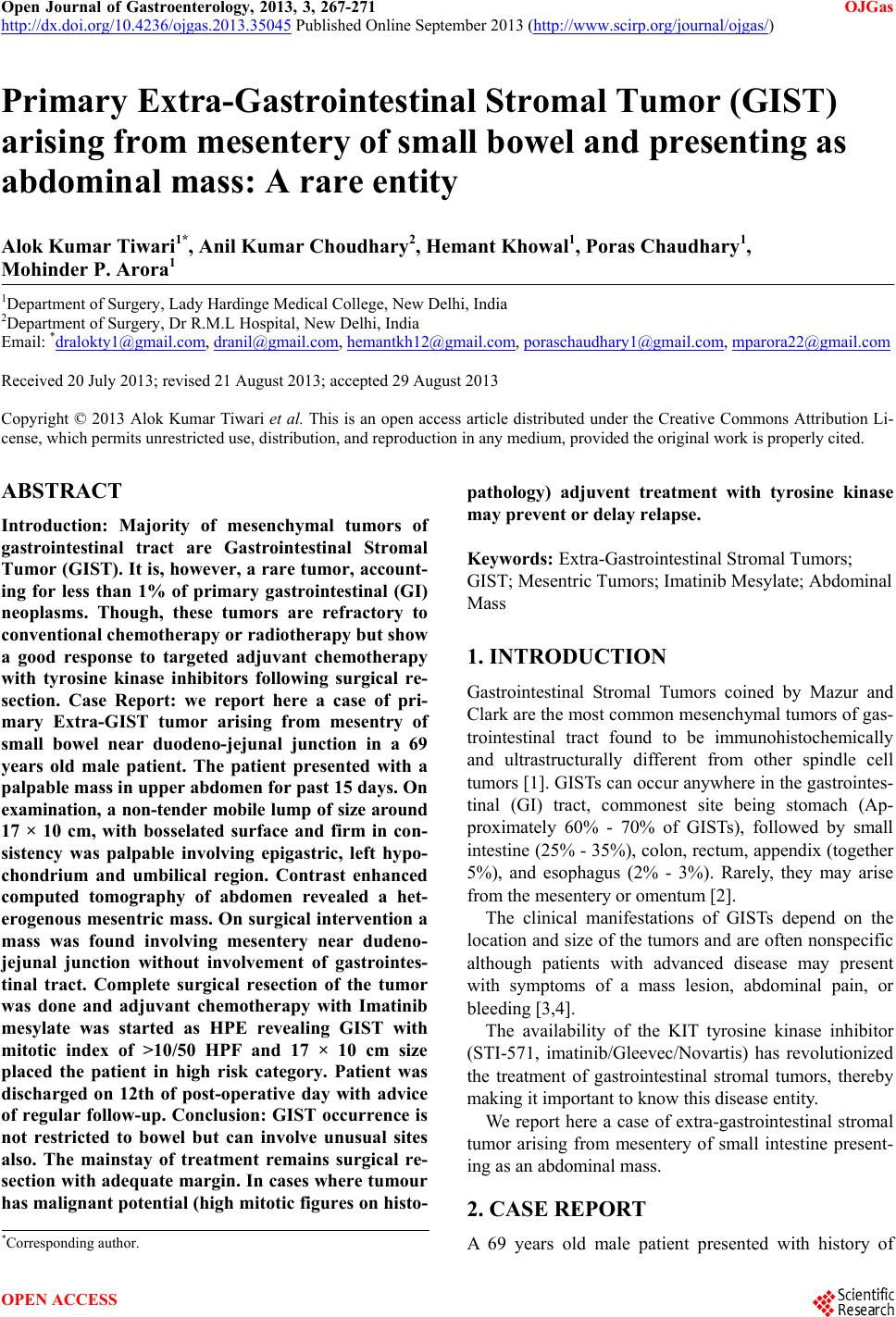
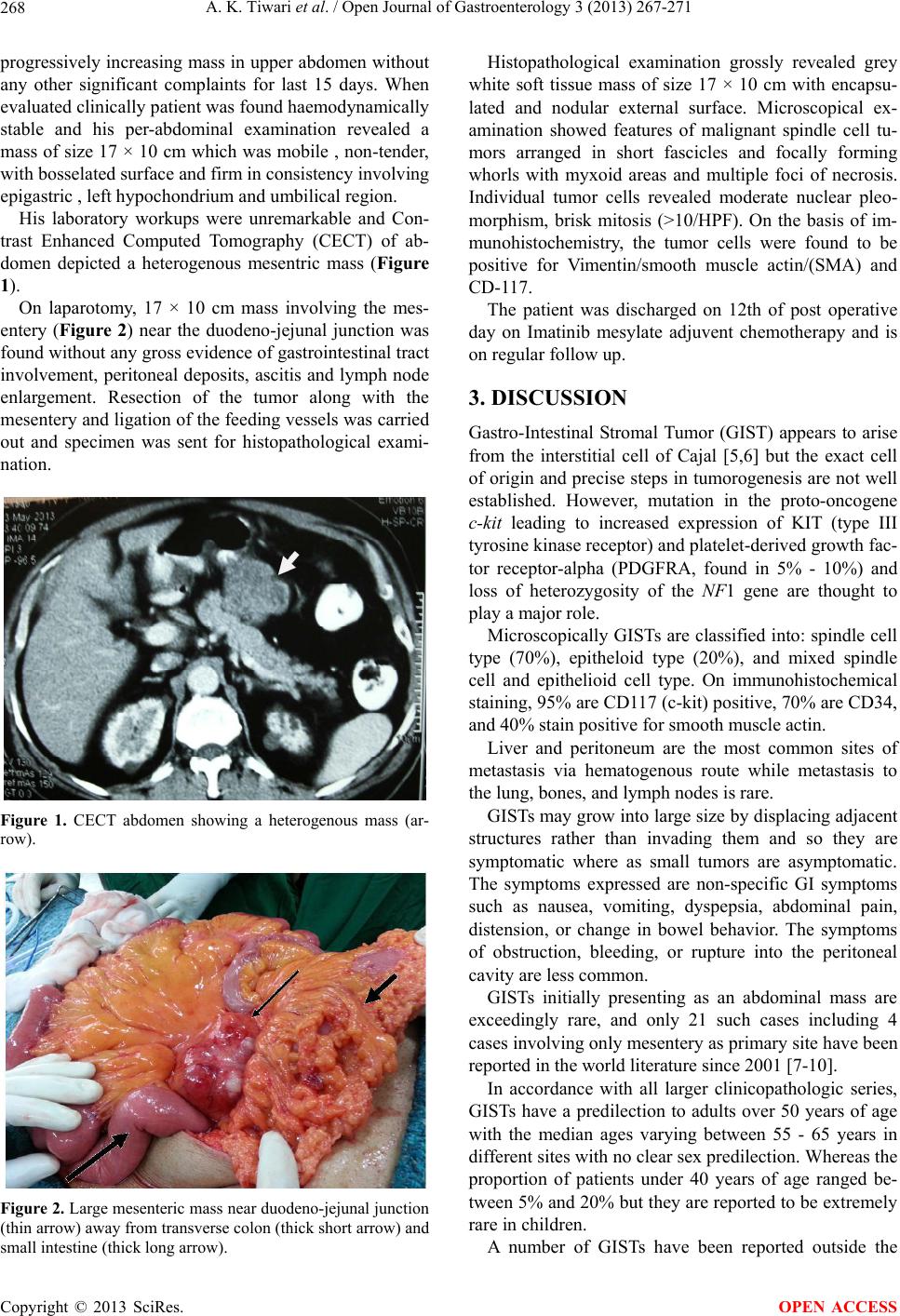
A. K. Tiwari et al. / Open Journal of Gastroenterology 3 (2013) 267-271
270
Most recurrences occurs within the first 2 years of re-
section necessitating the regular follow up of these pa-
tients.
A high risk patient should have a CT scan every 3 - 4
months for 3 years, then every 6 months to 5 years. For
low risk, a CT scan every 6 months for 5 years is ac-
ceptable [14].
GISTs of the jejunum and ileum treated surgically
have been shown to have a 39% tumor related mortality,
which was twice that of gastric GISTs [19].
Our case had mesenteric stromal tumor near duodeno-
jejunal junction, mitoses were >10 per 50 HPF and total
tumor diameter was 17 cm. The localization was unfa-
vorable but bad prognostic signs such as liver or lymph
node metastases were not seen.
Gene expression patterns in GISTs are assessed by
DNA microarray techniques. The technique revealed that
the gene FLJ10261 responsible for encoding the DOG1
protein is specifically expressed in GISTs, irrespective of
KIT or PDGFRA mutation status. However, its function
is not well understood, although it seems to be fairly
specific to GIST and rarely being expressed in other soft
tissue tumors. In future it may play a pivotal role in di-
agnosis of GISTs, especially in PDGFR mutants failing
to express the KIT antigen [20].
5. CONCLUSION
GIST occurrence is not restricted to bowel but can in-
volve unusual sites also and involvement of mesentery
near dudeno-jejunal junction is very rare. The mainstay
of treatment remains surgical resection with adequate
margin. In cases where tumor has malignant potential
based on high mitotic figures on histopathology adjuvent
treatment with tyrosine kinase may prevent or delay re-
lapse. DNA microarray technique may play a vital role in
identifying the gene encoding DOG1 protein in mutants
that does not express KIT antigen and thus may help in
its diagnosis.
6. AUTHOR’S CONTRIBUTIONS
All authors have contributed to patient management,
writing the case report, reviewing and have given the
final approval for publishing the manuscript.
REFERENCES
[1] Mazur, M.T. and Clark, H.B. (1983) Gastric stromal tu-
mours. Reappraisal of their histogenesis. American Jour-
nal of Surgical Pathology, 7, 507-519.
doi:10.1097/00000478-198309000-00001
[2] Miettinen, M. and Lasota, J. (2001) Gastrointestinal stro-
mal tumors: Definition, clinical, histological, immuno-
histochemical, and molecular genetic features and differ-
ential diagnosis. Virchows Arch, 438, 1-12.
doi:10.1007/s004280000338
[3] Filippou, D.K., Pashalidis, N., Skandalakis, P. and Rizos,
S. (2006) Malignant gastrointestinal stromal tumor of the
ampulla of Vater presenting with obstructive jaundice.
Journal of Postgraduate Medicine, 52, 204-206.
[4] Towu, E. and Stanton, M. (2006) Gastrointestinal stromal
tumour presenting with severe bleeding: A review of the
molecular biology. Pediatric Surgery International, 22,
462-464. doi:10.1007/s00383-006-1636-5
[5] Torihashi, S., Nishi, K., Tokutomi, Y., Nishi, T., Ward, S.
and Sanders, K.M. (1999) Blockade of kit signaling in-
duces transdifferentiation of interstitial cells of Cajal to a
smooth muscle phenotype. Gastroenterology, 117, 140-
148. doi:10.1016/S0016-5085(99)70560-3
[6] Young, H.M., Ciampoli, D. and Southwell Newgreen,
D.F. (1996) Origin of interstitial cells of Cajal in the
mouse intestine. Developmental Biology, 180, 97-107.
doi:10.1006/dbio.1996.0287
[7] Sinha, R., Verma, R. and Kong, A. (2004) Mesenteric
gastrointestinal stromal tumor in a patient with neurofi-
bromatosis. American Journal of Roentgenology, 183,
1844-1846. doi:10.2214/ajr.183.6.01831844
[8] Basile, A., Kettenbach, J., Mundo, E., et al. (2006) Erra-
tum: Primitive mesenteric gastrointestinal stromal tumor
with autonomic nerve/ganglionic differentiation present-
ing as a huge mass with small synchronous nodules.
European Radiology, 16, 519.
doi:10.1007/s00330-005-2845-3
[9] Gupta, N., Mittal, S., Lal, N., Misra, R., Kumar, L. and
Bhalla, S. (2007) A rare case of primary mesenteric gas-
trointestinal stromal tumor with metastasis to the cervix
uteri. World Journal of Surgical Oncology, 5, 137.
doi:10.1186/1477-7819-5-137
[10] Patil, S., Jain, S., Kaza, R.C.M. and Chamberlain, R.S.
(2011) Giant gastrointestinal stromal tumor presenting as
a palpable abdominal mass: An unusual presentation.
ISRN Surgery, 2011, Article ID: 894829.
[11] Miettinen, M., Monihan, J.M., Sarlomo-Rikala, M., Ko-
vatich, A.J., Carr, N.J., Emory, T.S. and Sobin, L.H.
(1999) Gastrointestinal stromal tumors/smooth muscle tu-
mors/GISTs in the omentum and mesentery—Clinicopa-
thologic and immunohistochemical study of 26 cases.
American Journal of Surgical Pathology, 23, 1109-1118.
doi:10.1097/00000478-199909000-00015
[12] Reith, J.D., Goldblum, J.R., Lyles, R.H. and Weiss, S.W.
(2000) Extragastrointestinal (soft tissue) stromal tumors.
An analysis of 48 cases with GIST 23 emphasis on histo-
logical predictors of outcome. Modern Pathology, 13,
577-585. doi:10.1038/modpathol.3880099
[13] Chourmouzi, D., Sinakos, E., Papalavrentios, L., Akrivia-
dis, E. and Drevelegas A. (2009) Gastrointestinal stromal
tumors: A pictorial review. Journal of Gastrointestinal
and Liver Diseases, 18, 379-383.
[14] Blay, J.Y., Bonvalot, S., Casali, P., et al. (2005) Consen-
sus meeting for the management of gastrointestinal stro-
mal tumors. Annals of Oncology, 16, 993.
[15] Cunningham, R.E., Federspiel, B.H., McCarthy, W.F.,
Sobin, L.H. and O’Leary, T.J. (1993) Predicting progno-
Copyright © 2013 SciRes. OPEN ACCESS