Open Journal of Orthopedics, 2013, 3, 220-223
http://dx.doi.org/10.4236/ojo.2013.35041 Published Online September 2013 (http://www.scirp.org/journal/ojo)
A Rare Case of Fish Bone Penetrating Injury to Radius
Causing Osteitis and Non-Healing Sinus*
Paragjyoti Gogoi1#, Krishanu Kakati1, Sunil Poonia1, Aditi Das2
1Department of Orthopaedics & Trauma, Silc har Medical College, Silchar, India; 2Department of Radio-Diagnosis, Silchar Medical
College, Silchar, India.
Email: #pggogoiparag@gmail.com
Received July 25th, 2013; revised August 21st, 2013; accepted August 28th, 2013
Copyright © 2013 Paragjyoti Gogoi et al. This is an open access article distributed under the Creative Commons Attribution License,
which permits unrestricted use, distribution, and reproduction in any medium, provided the original work is properly cited.
ABSTRACT
Penetrating injury to radius or any other bone by fish bone is a rare kind of injury. Commonly metallic sharp objects,
bullets, sharpnels, glass particles, hard wooden pieces are the causes of penetrating injury to bones. Radio-opaque ob-
jects are easy to detect and thus retrieval is relatively simpler than the radiolucent objects. Non-biological foreign bod-
ies do not incite any reaction in the host bone whereas biological foreign bodies of either plant or animal origin cause
hypersensitivity reaction. Osteitis along with excessive formation of new bones may be presented as a part of host reac-
tion to the foreign body. We are presenting here an interesting case of a fisherman boy who sustained penetrating injury
to radius by fish bone resulting in osteitis and a chronic non-healing sinus with watery discharge.
Keywords: Fish Bone; Foreign Body; Granuloma; Penetrating Injury; Radio-Opaque
1. Introduction
Foreign body retained in the soft tissues of extremities
causing sinus formation is not uncommon. Mostly the
working class people are affected. Wooden foreign body,
blackthorn injury, glass particle, metallic pieces are the
major causes of granuloma formation along with sinus
[1-3]. A case of fisherman penetrating injury by fish
bone along with granuloma formation is reported [4].
However, fish bone penetrating the bone causing osteitis
and granuloma formation is very rare. We are reporting a
very unusual case of fish bone injury to forearm where
the fish bone penetrated the radius and there were osteitis
and granuloma formations along with a sinus. Perhaps
this is the first case of this kind. The purpose of this
paper is to highlight the uniqueness and the rarity of this
case and to bring to the fore the necessary discussion in
management of medical and surgical oddities.
2. Case Report
A 14-year-old boy attended our outpatient department
with pain over left forearm and a discharging sinus on
the volar aspect. He gave a peculiar history of sustaining
trauma over his left forearm. He hailed from a fisherman
family and while helping his father out in transferring big
fishes from one container to another he fell down over a
big fish and sustained injury over his left forearm. There
was a pricking injury which healed after primary treat-
ment and some medications. He was normal for four
months except some catch in some rotational movement.
Gradually he started having pain over the forearm along
with slight swelling. With in two weeks of pain, he devel-
oped a sinus over the volar aspect of left forearm near the
old pricking site with some watery discharge.
On examination a scarred area was noted over the
volar aspect of mid-forearm with a small sinus with wa-
tery discharge (Figure 1). The sinus seemed to be ad-
hered to underlying soft tissues and mild tenderness on
palpation was noted. Range of movement of elbow and
wrist were within normal limits. There was mild pain at
the extremes of pronation and supination of the forearm.
X-rays of the forearm revealed a radio-opaque linear
foreign body impinging the radius and surrounded by
new bone formation and a lytic radio-lucent ream (Fig-
ures 2 and 3). Marked osteitis with extensive periosteal
reaction is noted both proximal and distal to the foreign
body. Musculo-skeletal ultrasonography using high fre-
quency probe confirmed the foreign body along with
formation of a granulomatous mass surrounding it (Fig-
*Conflict of Interests: The authors declare that they have no conflict o
f
interests. Consent: Informed consent was taken f o r this work.
#Corresponding author.
Copyright © 2013 SciRes. OJO
A Rare Case of Fish Bone Penetrating Injury to Radius Causing O s te i t i s a nd Non-Healing Sinus 221
Figure 1. Sinus formation in forearm.
Figure 2. AP view showing fish bone and osteitis of radius
with new bone formation.
Figure 3. Lateral view show ing r adio-luce ny around the fis h
bone and periosteal reaction of radius.
ures 4 and 5). There was no injury to the neuro-vascular
structures.
Blood parameters revealed leucocytosis with eosino-
philia and ESR was mildly elevated. Chest X-ray was
normal.
From the clinical history and radiological pictures the
diagnosis of retained foreign body impinging the radius
with osteitis was made and decision taken to explore the
lesion.
The lesion was addressed by anterior Henry’s ap-
proach. The sinus was excised. The area was exposed
well with due care and the fish bone iden tified impinging
the radius. There was necrotic area around the fish bone
covered by a fibrotic pseudo-capsule. The fish bone re-
moved in single piece and measured 28 mm with a sharp
pointing end (Figures 6 and 7). The fibous pseudocap-
sule surrounding the fish bone was excised (Figure 8).
Extensive new bone formation was noted in the radius
around the impinged area. There was a central crater
filled with necrotic material surrounded by new bones
(Figure 9). The new bones were chiseled out to get a
smooth contour of the radius (Figure 10). Thorough cu-
rettage of the area was done and the material sent for
histology. Histological picture of the curetted material
was consistent with those of a granuloma.
Figure 4. Ultrasonography of forearm.
Figure 5. USG showing embedded for eign body.
Figure 6. Fish bone removed in toto.
Copyright © 2013 SciRes. OJO
A Rare Case of Fish Bone Penetrating Injury to Radius Causing Osteitis and Non-Healing Sinus
222
Figure 7. Fish bone measuring 28 mm.
Figure 8. Excised fibrous pseud o c a psule .
Figure 9. Crater on the ra dius surr ounded by new bone s.
Figure 10. Excessive new bone chiseled out.
Post-operative X-rays confirmed thorough removal of
the foreign body an d removal of new bones (Figure 11).
The surgical wound healed well and the boy got rid of
the symptoms. He was followed up for six months and no
Figure 11. Post operative X-ray.
recurrence of sinus was there.
3. Discussion
Fishing is regarded as one of the oldest professions.
Many people derive their livelihood from fishing. It is
also seen as a great recreational activity. But this is also
not free from injury risks like many other professions.
Mostly injury occurs due to penetrating injury by the fins
to the extremities [4,5]. In some rare instances penetra-
ting injury to heart can occur by long stings of some
species [6]. Some fishes contain venom in their stings
and fish envenomatosis require special care and treat-
ment. Apart from broad spectrum antibiotics, tetanus
toxoid, analgesics, this condition may demand specific
antivenom administration [7].
There have been many case reports on fish bone inju-
ries to hands and forearm as is evident in the study car-
ried out by Imberg R. et al. [4] in which 116 patients
were followed up with injuries sustained by fish bone to
their hands and forearm. Strangely enough not a single
case reported embedding of the fish bone to bone. To the
best of our knowledge the above mentioned study is the
most extensive one carried out in this matter.
Diagnosis of retained fish bone is not difficult. There
is always a history of penetrating injury resulting a skin
wound. Irrespective of the history it should be presumed
that there is a part of fish bone inside the body until all
investigations are over. X-ray usually detects the radio-
opaque fish bones. In case of radio-lucent objects ultraso-
nography is helpful [1,8]. Normally CT scan or MRI scan
is not required.
Inoculation of polymicrobial flora following fish bone
injury is a concern. There are reports of wound contami-
nation by unusual species like vibrio, actino-mycosis etc.
[9,10]. Broad spectrum antibiotics should always be re-
commended for such injury. Tetanus toxoid also may
have a role in preventing life threaten ing toxaemia.
Retained foreign bodies in the soft tissues incite granu-
lomatous reaction. The host tries to isolate the biological
Copyright © 2013 SciRes. OJO
A Rare Case of Fish Bone Penetrating Injury to Radius Causing O s te i t i s a nd Non-Healing Sinus
Copyright © 2013 SciRes. OJO
223
foreign body by forming a granuloma around it [8]. In
the centre of the granuloma there lies the foreign body
and a layer of necrotic tissues surrounding it.
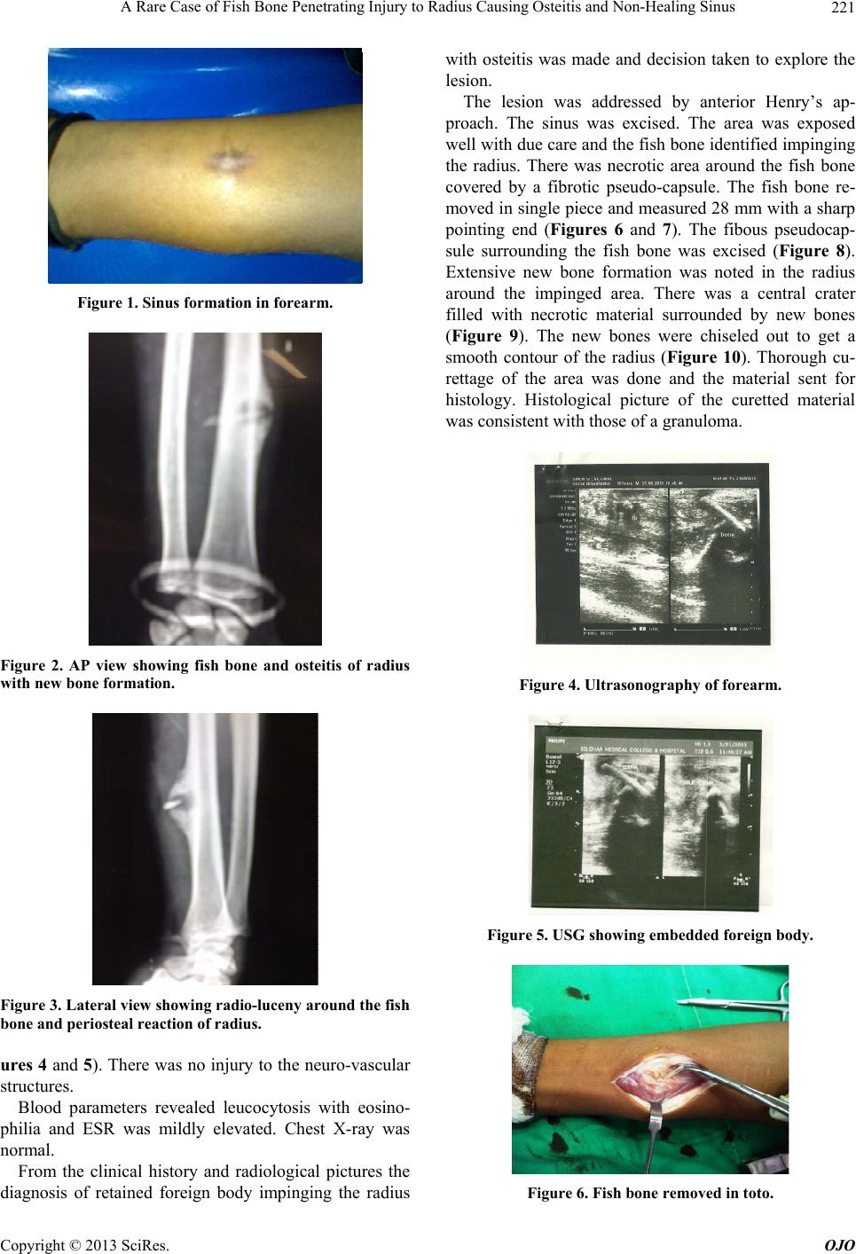
Penetration of the host bone by fish bone is a rare
event. There is a host reaction in the form of osteitis or
periosteal reaction. There is formation of excessive bone
surrounding the fish bone which is akin to granuloma
formation in soft tissues. In this particular case the fish
bone was long enough to have partly inside the radius
and partly in soft tissues of fo rearm. Th erefore w e found
excessive fibrous tissu es surrounding the fish bone in th e
soft tissue part and excessive bone formation around it in
the bony tract. The rareness of this case and the relative
dearth of literature in this matter compelled us to direct
our treatment towards normal protocol of granulamatous
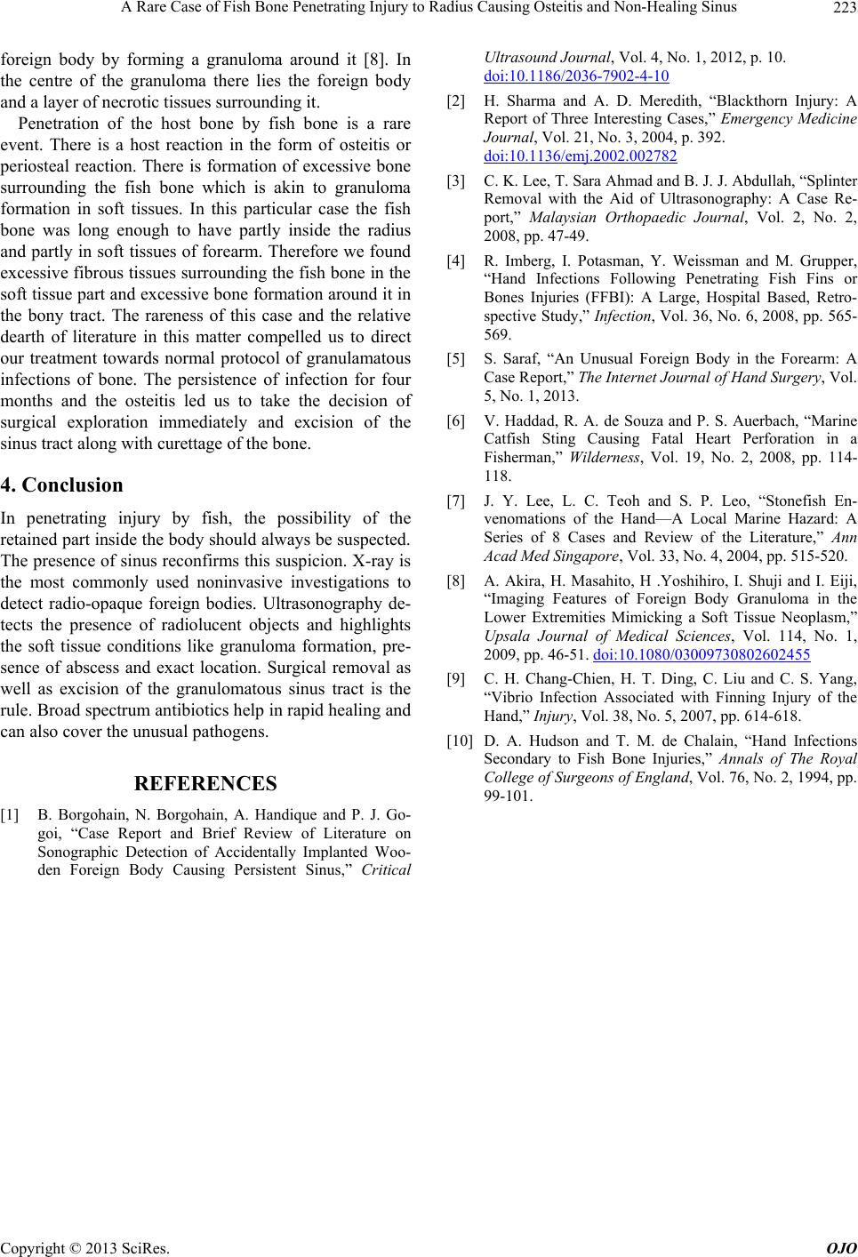
infections of bone. The persistence of infection for four
months and the osteitis led us to take the decision of
surgical exploration immediately and excision of the
sinus tract along with curettage of the bon e.
4. Conclusion
In penetrating injury by fish, the possibility of the
retained part inside the body should always be su spected.
The presence of sinus reconfirms this suspicion. X-ray is
the most commonly used noninvasive investigations to
detect radio-opaque foreign bodies. Ultrasonography de-
tects the presence of radiolucent objects and highlights
the soft tissue conditions like granuloma formation, pre-
sence of abscess and exact location. Surgical removal as
well as excision of the granulomatous sinus tract is the
rule. Broad spectrum antibiotics help in rapid healing and
can also cover the unusual pathogens.
REFERENCES
[1] B. Borgohain, N. Borgohain, A. Handique and P. J. Go-
goi, “Case Report and Brief Review of Literature on
Sonographic Detection of Accidentally Implanted Woo-
den Foreign Body Causing Persistent Sinus,” Critical
Ultrasound Journal, Vol. 4, No. 1, 2012, p. 10.
doi:10.1186/2036-7902-4-10
[2] H. Sharma and A. D. Meredith, “Blackthorn Injury: A
Report of Three Interesting Cases,” Emergency Medicine
Journal, Vol. 21, No. 3, 2004, p. 392.
doi:10.1136/emj.2002.002782
[3] C. K. Lee, T. Sara Ahmad and B. J. J. Abdullah, “Splinter
Removal with the Aid of Ultrasonography: A Case Re-
port,” Malaysian Orthopaedic Journal, Vol. 2, No. 2,
2008, pp. 47-49.
[4] R. Imberg, I. Potasman, Y. Weissman and M. Grupper,
“Hand Infections Following Penetrating Fish Fins or
Bones Injuries (FFBI): A Large, Hospital Based, Retro-
spective Study,” Infection, Vol. 36, No. 6, 2008, pp. 565-
569.
[5] S. Saraf, “An Unusual Foreign Body in the Forearm: A
Case Report,” The Internet Journal of Hand Surgery, Vol.
5, No. 1, 2013.
[6] V. Haddad, R. A. de Souza and P. S. Auerbach, “Marine
Catfish Sting Causing Fatal Heart Perforation in a
Fisherman,” Wilderness, Vol. 19, No. 2, 2008, pp. 114-
118.
[7] J. Y. Lee, L. C. Teoh and S. P. Leo, “Stonefish En-
venomations of the Hand—A Local Marine Hazard: A
Series of 8 Cases and Review of the Literature,” Ann
Acad Med Singapore, Vol. 33, No. 4, 2004, pp. 515-520.
[8] A. Akira, H. Masahito, H .Yoshihiro, I. Shuji and I. Eiji,
“Imaging Features of Foreign Body Granuloma in the
Lower Extremities Mimicking a Soft Tissue Neoplasm,”
Upsala Journal of Medical Sciences, Vol. 114, No. 1,
2009, pp. 46-51. doi:10.1080/03009730802602455
[9] C. H. Chang-Chien, H. T. Ding, C. Liu and C. S. Yang,
“Vibrio Infection Associated with Finning Injury of the
Hand,” Injury, Vol. 38, No. 5, 2007, pp. 614-618.
[10] D. A. Hudson and T. M. de Chalain, “Hand Infections
Secondary to Fish Bone Injuries,” Annals of The Royal
College of Surgeons of England, Vol. 76, No. 2, 1994, pp.
99-101.