Open Journal of Radiology, 2013, 3, 108-111
http://dx.doi.org/10.4236/ojrad.2013.33017 Published Online September 2013 (http://www.scirp.org/journal/ojrad)
The Pathognomic Radiologic Features of Gout in the
Fingers and Review of the Literature, Including the
Latest Drug Therapy
Mehdi Hossaini1*, Eric Tetteroo2, Frank M. A. Slaats3, Annechien Beumer4
1Department of Orthopaedic Surgery, Catharina Hospital, Eindhoven, The Netherlands
2Department of Radiology, Amphia Hospital Breda, Breda, The Netherlands
3Department of Rheumatolog y, Amphia Hospit al Breda, Breda, The Netherlands
4Department of Orthopaed ic Surgery, Amphia Hospital Breda, The Netherlands
Email: *hossaini.ariyan@gmail.com
Received June 27, 2013; revised July 27, 2013; accepted August 4, 2013
Copyright © 2013 Mehdi Hossaini et al. This is an open access article distributed under the Creative Commons Attribution License,
which permits unrestricted use, distribution, and reproduction in any medium, provided the original work is properly cited.
ABSTRACT
Gout is one of the most common inflamma tory arthritid es that pr edominantly affects the first metatarsophalang eal joint.
The underlying inflammatory process is elicited by the deposition of monosodium urate (MSU) crystals in the affected
joint that are termed tophi and wh ich may in the course of ch ronic gout progress into subcutaneous depositions of tophi.
In this case report, we present a patient (PID: 6925981) who was seen in our outpatient department with a first episode
of tophaceous gout in the finger, an infrequently seen condition that mostly affects older men with long-term gout. We
discuss the case in view of the current literature on diagnosis and treatment of tophaceous gou t.
Keywords: Gout; Tophi; Interphalang eal Joint; Finger; Monosodium Urate; Arthritis
1. Introduction
Gout is a form of arthritis and it is one of the earliest
recognized clinical entities in medical history. Gout was
first identified by the Egyptians in 2640 BC and later
described by Hippocrates and Galen [1]. The underlying
pathophysiology is an inflammatory process induced by
deposition of monosodium urate (MSU) crystals in ar-
ticular and periarticular tissues [2], and the presence of
these negatively birefringent crystals in the aspirated
specimen of the affected joint is a prerequisite for the
diagnosis of gout. The first sign of a gouty arthritis is
usually an acute painful attack in the first metatarsopha-
langeal joint of the foot, a condition termed podagra [3],
while other joints are less frequ ently affected [4]. Gou t is
strongly associated with an increased level of uric acid in
the blood but hyperuricaemia is not required for the de-
velopment of gout and patients with hyperuricaemia may
remain asymptomatic throughout their lives [5]. Further,
gout is often associated with obesity, metabolic syn-
dromes, chronic kidney and/or cardiovascular diseases [3,
6]. The use of certain drugs such as diuretics (Furose
mide, Hydrochlorothiazide), immunosuppressant (Cyc-
losporin) and low dose Aspirin may also be correlated
with hyperuricaemia and gout [7]. The incidence of gout
is rising worldwide and this may be related to aging and
the increased prevalence of obesity [8-10]. In addition,
inadequate treatment and sedentary lifestyles with high
intakes of alcohol, protein and fructose (the only carbo-
hydrate known to increase uric acid levels) may lead to
chronic forms of gout with periodic flare-ups [11-13].
2. Case Report
An eighty year old male patient with a long history of
gout visited our outpatient department complaining of
axial pain in the proximal interphalangeal (PIP) joint of
his left third finger that was present for three weeks. Soft
tissue swelling was seen on the dorsal and palmar sides
of PIP with slightly decreased range of motion. Radio-
graphic examination (Figure 1) showed sharply margi-
nated juxta-articular erosions and marked soft tissue
swelling. Blood examination showed an elevated uric
acid level of 0.58 mmol/L (normal < 0.30). Over the
years, the patient had painful flares of gout limited to his
pedal joints when going on vacations in Mediterranean
areas, likely due to dietary changes including alcohol
*Corresponding author.
C
opyright © 2013 SciRes. OJRad
M. HOSSAINI ET AL. 109
Figure 1. Posteroanterior (A) and lateral (B) third finger
radiographs show at the proximal interphalangeal joint
typical juxta-articular “punched-out” erosions with scle-
rotic borders and overhanging edges (open arrows). There
is marked periarticular hyperdense soft tissue swelling rep-
resenting monosodium urate crystal deposits in thickened
synovia. These so-called tophi are pathognomonic for gout.
Note joint space narrowing due to cartilage destruction,
leading to secondary degenerative osseous changes and
subluxation which can be observed in chronic gout (ar-
rows).
consumption. Further, the patient was treated with Col-
chicine by the rheumatologist and he was aware of the
dietary restrictions advised to control his uric acid levels.
Next to gout, the patient was diagnosed with Polycythe-
mia Vera and he suffered from polyglobulia, thrombocy-
tosis and microcytosis. The patient did not have any
symptomatic cardiovascular and gastrointestinal abnor-
malities except for mild diarrhea related to Colchicine
treatment. After we confirmed tophaceous gout Allo-
purinol was started in order to decrease the serum uric
acid levels that resulted in remission of the gouty attack
in the patient’s finger.
3. Discussion
3.1. Chronic Gout
In the chronic course of gout, densely packed MSU cry-
stals may deposit subcutaneously in (peri) articular tissue
with subsequent jo int damage and painful episodes years
after the first acute painful attack [14]. Subcutaneous
tophi deposition are usually visible in th e helix of the ear,
over the olecranon process and on the Achilles tendons,
and less frequently in the finger joints [14,15]. There are
several case reports on tophaceous gout in the fingers
[16,17] and in most of the patients the gouty attacks are
localized in the proximal interphalangeal joints. Further,
it seems that the incidence of gouty attacks is increasing
in the past decade, notably in men older than 60 years
with the foot and spine being the most affected sites [18].
In the present case, Polycythemia Vera induced blood
dyscrasia and increased cell turnover may produce hyper-
uricaemia leading to gout [19-21]. Further, poor com-
pliance with drug treatment [22] and high protein and
fructose intake [12,13] during the vacations may also
have contributed to the development of tophi deposition
in the finger joint of the pr esent patient. In case of symp-
tomatic gout, Colchicine is the mainstay therapy for pro-
phylaxis and treatment of acute attacks while urate-low-
ering drugs such as Allopurinol are employed for long-
term management and prophylaxis of gout [5,23]. It
should be noted that novel therapies such as anti-inter-
leukin-1 (Anakinra, Canakinumab, Rilonacept), a new
xanthine oxidase inhibitor (Febuxostat) and uricolytic
drugs (Rasburicase, Pegloticase) seem to be beneficial in
treating refractory chron ic gout. In addition , they seem to
improve health-related quality of life, particularly b y pre-
venting cardiovascular comorbidities [24-26]. Drug treat-
ment aimed at lowering uric acid levels will subsequently
lead to tophi resolution and regain of full range of motion.
If drug treatment is inadequate in mobilizing tophaceous
deposits, then operative tophectomy should be consid-
ered in order to avoid permanent joint deformity and loss
of function [27].
3.2. Radiologic Features of Gout
From a radiological point of view, characteristic changes
only occur in the chronic stage of gout. Only 45% of
patients with gout manifest radiographic bone changes,
and then only 6 to 8 years after the initial attack. Fur-
thermore, radiographic abnormalities alone are not suffi-
ciently sensitive and specific for the diagnosis of gout
[28]. Gouty arthritis usually has an asymmetrical polyar-
ticular distribution with a predilectio n for the small joints
of hands and feet. The radiographic hallmarks of gout are
well-defined “punched-out” erosions with sclerotic mar-
gins in a marginal and juxta-articular distribution, with
overhanging edges. Periarticular soft tissue swelling due
to tophi deposition in synovial tissue is common. Tophi
are also found to deposit in cartilage, bone, bursae and
other places throughout the body. The periarticular tophi
may be hyperdense due to the crystals and are pathog-
nomonic for gout. In addition, absence of periarticular
osteopenia (as seen in other common inflammatory ar-
thritides) and preservation of joint space until late stages
of the disease are other clues to the diagnosis. Because
Copyright © 2013 SciRes. OJRad
M. HOSSAINI ET AL.
110
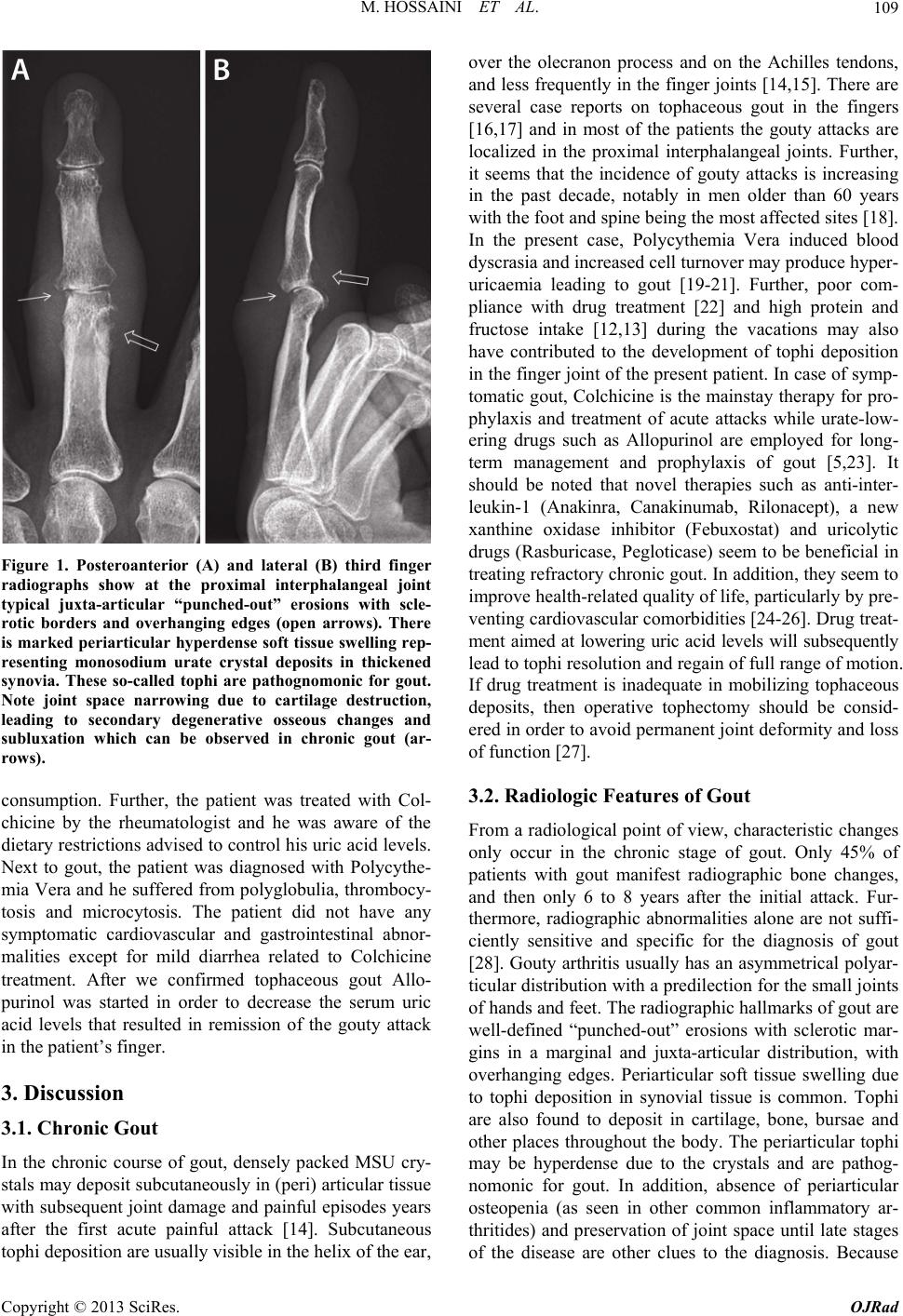
the radiographic findings may at times be confusing and
appear quite unusual, it may be helpful to remember,
“When in doubt, think gout” [29]. However, the refine-
ment in detecting tophi deposition using ultrasound [30,
31] and dual energy computed tomography (CT) has
made it easier to diagnose gout [32]. This will enable us
to more accurately determine the consequence of tophi
depositions on joint deformity and functional impair-
ments and to monitor the effect of urate-lowering drugs
on tophi formation. These outcomes might encourage
new therapeutic approaches in the management and fol-
low-up of patients with gout.
4. Conclusion
In conclusion, patients with chronic gout may complain
of pain in the interphalangeal joints as a result of tophi
deposition that may lead to (peri) articular changes visi-
ble on radiographic examinations. Adequate therapy in-
cludes dissuading the patient from high alcohol, protein
and fructose diets and treatment with up-to-date drugs.
REFERENCES
[1] G. Nuki and P. A. Simkin, “A Concise History of Gout
and Hyperuricemia and Their Treatment,” Arthritis Re-
search & Therapy, Vol. 8, Suppl. 1, 2006, pp. S1-S5.
[2] L. Punzi, A. Scanu, R. Ramonda and F. Oliviero, “Gout
as Autoinflammatory Disease: New Mechanisms for
More Appropriated Treatment Targets,” Autoimmunity
Reviews, Vol. 12, No. 1, 2012, pp. 66-71.
doi:10.1016/j.autrev.2012.07.024
[3] W. Zhang, M. Doherty, E. Pascual, T. Bardin, V.
Barskova, P. Conaghan, J. Gerster, J. Jacobs, B. Leeb, F.
Liote, G. McCarthy , P. Netter, G. Nuki, F. Perez-Ruiz, A.
Pignone, J. Pimentao, L. Punzi, E. Roddy, T. Uhlig and I.
Zimmermann-Gorska, “EULAR Evidence Based Rec-
ommendations for Gout. Part I: Diagnosis. Report of a
Task Force of the Standing Committee for International
Clinical Studies Including Therapeutics (ESCISIT),” An-
nals of the Rheumatic Diseases, Vol. 65, No. 10, 2006, pp.
1301-1311. doi:10.1136/ard.2006.055251
[4] E. Roddy, W. Zhang and M. Doherty, “Are Joints Af-
fected by Gout Also Affected by Osteoarthritis?” Annals
of the Rheumatic Diseases, Vol. 66, No. 10, 2007, pp.
1374-1377. doi:10.1136/ard.2006.063768
[5] P. Richette and T. Bardin, “Gout,” Lancet, Vol. 375, No.
9711, 2010, pp. 318-328.
doi:10.1016/S0140-6736(09)60883-7
[6] H. K. Choi and E. S. Ford, “Prevalence of the Metabolic
Syndrome in Individuals with Hyperuricemia,” The
American Journal of Medicine, Vol. 120, No. 5, 2007, pp.
442-447. doi:10.1016/j.amjmed.2006.06.040
[7] M. Marangella, “Uric Acid Elimination in the Urine.
Pathophysiological Implications,” Contributions to Ne-
phrology, Vol. 147, No. 2005, pp. 132-148.
[8] K. L. Wallace, A. A. Riedel, N. Joseph-Ridge and R.
Wortmann, “Increasing Prevalence of Gout and Hyperu-
ricemia over 10 Years among Older Adults in a Managed
Care Population,” The Journal of Rheumatology, Vol. 31,
No. 8, 2004, pp. 1582-1587.
[9] P. C. Robinson, T. R. Merriman, P. Herbison and J.
Highton, “Hospital Admissions Associated with Gout and
Their Comorbidities in New Zealand and England 1999-
2009,” Rheumatology, Vol. 52, No. 1, 2013, pp. 118-126.
doi:10.1093/rheumatology/kes253
[10] G. Trifiro, P. Morabito, L. Cavagna, C. Ferrajolo, S. Pe-
cchioli, M. Simonetti, E. Bianchini, G. Medea, C. Cricelli,
A. P. Caputi and G. Mazzaglia, “Epidemiology of Gout
and Hyperuricaemia in Italy during the Years 2005-2009:
A Nationwide Population-Based Study,” Annals of the
Rheumatic Diseases, Vol. 72, No. 5, 2013, pp. 694-700.
doi:10.1136/annrheumdis-2011-201254
[11] H. K. Choi, K. Atkinson, E. W. Karlson, W. Willett and
G. Curhan, “Alcohol Intake and Risk of Incident Gout in
Men: A Prospective Study,” Lancet, Vol. 363, No. 9417,
2004, pp. 1277-1281.
doi:10.1016/S0140-6736(04)16000-5
[12] H. K. Choi, K. Atkinson, E. W. Karlson, W. Willett and
G. Curhan, “Purine-Rich Foods, Dairy and Protein Intake,
and the Risk of Gout in Men,” The New England Journal
of Medici ne, Vol. 350, No. 11, 2004, pp. 1093-1103.
doi:10.1056/NEJMoa035700
[13] H. K. Choi and G. Curhan, “Soft Drinks, Fructose Con-
sumption, and the Risk of Gout in Men: Prospective Co-
hort Study,” British Medical Journal, Vol. 336, No. 7639,
2008, pp. 309-312. doi:10.1136/bmj.39449.819271.BE
[14] T. Gibson, “Clinical Features of Gout,” In: M. Hochberg,
Ed., Rheumatology, Mosby, Edinburg, 2003, pp. 1919-
1928.
[15] R. Wortmann, “Gout and Hyperuricaemia,” In: G. Fire-
stein, Ed., Kelleys Textbook of Rheumatology, Saunders
Elseviers, Philadelphia, 2008, pp. 1481-1524.
[16] A. Alexandroff, N. Kirkham and N. Nayak, “A Painless,
Swollen Finger (for 20 Years),” Lancet, Vol. 371, No.
9618, 2008, p. 1114.
doi:10.1016/S0140-6736(08)60487-0
[17] J. M. Geiderman, “An Elderly Woman with a Warm,
Painful Finger,” The Western Journal of Medicine, Vol.
172, No. 1, 2000, pp. 51-52. doi:10.1136/ewjm.172.1.51
[18] F. Bolzetta, N. Veronese, E. Manzato and G. Sergi, “To-
phaceous Gout in the Elderly: A Clinical Case Review,”
Clinical Rheumatology, Vol. 31, No. 7, 2012, pp. 1127-
1132. doi:10.1007/s10067-012-1956-x
[19] A. M. Denman, L. Szur and B. M. Ansell, “Joint Com-
plaints in Polycythaemia Vera,” Annals of the rheumatic
diseases, Vol. 23, No. 1964, pp. 139-144.
[20] M. Denman, L. Szur, and B. M. Ansell, “Hype ruricaemia
in Polycythaemia Vera,” Annals of the Rheumatic Dis-
eases, Vol. 25, No. 4, 1966, pp. 340-344.
[21] L. R. Wasserman, “Polycythemia Vera: Its Course and
Treatment; Relation to Myeloid Metaplasia and Leuke-
mia,” Bulletin of the New York Academy of Medicine, Vol.
30, No. 5, 1954, pp. 343-375.
[22] B. A. Briesacher, S. E. Andrade, H. Fouayzi and K. A.
Copyright © 2013 SciRes. OJRad
M. HOSSAINI ET AL.
Copyright © 2013 SciRes. OJRad
111
Chan, “Comparison of Drug Adherence Rates among Pa-
tients with Seven Different Medical Conditions,” Phar-
macotherapy, Vol. 28, No. 4, 2008, pp. 437-443.
doi:10.1592/phco.28.4.437
[23] A. T. Eggebeen, “Gout: An Update,” American Family
Physician, Vol. 76, No. 6, 2007, pp. 801-808.
[24] K. M. Jordan, “Up-to-Date Management of Gout,” Cur-
rent Opinion in Rheumatology, Vol. 24, No. 2, 2012, pp.
145-151.
[25] F. Perez-Ruiz, L. Martinez-Indart, L. Carmona, A. M.
Herrero-Beites, J. I. Pijoan and E. Krishnan, “Tophaceous
Gout and High Level of Hyperuricaemia are Both Asso-
ciated with Increased Risk of Mortality in Patients with
Gout,” Annals of the Rheumatic Diseases, 2013, in Press.
[26] E. Suresh and P. Das, “Recent Advances in Management
of Gout,” Monthly Journal of the Association of Physi-
cians, Vol. 105, No. 5, 2012, pp. 407-417.
[27] R. H. Gelberman, D. H. Doty and M. L. Hamer, “Topha-
ceous Gout Involving the Proximal Interphalangeal Joint,”
Clinical Orthopaedics and Related Research, Vol. 147,
1980, pp. 225-227.
[28] A. Brower and D. Flemming, “Gout”, In: Arthritis: In
Black and White, WB Saunders, Philadelphia, 1997, pp.
325-341.
[29] J. A. Jacobson, G. Girish, Y. Jiang and B. J. Sabb, “Ra-
diographic Evaluation of Arthritis: Degenerative Joint
Disease and Variations,” Radiology, Vol. 248, No. 3,
2008, pp. 737-747. doi:10.1148/radiol.2483062112
[30] P. V. Chowalloor and H. I. Keen, “A Systematic Review
of Ultrasonography in Gout and Asymptomatic Hyperu-
ricaemia,” Annals of the Rheumatic Diseases, Vol. 72, No.
5, 2013, pp. 638-645.
doi:10.1136/annrheumdis-2012-202301
[31] E. De Miguel, J. G. Puig, C. Castillo, D. Peiteado, R. J.
Torres and E. Martin-Mola, “Diagnosis of Gout in Pa-
tients with Asymptomatic Hyperuricaemia: A Pilot Ul-
trasound Study,” Annals of the Rheumatic Diseases, Vol.
71, No. 1, 2012, pp. 157-158.
doi:10.1136/ard.2011.154997
[32] H. K. Choi, L. C. Burns, K. Shojania, N. Koenig, G. Reid,
M. Abufayyah, G. Law, A. S. Kydd, H. Ouellette and S.
Nicolaou, “Dual Energy CT in Gout: A Prospective Vali-
dation Study,” Annals of the Rheumatic Diseases, Vol. 71,
No. 9, 2012, pp. 1466-1471.
doi:10.1136/annrheumdis-2011-200976