American Journal of Plant Sciences, 2013, 4, 1765-1773
http://dx.doi.org/10.4236/ajps.2013.49217 Published Online September 2013 (http://www.scirp.org/journal/ajps)
Variation of Calcium Oxalate (CaOx) Crystals in Porang
(Amorphophallus muelleri Blume)
Nurul Chairiyah*, Nunung Harijati, Retno Mastuti
Biology Department, Faculty of Mathematic and Natural Sciences, Brawijaya University, Malang, Indonesia.
Email: *nchairiyah@gmail.com
Received June 25th, 2013; revised July 25th, 2013; accepted August 15th, 2013
Copyright © 2013 Nurul Chairiyah et al. This is an open access article distributed under the Creative Commons Attribution License,
which permits unrestricted use, distribution, and reproduction in any medium, provided the original work is properly cited.
ABSTRACT
This research had aimed to determine variations in form of calcium oxalate (CaOx) crystals in porang. Slides for mi-
croscopic observation were prepared from sliced leaf, petiole, and tuber of porang. Sliced organs were cleared by using
modified clearing method. The parameters that were observed included form and variety of morphology type of CaOx
crystals. Microscopic observation showed there were four basic forms of crystals, i.e., druse, raphide, prism and styloid
with some variation of diverse types, each of which had 49, 10, 8, and 5 variations. CaOx crystals, based on the size,
were grouped into large (20 - 250 µm) and small (1 - 15 µm) crystals.
Keywords: Porang; Variation; Shape; CaOx Crystals
1. Introduction
Calcium oxalate (CaOx) crystals can be found in all pho-
tosynthetic organisms including algae. The production of
CaOx crystal in plants is a normal physiological process
and as a potential defense mechanism. These crystals can
be found in the vegetative, reproductive, storage, and de-
veloping organs, and also in photosynthetic and non-pho-
tosynthetic tissues [1,2].
CaOx crystals are formed from calcium (Ca) that de-
rived from the environment and oxalic acid (2-carbon di-
carboxylic acid). CaOx crystals’ formation through sev-
eral metabolic processes from different biochemical path-
ways, i.e. glyoxylate and ascorbic acid pathways [3]. Ac-
cording to Franceschi and Nakata [2], crystal formation
required coordination of several different mechanisms.
These mechanisms include expense of Ca from the apop-
last. Ca transfer from the cytosol into the vacuole, and
then it was transferred into the crystal chamber. Silmu-
taneously, the oxalic acid was synthesized in the cyto-
plasm and transferred to the vacuole and the chamber.
CaOx crystal forms which were often found in plants
were raphide (needle-shaped), prism, druse, styloid and
sand crystals [1,2]. Prism crystal shaped like a rhombo-
hedral box, which was generally found in the single form
crystals per cell or more. Raphide crystal was a rectan-
gular styloid crystal which had been thickened and leng-
thened. It could be found in the form of single crystal or
bundle crystal per cell in plant. Sand crystal was derived
from an association of a small angular crystal. Druse
crystals were derived from the combination of multifacet
crystals. Druse crystal could consist of one or more per
cell [2]. Styloid or pseudo-raphide crystal had a shape
thicker than common raphide crystal and is usually soli-
tary in the cells [4]. CaOx crystals which were found in
the cell walls were generally rhombohedral or prismatic
crystals, whereas crystals in the cells could be raphide,
druse, and sands crystals [2].
Each form of CaOx crystals has varieties of morpho-
logical crystal types. This statement was supported by the
researches of Keating [5], Cote [6], and Crowther [7] by
using Araceae Family. They stated that raphide and druse
crystals had varieties of morphology types, such as sim-
ple raphide, elongated raphide, druse, very small druse,
biforine, and varieties of ends raphide crystals. Varia-
tions in the type of crystal morphology could be found
throughout the plant organs.
Porang (Amorphophallus muelleri Blume) is a bulbous
plant, which is native to tropical areas. Porang can cause
irritation and itchy when it consumed because it contains
calcium oxalate crystals [8]. Based on the results of re-
search conducted by Prychid et al. [9], plant of the genus
Amorphophallus, which was a member of Family Araceae,
accumulated CaOx crystals, generally in raphide and
druse crystal form. However, it was possible to find other
Copyright © 2013 SciRes. AJPS
Variation of Calcium Oxalate (CaOx) Crystals in Porang (Amorphophallus muelleri Blume)
1766
crystal types in Amorphophallus because some species
which were grouped in Family Araceae could accumulate
other crystal types in their tissue [1,6,10-14]. Therefore,
it was interesting to observe the possibility of a variety of
shapes and types of CaOx crystals in porang (Amorpho-
phallus muelleri Blume). In different organs thought to
have a variation of different forms of CaOx crystals, ob-
servations of a number of variations type of CaOx crys-
tals in the porang leaves, petiole, and tuber were appar-
ently valuable.
2. Materials and Methods
2.1. Preparation Microscopy Material
Leaf, petiole and tuber of Porang were used as micros-
copy material. The materials were harvested from eight
weeks old of Porang
2.2. Preparation of Microscopy Slide
Preparation of microscopy slide from leaves sample were
conducted by cutting 1 × 1 cm2 at three locations per
intact leave using razor blade. Different with leaves, pe-
tiole samples were obtained from skin and central part of
petiole. Petiole sampling included top-, middle- and
base-part of petiole, whereas tuber sampling taken from
the edge and center of the tuber. Sample slices of petiole
and tuber were prepared using hand clamp sliding micro-
tome.
The cut and sliced sample was cleared according to
Ilarslan et al. [15] with some modification as followed:
all samples were soaked in 5% NaOH at 37˚C for 24
hours. After incubation, the sample transferred to 50%
sodium hypoclorite commercial at room temperature for
1 hour. Rinse in running tap water was required after
soaked in sodium hypoclorite and followed by dehydra-
tion in alcohol series 30%, 50%, 70%, 80% for 10 minute
to each sample. The last step of dehydration was con-
ducted in 100% EtOH for 5 minutes. The cut or sliced
object were place on slide glass, mounted with Hoyer so-
lution and covered using cover glass. Now all slide ready
to observe under light microscope (Olymphus CX31) at
100, 400, and 1000 times magnification. The observed
objects were documented using a digital camera 7.2 MP.
3. Results and Discussion
3.1. The Forms and Types of Caox Crystals in
Leaf, Petiole, and Tuber of Porang
(Amorphophallus muelleri Blume)
In general we found four basic forms of CaOx crystals in
Porang (A. muelleri). They were 1) druse (large and small
size), 2) raphide (bundle and single), 3) prism, and 4) sty-
loid crystals. These results were different with Prychid et
al. [9] which stated Amorphophallus plants only accu-
mulated raphide and druse crystals. However, Cote [6]
found three basic forms of CaOx crystal in Dieffenbachia
seguine, species that have same family with A.muelleri,
Araceae family. The crystals included druses, rhapides,
and prisms. In detail the CaOx crystals that we found as
below.
3.2. Druse Crystal
Druse crystals had a variety of sizes, types, globular forms
and its constituent units. Such crystals were grouped into
two categories, namely small druse which had 5 - 13 μm
in diameter and large druse with 20 - 135 μm in diameter
(Figure 1). Based on druse diversity type, we found four
types, i.e. solid druse, semisolid druse, and loose druse
(Table 1).
The classification three types of druse crystal were
based on differences in the composition of globular crys-
tal that made up those crystals. We found druse crystal
which had morphology thick ring in the edge and empty
in center under observation at 100× magnification. How-
ever, when we altered focus by move up and down of
microscope knob, we found center part actually not abso-
Figure 1. The difference between large and small CaOx
crystals in Porang (red circle): Inset shows the difference
between large and small druse crystal in leaf.
Table 1. Crystal druse types in porang (A. muelleri).
Type Diameter
(µm) Globular Forms Constituent
Unit
Solid Druse Densely Like Rose
Petals
Semisolid
Druse Only on the Edges Like Rose
Petals
Loosely
Druse
20 - 135
There Is a
Distance/Tenuous
Like Glass
Flakes
Prism
Stiloid
Small Druse5 - 13 Densely
Prism and
Stiloid
Copyright © 2013 SciRes. AJPS
Variation of Calcium Oxalate (CaOx) Crystals in Porang (Amorphophallus muelleri Blume)
Copyright © 2013 SciRes. AJPS
1767
which were found in petiole were solid druse, semisolid
druse, and loose druse. Small druses were not found in
this organ. Just the same as druse crystals which were
found in the leaves, druse crystals which were found in
petiole were located very close. The close form was two
or three crystals coincide, or stuck together.
lutely empty, it had very thin structure. We named semi-
solid for such entire feature (Figure 2(d)). We called
solid when in the center of crystal full with constituent of
crystal and observation in detail showed that look like
roses. The entire morphology of that crystal formed a
perfect sphere and no concave on its structure (Figure
2(c)). The loose type was given to druse crystal which
had less perfect of globular structure, in between consti-
tuen unit had tiny distance (Figure 2(e)). Distribution of
big size crystal include leaves which had size 20 - 60 μm,
petiol and tuber with size 20 - 40 μm and 30 - 135 μm re-
spectively. Apparently those variation sizes were caused
by genetic variation.
Druse crystal types which were found in tuber i.e.
semisolid and loose druse crystals. As in the leaf and pe-
tiole, druse crystals were also found in pairs but not as
much as in the leaf. And the crystals, especially the solid
one had darker color than it found in the leaf. Generally
druse crystals had form like roses. However, druse crys-
tal in tuber had forms like a solid lump of noodles if ob-
served under microscope with low magnification (Figure
2(g)). However, if it observed under the highest magni-
fication, the forms of lump noodles were compact rather
than scarce. The group of lump noodle form consisted of
2 - 5 druse crystals that were closely (Figure 2(h)).
Noodless group crystals had a diameter 80 - 135 μm in
size. Some varies types of druse crystal that we found in
line with Prychid et al. [9] statements. They mentioned,
druse crystals were found in all of Amorphophallus spe-
cies have variation of crystal number and size, ranging
from one or several small crystals. Each druse was a
group of various forms of inter-crystalline closed toge-
ther each other.
Small druse crystals had diameter between 5 - 13 μm,
only could be observed under microscope with 1000 ×
magnification. These small druse crystals were grouped
into three types based on the composition of crystals con-
stituent. Small druse crystals were composed of styloid
crystals only (Figure 2(b)), prism crystals only (Figure
2(a)), and mixed prism and styloid crystals. These small
druse crystals had a transparent white color.
In leaf, druse crystals commonly were found in the
adaxial site, which was the most frequently exposed to
sunlight (Figure 1). Solid druse crystals were abundant
in the leaf. Solid druse crystals generally were found in
pairs with another solid druse crystal. Semisolid (Figure
2(d)) and loose (Figure 2(e)) druse crystals were rarely
found in leaf. Small druse crystals that were found in leaf
had several different forms, namely small druse crystals
were composed of styloid crystals only (Figure 2(b)),
prism crystals only (Figure 2(a)), and combination of
prism and styloid crystals.
3.3. Raphide Crystal
In addition to having variations in size and type, raphide
crystals that were found in porang also had color variation
(Table 2). Raphide crystals were found in the form of a
single and bundle crystal. The size of crystal length grouped
into long and short size. The long one had ranged from
In petiole, druse crystals were found align vertically
and sometimes in pair form. The types of druse crystal
(h)
(g)(f)
(c) (d)
(b) (a)
(e)
Figure 2. Variation of druse crystal types in Porang: (a) Small druse crystal was composed of prism crystals; (b) Small druse
crystal were composed of styloid crystals; (c) Solid druse crystal; (d) Semisolid druse crystal; (e) Loose druse crystal; (f) Solid
druse crystals that coincide; (g) Noodles solid druse crystals; (h) Noodles druse crystal was composed of five solid druse crys-
tals.
Variation of Calcium Oxalate (CaOx) Crystals in Porang (Amorphophallus muelleri Blume)
1768
135 - 250 μm in length and 37 - 80 μm in length for short
size. The classification was based on differences in shape,
color, size, neatness of crystal edge, whole crystaline or-
ganization.
Bundle straight edge raphide crystals which had size
37 - 65 μm in length was composed of a group of needle
shape raphide crystals, well-organized and had straight
edge. Bundle raphide which had size 37 - 65 μm in
length could be classified into short raphide crystals. This
was because the length of the single raphide crystals that
made up the bundles tends to be shorter than the long one.
Not straight edges short raphide crystal was composed of
a group of short raphide crystal, well-organized but
didn’t have straight edge. Not neatly organized short
raphide crystal was composed of a group of short raphide
crystals and the arrangement of the crystals was not ir-
regular but still organized. Not organized short raphide
crystal was composed of a group of unorganized short
raphide crystals and it was not irregular but still formed a
group. Organized bundle short raphide crystal and each
Table 2. Types of crystals raphide in porang (A. muelleri).
Whole Crystalline Organization
Type Colour Size (µm) Neatness of Crystal Edges
Organized neatness
Additional Information
straight Oa N
c -
*
Not straight Oa N
c -
*
Not straight Oa nNd -
*
Not straight Oa nNd Puncture each other
135 - 250
Not straight nOb nNd -
*
straight Oa N
c -
*
Not straight Oa N
c -
*
Not straight Oa nNd -
*
Not straight Oa nNd Puncture each other
Black
37 - 80
Not straight nOb nNd -
*
straight Oa N
c -
*
Not straight Oa N
c -
*
Not straight Oa nNd -
*
Not straight Oa nNd Puncture each other
Brown 37 - 80
Not straight nOb nNd -
*
straight Oa N
c -
*
Not straight Oa N
c -
*
Not straight Oa nNd -
*
Not straight Oa nNd Puncture each other
Dark Brown 37 - 80
Not straight nOb nNd -
*
Reddish Brown 37 - 80 Not straight Oa nNd -
*
straight Oa N
c -
*
Yellow 37 - 80
Not straight Oa N
c -
*
Bundle
Slightly Greenish Yellow37 - 80 Not straight Oa N
c -
*
135 - 210 -* nOb -
* -*
Single Transparent
37 - 80 -* nOb -
* -*
*
Can not be observed clearly; aOrganized; bNot organized; cNeat; dNot neat.
Copyright © 2013 SciRes. AJPS
Variation of Calcium Oxalate (CaOx) Crystals in Porang (Amorphophallus muelleri Blume) 1769
puncture was composed of two or more short raphide
crystals that were laid very close so that groups were in-
tersected (stabbed each other) (Figures 3(h) and (j)). In
addition to bundle form, raphide also had single form.
The crystal which had 37 - 80 μm in size was single short
raphide crystal which was not form a group and it was
spreading. Generally single raphide crystal which had 37
- 80 μm in size based on the observation was not con-
tained in idioblast cell.
By focus on individual crystal, we found three types of
crystal end morphology. They include pointed end (Fig-
ure 3(f)), blunt end, and hook end (Figure 3(g)).
Raphide crystals that were found in porang had several
different colors. The crystals colors were grouped into
dark and bright colors. Crystal colors which were grouped
into dark colors i.e. dark brown (Figures 3(h) and (i)),
reddish brown and black colors. Crystal colors which
were grouped into bright colors i.e. transparent white,
yellow (Figure 3(k)), greenish yellow (Figure 3(l)), and
brown (Figures 3(j) and (n)) colors. Long black and
short black raphide crystal in the bundle-shaped had
black color. Single long and short raphide crystal had a
transparent white color. Short brown raphide crystal had
several colors i.e. greenish yellow, yellow, and light
brown colors. Short dark brown raphide crystal also had
two colors i.e. deep brown and reddish brown colors.
Raphide crystal formation on porang occurs in the
early stages of leaf development of organs and tissue dif-
ferentiation. Generally raphide idioblast was gradually
formed and organized, and initiated the formation raph-
ide continuously throughout life of plant tissues. Raphide
crystals in Amorphophallus could as defend weapon to
attacks from herbivorous animals, and contribute to the
accumulation of Ca and reabsorption under certain envi-
(m) (n) (o)
(d)
(g)
(b)
(a)
(h)
(f)
(e)
(l)
(k)
(i) (j)
(c)
(p)
Figure 3. Variation of raphide crystal types in porang: (a) Straight edge long black raphide crystal; (b) Not Stright edge long
black raphide crystal; (c) Not neatly organized long black raphide crystal; (d) Long black raphide crystal with puncture each
other structure; (e) Not organized long black raphide crystal; (f) Single raphide crystal with pattern central line and pointed
end; (g) Single raphide crystal with pattern central line and hooked end; (h) Short dark brown raphide crystal with puncture
each other structure (formed a fold); (i) Short perfect block shaped dark brown raphide crystals; (j) Short brown raphide
crystals with puncture each other structure (formed a belt pattern (arrow)); (k) Not straight edge short yellow raphide crys-
tals; (l) Straight edge short slightly greenish yellow raphide crystal; (m) Spherical Shape of short bundle raphide crystal; (n)
Short imperfect block shaped brown raphide crystal; (o) Sequenced of five short bundle raphide crystals; (p) Brittle raphide
rystal. c
Copyright © 2013 SciRes. AJPS
Variation of Calcium Oxalate (CaOx) Crystals in Porang (Amorphophallus muelleri Blume)
1770
ronmental conditions, while the druse crystals only play a
role in the buildup process for the exiled (sequestration)
[2]. Raphide crystals had a variety of sizes and number,
per bundle 100 to 800 single raphide crystal. Single rap-
hide crystals had a constant thickness in the middle and
tapered at the edges. Each crystal had two opposing
grooves, cavities in almost all parts of the crystal that
comes from the center of the crystal. Latch or hook at the
end of raphide crystals could be found in some of Amor-
phophallus species [9], one of them was Porang (Amor-
phophallus muelleri).
Raphide crystals that were found in leaf were grouped
into bundle and single form. The bundle had two size,
namely short (around 75 - 80 μm in size) and long (135 -
250 µm in size). And the single had size 37 - 80 µm. In
the leaf, we found unique form i.e. sequenced five short
crystals (Figure 3(o)). The color of raphide crystals in
petiole was generally black, dark brown, reddish brown,
brown, and transparent.
Raphide crystals that were found in petiole had some
similarity form with leaf’s crystals. Some crystals seemed
broken so that the crystal looks rough, brittle, and frizzy
(Figure 3(p)). The color of crystal included black, dark
brown, brown, and transparent. Such as in leaves, petiole
also had varied form and size of crystal. The bundle ra-
phide was categorized short and long. Example short size
was in Figure 3(j). The crystal had pretty morphology,
i.e. flag shape with brown color.
Raphide crystals in tuber were dominated by short-
bundle form and both black and brown colors. The bun-
dle had conspicuous morphology, block and spherical
shape. The block shape was divided into two types,
namely perfectly block-shaped (Figure 3(i)) and imper-
fectly block-shaped (Figure 3(n)). The spherical shape
could not divide into certain category because they didn’t
have various shapes. Example of spherical shape was like
“compresed rugby ball” (Figure 3(m)). Raphide crystal
had some varies color. They were black, dark brown,
reddish brown, brown, yellow, slightly greenish yellow
and transparent colors. Single raphide crystal in tuber had
patterns that were not much different from that were
found in leaf and petiole. Although rarely found in tuber,
it was recognised single raphide crystals which has a
curved shape.
Variations of raphide crystals that were found in this
study presumably were caused by genetic influences.
Franceschi and Nakata [2] explained that the crystal size
variation within a species was influenced by intrinsic fac-
tors (genetic factors). In addition to genetic factor, envi-
ronmental factors also influenced crystal formation. The
availability of calcium was example of environmental
factor.
3.4. Prism Crystal
Prism crystals which were found in porang were trans-
parent white, rectangular shape and composed of six as
well as single (Figure 4(a)) or in groups (Figures 4(b)-
(d) and (f)) with size 2 - 20 μm (Table 3). It was also
found that short agglomeration that had 2 - 10 μm in size.
Based on observation, prism crystals that were found
in leaf were divided into groups and single prism crystal.
it could be composed of single prism crystal with the
same shape (Figure 4(b)) or different, like a prism crystal
group which was composed of single prism crystal 11 -
20 μm in size and single prism 2 - 10 μm in size (Figure
4(c)). Single prism crystals that were found in leaf, i.e.
single prism which had 2 - 10 μm in size (Figure 4(a)),
(f)
(e)
(d)
(c)
(b)
(a)
Figure 4. Variation of prism crystal types in porang: (a) single prism crystal with size about 2 - 10 μm; (b) Prism crystal
group that were composed of single crystal with size about 2 - 10 μm; (c) Prism crystal group that were composed of single
crystal with size about 2 - 10 μm and single prisms crystal with size about 11 - 20 μm; (d) Prism crystal group that were
composed of single prism crystal with size about 11 - 20 μm; (e) Hexagon prism crystal; (f) Prism crystal group that were
omposed of two hexagon crystals. c
Copyright © 2013 SciRes. AJPS
Variation of Calcium Oxalate (CaOx) Crystals in Porang (Amorphophallus muelleri Blume) 1771
single prism which had 11 - 20 μm in size, and hexagon
prism crystal (Figures 4(e) and (f)).
Prism crystals that were found in petiole were divided
into single and group of crystal. Single prism crystals
that were found in petiole had several different forms,
such as single prism crystal which had size 11 - 20 μm
and 2 - 10 μm. Prism crystal group that were found in
petiole also had several different forms, such as a prism
crystal group which was composed of size 2 - 10 μm
prism crystals, a prism crystal group which was com-
posed of hexagon prism crystals, and prism crystal groups
which was a combination of hexagon prism crystals and
size 2 - 10 μm prism crystal.
Prism crystals that were found in tuber had similar
variation with the one in petiole and leaf. Therefore it
was divided into 3 types of prism crystals, i.e. single
prism crystal which had 11 - 20 μm in size, slim single
prism crystal, and single prism crystal which had 2 - 10
μm in size. Prism crystal group that were found could be
composed of single prism crystal with a particular form.
The form of prism crystal groups were composed of a
slim single prism crystals which had 11 - 20 μm in size
and slim single prism crystals which had 2 - 10 μm in
size.
Prism crystal that had found in porang had different
size with the one in another species from Family Araceae.
Some of species from Family Araceae had longer size of
prism crystal, e.g. prism crystal in Dieffenbachia seguine
had size of about 48 μm and about 65 μm in Caladium
bicolor and Xanthosoma sp. Prism crystal in Dieffenba-
chia seguine, Caladium bicolor and Xanthoso ma sp
could found in inside the thecae, mixed in with the pollen
grains [14].
There was also a group called agglomeration prism
crystal (Figures 5(e) and (f)) [16]. Named agglomeration
prism crystal based on irregular crystalline forms. These
crystals were one of the types of crystals that could be
found in all plant organs has been observed. Although it
could be found throughout of the plant organs, these
crystals were present in small amounts. It could be found
in a single or a group of crystal. Agglomeration prism
crystal had a translucent white color.
3.5. Styloid Crystal
Styloid crystals that contained in porang were transparent
white which had a size range of 2 - 13 μm and composed
of single or group (Table 4). Styloid crystals which were
arranged regularly and lined up neatly were generally
found in groups. Otherwise, single styloids that were
spread were grouped in an irregular arrangement. Styloid
crystals that were found in leaf usually present in group.
Styloid crystal groups could be found in the form of
Table 3. Types of crystal prisms in porang (A. muelleri).
Type Size (µm) Angles/ Side
11 - 20 Four
2 - 10 Four
Single
11 - 20 Six
11 - 20 Four
2 - 10 Four
2 - 10 Six
Group
2 - 20 -*
Agglomeration 2 - 10 -*
*Could not be observed clearly
(c)(a)
(e) (f)
(b)
(d)
Figure 5. Other Variation of small crystal types in porang: (a) Regular styloid crystal group with size about 2 - 10 µm; (b)
Styloid crystal group that were composed of styloid with size about 11 - 13 µm and the smaller one (2 - 10 µm); (c) Irregular
styloid crystal group with size about 11 - 13 µm; (d) Irregular styloid crystal group with size about 2 - 10 µm; (e) Agglomera-
ion prism crystal group; (f) Single agglomeration prism crystal. t
Copyright © 2013 SciRes. AJPS
Variation of Calcium Oxalate (CaOx) Crystals in Porang (Amorphophallus muelleri Blume)
1772
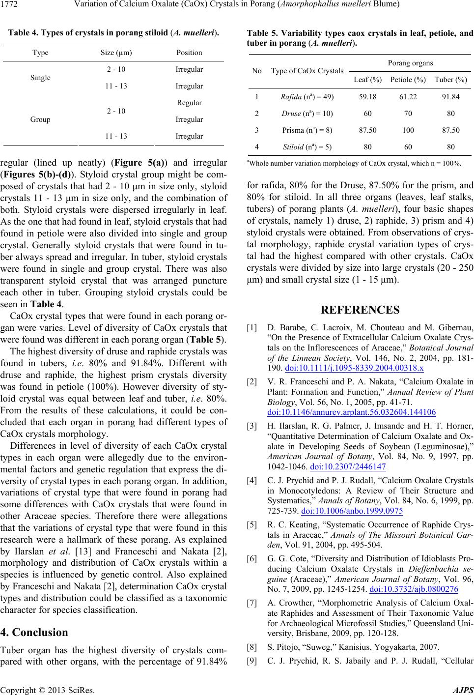
Table 4. Types of crystals in porang stiloid (A. muelleri).
Type Size (µm) Position
2 - 10 Irregular
Single
11 - 13 Irregular
Regular
2 - 10
Irregular Group
11 - 13 Irregular
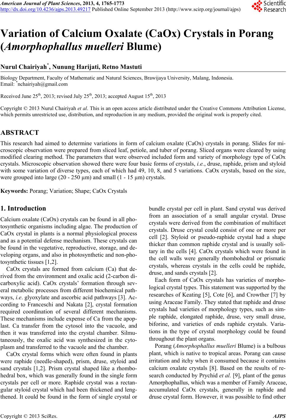
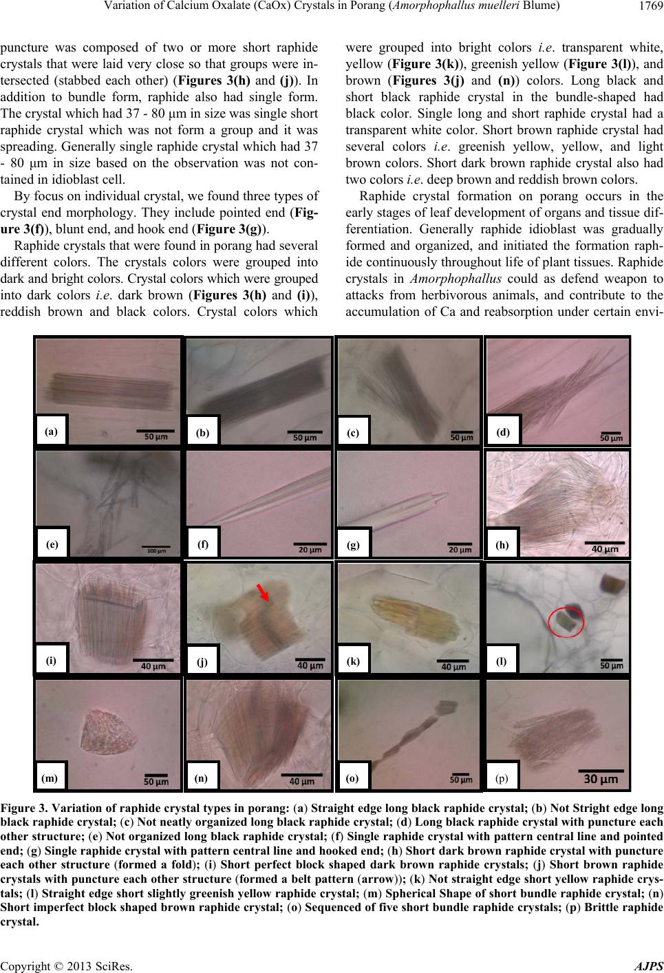
regular (lined up neatly) (Figure 5(a)) and irregular
(Figures 5(b)-(d)). Styloid crystal group might be com-
posed of crystals that had 2 - 10 μm in size only, styloid
crystals 11 - 13 μm in size only, and the combination of
both. Styloid crystals were dispersed irregularly in leaf.
As the one that had found in leaf, styloid crystals that had
found in petiole were also divided into single and group
crystal. Generally styloid crystals that were found in tu-
ber always spread and irregular. In tuber, styloid crystals
were found in single and group crystal. There was also
transparent styloid crystal that was arranged puncture
each other in tuber. Grouping styloid crystals could be
seen in Table 4.
CaOx crystal types that were found in each porang or-
gan were varies. Level of diversity of CaOx crystals that
were found was different in each porang organ (Table 5).
The highest diversity of druse and raphide crystals was
found in tubers, i.e. 80% and 91.84%. Different with
druse and raphide, the highest prism crystals diversity
was found in petiole (100%). However diversity of sty-
loid crystal was equal between leaf and tuber, i.e. 80%.
From the results of these calculations, it could be con-
cluded that each organ in porang had different types of
CaOx crystals morphology.
Differences in level of diversity of each CaOx crystal
types in each organ were allegedly due to the environ-
mental factors and genetic regulation that express the di-
versity of crystal types in each porang organ. In addition,
variations of crystal type that were found in porang had
some differences with CaOx crystals that were found in
other Araceae species. Therefore there were allegations
that the variations of crystal type that were found in this
research were a hallmark of these porang. As explained
by Ilarslan et al. [13] and Franceschi and Nakata [2],
morphology and distribution of CaOx crystals within a
species is influenced by genetic control. Also explained
by Franceschi and Nakata [2], determination CaOx crystal
types and distribution could be classified as a taxonomic
character for species classification.
4. Conclusion
Tuber organ has the highest diversity of crystals com-
pared with other organs, with the percentage of 91.84%
Table 5. Variability types caox crystals in leaf, petiole, and
tuber in porang (A. muelleri).
Porang organs
NoType of CaOx Crystals
Leaf (%) Petiole (%) Tuber (%)
1Rafida (na) = 49) 59.18 61.22 91.84
2Druse (na) = 10) 60 70 80
3Prisma (na) = 8) 87.50 100 87.50
4Stiloid (na) = 5) 80 60 80
aWhole number variation morphology of CaOx crystal, which n = 100%.
for rafida, 80% for the Druse, 87.50% for the prism, and
80% for stiloid. In all three organs (leaves, leaf stalks,
tubers) of porang plants (A. muelleri), four basic shapes
of crystals, namely 1) druse, 2) raphide, 3) prism and 4)
styloid crystals were obtained. From observations of crys-
tal morphology, raphide crystal variation types of crys-
tal had the highest compared with other crystals. CaOx
crystals were divided by size into large crystals (20 - 250
μm) and small crystal size (1 - 15 μm).
REFERENCES
[1] D. Barabe, C. Lacroix, M. Chouteau and M. Gibernau,
“On the Presence of Extracellular Calcium Oxalate Crys-
tals on the Inflorescences of Araceae,” Botanical Journal
of the Linnean Society, Vol. 146, No. 2, 2004, pp. 181-
190. doi:10.1111/j.1095-8339.2004.00318.x
[2] V. R. Franceschi and P. A. Nakata, “Calcium Oxalate in
Plant: Formation and Function,” Annual Review of Plant
Biology, Vol. 56, No. 1, 2005, pp. 41-71.
doi:10.1146/annurev.arplant.56.032604.144106
[3] H. Ilarslan, R. G. Palmer, J. Imsande and H. T. Horner,
“Quantitative Determination of Calcium Oxalate and Ox-
alate in Developing Seeds of Soybean (Leguminosae),”
American Journal of Botany, Vol. 84, No. 9, 1997, pp.
1042-1046. doi:10.2307/2446147
[4] C. J. Prychid and P. J. Rudall, “Calcium Oxalate Crystals
in Monocotyledons: A Review of Their Structure and
Systematics,” Annals of Botany, Vol. 84, No. 6, 1999, pp.
725-739. doi:10.1006/anbo.1999.0975
[5] R. C. Keating, “Systematic Occurrence of Raphide Crys-
tals in Araceae,” Annals of The Missouri Botanical Gar-
den, Vol. 91, 2004, pp. 495-504.
[6] G. G. Cote, “Diversity and Distribution of Idioblasts Pro-
ducing Calcium Oxalate Crystals in Dieffenbachia se-
guine (Araceae),” American Journal of Botany, Vol. 96,
No. 7, 2009, pp. 1245-1254. doi:10.3732/ajb.0800276
[7] A. Crowther, “Morphometric Analysis of Calcium Oxal-
ate Raphides and Assessment of Their Taxonomic Value
for Archaeological Microfossil Studies,” Queensland Uni-
versity, Brisbane, 2009, pp. 120-128.
[8] S. Pitojo, “Suweg,” Kanisius, Yogyakarta, 2007.
[9] C. J. Prychid, R. S. Jabaily and P. J. Rudall, “Cellular
Copyright © 2013 SciRes. AJPS
Variation of Calcium Oxalate (CaOx) Crystals in Porang (Amorphophallus muelleri Blume) 1773
Ultrastructure and Crystal Development in Amorphophal-
lus (Araceae),” Annals of Botany, Vol. 101, No. 7, 2008,
pp. 983-995. doi:10.1093/aob/mcn022
[10] J. M. Genua and C. J. Hillson, “The Occurrence, Type
and Location of Calcium Oxalate Crystals in the Leaves
of Fourteen Species of Araceae,” Annals of Botany, Vol.
56, 1985, pp. 351-361.
[11] M. H. Grayum, “Evolution and Phylogeny of the Ara-
ceae,” Annals of the Missouri Botanical Garden, Vol. 77,
1990, pp. 628-697. doi:10.2307/2399668
[12] S. J. Mayo, J. Bogner and P. C. Boyce, “The Genera of
Araceae,” The Trustees, Kew, 1997.
[13] R. C. Keating, “Leaf Anatomical Characters and Their
Value in Understanding Morphoclines in the Araceae,”
The Botanical Review, Vol. 68, No. 4, 2003, pp. 510-523.
doi:10.1663/0006-8101(2002)068[0510:LACATV]2.0.C
O;2
[14] G. G. Cote, “Distribution of Calcium Oxalate Crystals in
Floral Organs of Araceae in Relation to Pollination Stra-
tegy,” American Journal of Botany, Vol. 99, No. 7, 2012,
pp. 1-12. doi:10.3732/ajb.1100499
[15] H. Ilarslan, R. G. Palmer and H. T. Horner, “Calcium
Oxalate Crystals in Developing Seeds of Soybean,” An-
nals of Botany, Vol. 88, No. 2, 2001, pp. 243-257.
doi:10.1006/anbo.2001.1453
[16] C. Meric, “Calcium Oxalate Crystals in Aster squamatus
and Bellis perennis (Asteraceae: Astereae),” Phytologia
Balcanica, Vol. 15, No. 2, 2009, pp. 255-259.
Copyright © 2013 SciRes. AJPS