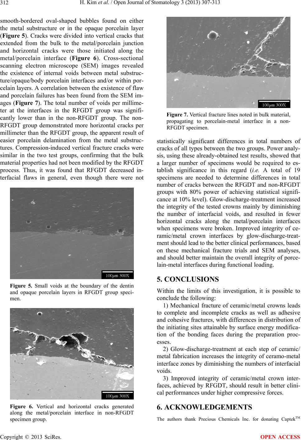
H. Kim et al. / Open Journal of Stomatology 3 (2013) 307-313
Copyright © 2013 SciRes.
313
materials for this study, and Mr. Peter J. Bush in the UB South Campus
Instrument Center for his assistance with Scanning Electron Micros-
copy.
OPEN ACCESS
REFERENCES
[1] Brecker, C.S. (1956) Porcelain baked to gold: A new
medium in prosthodontics. Journal of Prosthetic Den-
tistry, 6, 801. doi:10.1016/0022-3913(56)90077-4
[2] Johnston, J., Dykema, R. and Cunningham, D. (1956)
The use and construction of gold crowns with a fused
porcelain veneer—A progress report. Journal of Pros-
thetic Dentistry, 6, 811-821.
doi:10.1016/0022-3913(56)90078-6
[3] Anusavice, K.J. (1985) Noble metal alloys for metal-
ceramic restorations. The Dental Clinics of North Amer-
ica, 29, 789-803.
[4] Anusavice, K.J. (1983) Screening tests for metal-ceramic
systems. In: McLean, J.W., Ed., Dental Ceramics Pro-
ceedings of the First International Symposium on Ce-
ramics, Quintessence, Chicago, 371-414.
[5] Campbell, S.D. and Sirakian, A. (1995) Effect of firing
cycle and surface finishing on distortion of metal ceramic
castings. Journal of Prosthetic Dentistry, 74, 476-481.
doi:10.1016/S0022-3913(05)80348-8
[6] Campbell, S.D. and Pelletier, L. (1992) Thermal cycling
distortion of metal ceramics: Part I- Metal collar width.
Journal of Prosthetic Dentistry, 67, 603-608.
doi:10.1016/0022-3913(92)90155-4
[7] Campbell, S.D. and Pelletier, L. (1992) Thermal cycling
distortion of metal ceramics: Part II- Etiology. Journal of
Prosthetic Dentistry, 68, 284-289.
doi:10.1016/0022-3913(92)90331-4
[8] Bridger, D.V. and Nicholls, J.I. (1981) Distortion of ce-
ramometal fixed partial dentures during the firing cycle.
Journal of Prosthetic Dentistry, 45, 507-514.
doi:10.1016/0022-3913(81)90036-6
[9] Wiltshire, W.A., Ferreira, M.R. and Ligthelm, A.J. (1996)
Allergies to Dental Materials. Quintessence International,
27, 513-520.
[10] Pierce, L.H. and Goodkind, R.J. (1989) A status report of
possible risks of base metal alloys and their component.
Journal of Prosthetic Dentistry, 62, 234-237.
doi:10.1016/0022-3913(89)90320-X
[11] Della Bona, A. and Kelly, J.R. (2008) The clinical suc-
cess of all-ceramic restorations. Journal of the American
Dental Association, 139, 85-135.
[12] Wall, J.G. and Cipra, D.L. (1992) Alternative crown sys-
tems. Is the metal-ceramic crown always the restoration
of choice? The Dental Clinics of North America, 36,
765-782.
[13] Erpenstein, H., Borchard, R. and Kerschbaum, T. (2000)
Long-term clinical results of galvano-ceramic and glass-
ceramic individual crowns. Journal of Prosthetic Den-
tistry, 83, 530-534. doi:10.1016/S0022-3913(00)70010-2
[14] Shoher, I. and Whiteman, A. (1995 ) Captek—A new ca-
pillary casting technology for ceramometal restorations.
Quintessence of Dental Technology, 18, 9-20.
[15] Zappala, C., Shoher, I. and Battaini, P. (1996) Microstru-
ctural aspects of the Captek alloy for Porcelain- fused-to-
metal restorations. Journal of Esthetic Dentistry, 8, 151-
156. doi:10.1111/j.1708-8240.1996.tb00419.x
[16] Shoher, I. (1998) Vital tooth esthetics in Captek restora-
tions. The Dental Clinics of North America, 42, 713-718.
[17] Baier, R.E. and DePalma, V.A. (1970) Electrodeless glow
discharge cleaning and activation of high-energy sub-
strates to insure their freedom from organic contamina-
tion and their receptivity for adhesives and coatings.
Calspan Advanced Technology Center, New York.
[18] Baier, R.E. (1990) Glow-Discharge-Treatment techniques,
improvement of adhesion in the intraoral environment by
glow-discharge-treatment (GDT) Techniques. Transac-
tions of the Academy of Dental Materials, 3, 6-29.
[19] Tran, H.S., Puc, M.M., Hewitt, C.W., Soll, D.B., Marra,
S.W., Simonetti, V.A., Cilley, J.H. and DelRossi, A.J.
(1999) Diamond-like carbon coating and plasma or glow
discharge treatment of Mechanical heart valves. Journal
of Investigative Surgery, 12, 133-140.
doi:10.1080/089419399272520
[20] Ramsey, W.S., Hertl, W., Nowlan, E.D. and Binkowski,
N.J. (1984) Surface treatments and cell attachment, Tis-
sue Culture Association, Inc. IN VITRO, 20, 802-808.
doi:10.1007/BF02618296
[21] Bertolotti, R. (1983) Porcelain-to-metal bonding and com-
patibility. In: McLean, J.W., Ed., Dental Ceramics Pro-
ceedings of the First International Symposium on Ce-
ramics, Quintessence, Chicago, 415-429.
[22] Kelly, J.R. (1995) Perspectives on strength. Dental Mate-
rials, 11, 103-110. doi:10.1016/0109-5641(95)80043-3
[23] Kelly, J.R., Giordano, R., Pober, R. and Cima, M.J. (1990)
Fracture surface analysis of dental ceramics: Clinically
failed restorations. International Journal of Prosthodon-
tics, 3, 430-440.
[24] Southan, D.E. (1983) The Porcelain jacket crown. In:
McLean, J.W., Ed., Dental Ceramics Proceedings of the
First International Symposium on Ceramics, Quintes-
sence, Chicago, 207-230.
[25] Kelly, J.R. (1999) Clinically relevant approach to failure
testing of all-ceramic restorations. Journal of Prosthetic
Dentistry, 81, 652-661.
doi:10.1016/S0022-3913(99)70103-4
[26] Kelly, J.R., Tesk, J.A. and Sorensen, J.A. (1995) Failure
of all-ceramic fixed partial dentures in in vitro and in vivo:
Analysis and modeling. Journal of Dental Research, 74,
1253-1258. doi:10.1177/00220345950740060301