 Advances in Bioscience and Biotechnology, 2013, 4, 860-865 ABB http://dx.doi.org/10.4236/abb.2013.49114 Published Online September 2013 (http://www.scirp.org/journal/abb/) Cytokine release in sepsis Ian Burkovskiy1*, Joel Sardinha1*, Juan Zhou2,3, Christian Lehmann1,2,3# 1Department of Pharmacology, Dalhousie University, Halifax, Canada 2Department of Anesthesia, Dalhousie University, Halifax, Canada 3Department of Microbiology & Immunology, Dalhousie University, Halifax, Canada Email: #chlehmann@dal.ca Received 1 June 2013; revised 1 July 2013; accepted 1 August 2013 Copyright © 2013 Ian Burkovskiy et al. This is an open access article distributed under the Creative Commons Attribution License, which permits unrestricted use, distribution, and reproduction in any medium, provided the original work is properly cited. ABSTRACT Despite the advances in the therapeutic approaches, health care protocols and policies, sepsis continues to be an important problem in clinical medicine. High lethality of sepsis cases calls for a detailed and criti- cally important study of the pathophysiology of sepsis. In this review, we discuss pathomechanisms of sepsis and the role of cytokines that are released in sepsis. We propose that the systemic levels of cytokines are not always reflecting the pathological picture and the immune status of the patient. One of the emerging approaches which may bring an effective treatment strategy exploits the endocannabinoid system for its immunomodulatory properties. Following from that, the research in this particular field is very important as it can bring understanding behind the complicated pathophysiology of sepsis. Keywords: Sepsis; Cytokine; Inflammation; Immunology 1. INTRODUCTION Sepsis is the systemic inflammatory resp onse to an infec- tion that is usually associated with tissue hypoperfusion and multi-organ dysfunction [1]. According to generally accepted definitions, there are three clinical stages of sepsis-sepsis, severe sepsis and septic shock. Each indi- vidual stage is based on the complexity and the severity of the symptoms. Prevalence of septic shock and severe sepsis in intensive care units and emergency rooms con- tinues to be high, despite the advances in surgical critical care and techniques, therapeutic approaches, establish- ment of new health care protocols and policies and de- velopment of new drugs [2,3]. One of the fundamental pathological characteristics of sepsis is the inability to maintain the balance between excessive and inadequate inflammation [4]. Sepsis is considered to be the 10th leading cause of death in the United States, with approximately a million severe sepsis cases each year in the United States and an estimated 18 million cases of sepsis globally [5,6]. Mod- ern therapeutic approach es are more frequent in utilizin g aggressive immunosuppressive and chemotherapeutic treatments that compromise normal defense mechanisms and contribute to the increase in sepsis case numbers [2]. In addition to being a critical health condition, severe sepsis also puts a heavy socio-economic strain on the health care system [7]. Literature reports that the care of patients with sepsis brings an annual cost of the care to $17 billion, in the United States alone [5]. In Canada, a study conducted by Letarte et al. [7] investigated the costs of severe sepsis and septic shock in the province of Quebec. The study reported a cost estimate between $36.4 and $72.9 millio n per year and co ncluded that the co st of severe sepsis is a significant economical burden for the Quebec health care system [7]. Finally, the patients that survive severe sepsis have a substantial reduction in their overall quality of life [8,9]. 2. ETIOLOGY AND PATHOMECHANISMS OF SEPSIS Etiologically, any infectious agent that enters the blood- stream of a patient and generates a systemic immune response can cause sepsis. However, the presence of the pathogen in the blood is not a requirement for th e clinical symptoms of sepsis to manifest themselves—as the pres- ence of pathogen’s signaling molecules or inflammatory mediators, which are released into the circulation from the source of infection, is sufficient for sepsis induction [10]. While sepsis can potentially be caused by many infectious agents such as bacteria, fungi, parasites or vi rus [11], the majority of cases of sepsis are reported to be due to bact er i a [10]. *These authors contributed equally. #Corresponding author. OPEN ACCESS  I. Burkovskiy et al. / Advances in Bio science and Biotechnology 4 (2013) 860-865 861 The detection of the bacteria that causes sepsis st ron gly depends on the severity of the clinical picture with bacte- ria being detected in approximately 20% to 40% of pa- tients with severe sepsis and in 40% to 70% of cases with septic shock [12]. One European study reported that the most frequent infectious agent that was associated with sepsis was Escherichia coli, with 22.7% share of de- tected cases [13]. A more detailed study by Vincent et al. [14] surveyed the prevalence and outcomes of infection in intensive care units from 75 countries. The results revealed that the microbiological culture results were positive in 70% of the infected patients with 62% of the positive isolates being gram-negative organisms, 47% be- ing gram-positive, and 19% being fungal in [14]. The first step in host defense is the recognition of mi- croorganisms via binding of pattern-recognition recap- tors, such as the Toll-like receptor family (TLRs) to highly conserved molecules called pathogen-associated molecu- lar proteins (PAMPs) [15]. TLRs then recruit adaptor proteins to the cell surface and the adaptor proteins, in turn, activate a series of cytoplasmic kinases, initiating cascades that activate various transcription factors [15]. Genes that are induced by these transcription factors pre- pare the cell to undergo one of the potential main re- sponses—rapidly undergoing apoptosis if the cell is over- whelmingly infected, fighting the infection with pro-in- flammatory molecules and proliferating or suppressing the inflammatory reaction if no pathogens are longer de- tected [15]. First responding cells are monocytes and macrophages, which through induction of early response genes such as TNFα and IL-6 rapidly produce inflammatory cytokines and chemokines, amplifying the inflammatory response [15]. This is followed by an activation of lymphocytes as an adaptive immune response, as well as the coordination of later phases of the immune response [15]. Addition- ally, nitric oxide (NO) that is produced by endothelial cells induces vasodilation and an increase in leukocyte delivery to the sites of immune-activity [15]. 3. ENDOCANNABINOID SYSTEM The endocannabionoid system has emerged as a potential therapeutic target in sepsis treatment due to its immune modulatory functions. This endogenous system derives its name from the plant Cannabis sativa which contains many phytocannabinoids that activate endocannabinoid receptors within our bodies. The two most well charac- terized endocannabinoid receptors are cannabinoid recap- tor 1 (CB1) and cannabinoid receptor 2 (CB2). The CB1 receptors are found throughout our bodies, but are highly concentrated in our central nervous system where they are implicated in modulating neurotransmitter release. Alternatively, the CB2 receptors are mainly localized on the surface of our immune cells, thus implicating a role within our immune sy stem. Exploiting the activi ty of these receptors may prove to be beneficial in sepsis treatment. It has been well established that activation of the CB2 receptors initiates immunosuppressive mechanisms. One component of this suppressive mechanism involves al- tering the cytokines released by immune cells. CB2 re- ceptor activation results in a reduction of pro-inflamma- tory cytokine release from leukocytes as well as an in- creased secretion of immunosuppressive cytokines. This outcome may hav e a beneficial effect if initiated during a pro-inflammatory phase of the septic cascade. However, due to the variability of the immune state during septic progression, immunosuppressive therapeutics can be det- rimental, leaving the patient vulnerable to infections. As a result, measurement of cytokine levels in the blood was suggested as a method for detecting the condition of the patient’s immune system. A proper understanding of the patient’s immune state during sepsis is vital for appropri- ate treatments. 4. CYTOKINE RELEASE Cytokines are low molecular weight signaling molecules secreted by a variety of cells that are used for cellular communication. Cytokines exist as proteins, glycopro- teins, or peptides, and have functional roles in immuno- modulation as well as development. They are involved in cellular activation, trafficking, signaling events, prolif- eration, differentiation and migration [16]. Cy t oki nes exert their effects on targets by binding to cell surface recap- tors. Receptor activation subsequently leads to modula- tion of intracellular cascades, eventually causing a bio- logical effect. A few cytokines are constit utively expresses, however a majority of cytokines is produced in response to antigens, other cytokines, or cellular stressors. Due to the facultative expression of these molecules, gene ex- pression of cytokines is tightly regulated, with rapid tran- scription and translation of proteins on demand. As an example, activation of transcription factor nuclear factor κB (NF-κB) causes the rapid production and release of pro-inflammatory cytokines such as IL-1 [16]. Cytokines exert their effects in a dose dependent man- ner and have relatively short half lives in the extracellu- lar environment. Therefore, cytokine levels can fluctuate drastically in microenv ironments during their short dura- tion of action. Once cytokines are released, they can act on their targets in either an autocrine or paracrine fashion. Multiple cytokines can also interact in the extracellular environment, causing a plethora of different effects. All cytokines are pleiotropic, which indicates that they can have variable effects based on the receptors they bind to. Alternatively, many cytokines can be redundant because they can have the same functional outcome. Different Copyright © 2013 SciRes. OPEN ACCESS 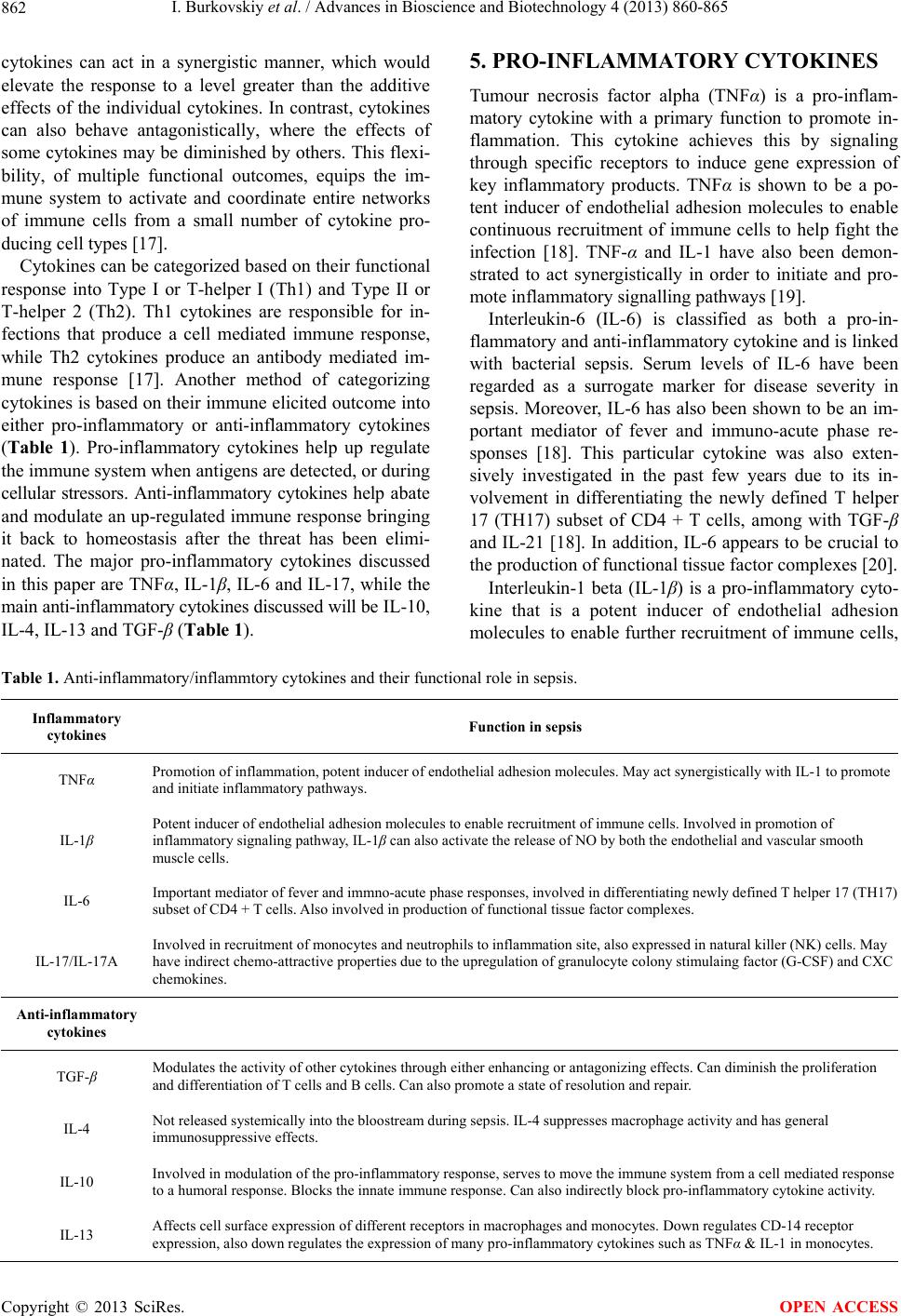 I. Burkovskiy et al. / Advances in Bio science and Biotechnology 4 (2013) 860-865 Copyright © 2013 SciRes. 862 OPEN ACCESS cytokines can act in a synergistic manner, which would elevate the response to a level greater than the additive effects of the individual cytokines. In contrast, cytokines can also behave antagonistically, where the effects of some cytokines may be diminished by others. This flexi- bility, of multiple functional outcomes, equips the im- mune system to activate and coordinate entire networks of immune cells from a small number of cytokine pro- ducing cell types [17]. Cytokines can be categorized based on their functional response into Type I or T-helper I (Th1) and Type II or T-helper 2 (Th2). Th1 cytokines are responsible for in- fections that produce a cell mediated immune response, while Th2 cytokines produce an antibody mediated im- mune response [17]. Another method of categorizing cytokines is based on their immune elicited outcome into either pro-inflammatory or anti-inflammatory cytokines (Table 1). Pro-inflammatory cytokines help up regulate the immune system when antigens are detected, or during cellular stressors. Anti-inflammatory cytokines help abate and modulate an up-regulated immune response bringing it back to homeostasis after the threat has been elimi- nated. The major pro-inflammatory cytokines discussed in this paper are TNFα, IL-1β, IL-6 and IL-17, while the main anti-inflammatory cytokines discussed will be IL-10, IL-4, IL-13 and TGF-β (Table 1). 5. PRO-INFLAMMATORY CYTOKINES Tumour necrosis factor alpha (TNFα) is a pro-inflam- matory cytokine with a primary function to promote in- flammation. This cytokine achieves this by signaling through specific receptors to induce gene expression of key inflammatory products. TNFα is shown to be a po- tent inducer of endothelial adhesion molecules to enable continuous recruitment of immune cells to help fight the infection [18]. TNF-α and IL-1 have also been demon- strated to act synergistically in order to initiate and pro- mote inflammatory signalling pathways [19]. Interleukin-6 (IL-6) is classified as both a pro-in- flammatory and anti-inflammatory cytok ine and is linked with bacterial sepsis. Serum levels of IL-6 have been regarded as a surrogate marker for disease severity in sepsis. Moreover, IL-6 has also been shown to be an im- portant mediator of fever and immuno-acute phase re- sponses [18]. This particular cytokine was also exten- sively investigated in the past few years due to its in- volvement in differentiating the newly defined T helper 17 (TH17) subset of CD4 + T cells, among with TGF-β and IL-21 [18]. In addition, IL-6 appears to b e crucial to the production of functional tissue factor complexes [20]. Interleukin-1 beta (IL-1β) is a pro-inflammatory cyto- kine that is a potent inducer of endothelial adhesion molecules to enable further recruitment of immune cells, Table 1. Anti-inflammatory/inflammtory cytokines and their functional role in sepsis. Inflammatory cytokines Function in sepsis TNFα Promotion of inflammation, potent inducer of endothelial adhesion molecules. May act synergistically with IL-1 to promote and initiate inflammatory pathways. IL-1β Potent inducer of endothelial adhesion molecules to enable recruitment of immune cells. Involved in promotion of inflammatory signaling pathway, IL-1β can also activate the release of NO by both the endothelial and vascular smooth muscle cells. IL-6 Important mediator of fever and immno-acute phase responses, involved in differentiating newly defined T helper 17 (TH17) subset of CD4 + T cells. Also involved in production of functional tissue factor complexes. IL-17/IL-17A Involved in recruitment of monocytes and neutrophils to inflammation site, also expressed in natural killer (NK) cells. May have indirect chemo-attractive properties due to the upregulation of granulocyte colony stimulaing factor (G-CSF) and CXC chemokines. Anti-inflammatory cytokines TGF-β Modulates the activity of other cytokines through either enhancing or antagonizing effects. Can diminish the proliferation and differentiation of T cells and B cells. Can also promote a state of resolution and repair. IL-4 Not released systemically into the bloostream during sepsis. IL-4 suppresses macrophage activity and has gen eral immunosuppressive effects. IL-10 Involved in modulation of the pro-inflammatory response, ser ves to move the immune system from a cell mediated response to a humoral response. Blocks the innate immune response. Can also indirectly block pro-inflammatory cytokine activity. IL-13 Affects cell surface expression of different receptors in macrophages and monocytes. Down regulates CD-14 receptor expression, also down regulates the expression of many pro-inflammatory cytokines such as TNFα & IL-1 in monocytes.  I. Burkovskiy et al. / Advances in Bio science and Biotechnology 4 (2013) 860-865 863 involved in promotion of inflammatory signaling path- ways [18]. IL-1β can also activate the release of NO by both the endothelial and vascular smooth muscle cells, via increased transcription and activity of the inducible form of NO synthase [18]. Interleukin-17 or Interleukin-17A (IL-17/IL-17A) is a pro-inflammatory cytokine that is produced by T helper 17, a subset of CD4 + T cells. IL-17 is involved in re- cruitment of monocytes and neutrophils to inflammation site [18]. IL-17 is also expressed in natural killer (NK) cells and this pro-inflammato ry cytokin e has been shown to be detrimental in the survival outcome of common lymphoid progenitor (CLP), the murine model of po- lymicrobial sepsis [21]. Literature reports that IL-17A may also be considered to have indirect chemoattractive properties due to the up-regulation of granulocyte col- ony-stimulating factor (G-CSF) and CXC chemokines, resulting in enhanced recruitment of neutrophils to the site of infection, resulting in an efficient bacterial clear- ance [22]. 6. ANTI-INFLAMMATORY CYTOKINES One of the main anti-inflammatory cytokines is IL-10. IL-10 helps modulate the pro-inflammatory response during early stages of sepsis by limiting exaggerated pro- inflammatory effects as well as making targets more tol- erant to repeated pro-inflammatory stimuli [15]. How- ever, in later stages, more sustained levels of IL-10 serve to move the immune system from a cell mediated re- sponse (Th1; innate immunity) to a humoral response (Th2; adaptive immunity). Known cellular sources of IL-10 include macrophages, dendritic cells, B-lympho- cytes, T-regulatory cells (TREGS), and natural killer T cells (NKT cells) [23]. IL-10 blocks the innate immune response by inhibiting development and cytokine release by Th1 cells, as well as blocking production of certain pro-inflammatory cytokines. Apart from blocking pro- inflammatory cytokine release, IL-10 can also indirectly block pro-inflammatory cytokine activity by inducing the expression of soluble antagonistic receptors such as IL-1 receptor antagonist (IL-1RA) and soluble TNF receptors (sTNFR). These soluble antagonistic receptors bind to the pro-inflammatory cytokines and prevent them from activating their targets, effectively diminishing the extent of pro-inflammatory stimulation. The role of IL-10 in sepsis pathogenesis is not well understood. Conflicting results have been shown by different groups using IL-10 in survival outcome studies [24]. One study pretreated mice with anti-IL-10 antibody before inducing a septic state and found a decreased ability to survive in com- parison to controls [25]. Conversely, another study tested mice lacking the IL-10 gene and observed survival wh en anti-IL-10 antibodies were administered at different time points [26]. These IL-10 deficient mice showed impro ved survival only if anti-IL-10 antibodies were given after a delay of initiating the septic challenge, presumably fol- lowing the initial pro-inflammatory cascade, but not when anti-IL-10 antibodies were administered directly after the septic challenge. The results of these, as well as many other studies, indicate that the role of IL-10 in sepsis may be dependent of immune state of the patient, and effec- tive treatments involving this cytokine are time critical during sepsis pathogenesis [23]. Interleukin 4 (IL-4) is another cytokine in the immune system arsenal that has immunosuppressive effects. This cytokine is unique because during sepsis it is not released systemically into the bloodstream. Studies have not been able to detect changes in IL-4 plasma levels of septic patients, however isolated splenocytes from septic mice did secrete higher levels of this cytokine when stimulated ex-vivo [23]. These results suggest a local effect of IL-4 immune suppression during a septic state, limiting its therapeutic potential. IL-4 works by inhibiting Th1 cells from polarizing and differentiating, as well as playing an important role in B cell differentiation, thereby promot- ing a Th2 mediated response [23]. IL-4 suppresses mac ro- phage activity by preventing their cytotoxic activity, parasite killing, and nitric oxide production [27,28]. Interleukin 13 (IL-13) shares many similar character- istics to IL-4, from its gene location (close proximity to IL-4 gene) to the receptor it activates (IL-4 type I recap- tor) [27]. However, unlike IL-4, IL-13 has negligible effects on Th2 cell function. IL-13 has been shown to affect cell surface expression of different receptors in macrophages and monocytes, such as up regulating the expression of some integrins, while leaving other cell- cell interacting receptors unaffected [29]. An important mechanism by which IL-13 exerts its anti-inflammatory properties is through the down regulation of CD-14 re- ceptor expression. CD-14 is an important co-receptor wi th Toll Like Receptor 4 (TLR4) that recognises lipopoly- saccharide (LPS) and elicits a strong immune response [29]. Furthermore, IL-13 also down regulates the expres- sion of many pro-inflammatory cytokines such as TNFα & IL-1 in monocytes [27]. Investig ations into the role of IL-13 during sepsis indicate that mice show elevated lev- els of IL-13, and anti-IL-13 antibody treatment reduces their survival [27]. Interestingly, the anti-IL-13 antibody treated mice showed an increased neutrophil influx into tissues, but no effect on bacterial load and other leuko- cyte infiltration. The excess neutrophil influx into tissues showed an increase in organ damage indicating progres- sion of the septic pathophysiology [27]. Transforming Growth Factor-β (TGF-β) has many reg u- latory properties, therefore can have both pro and anti- inflammatory properties. TGF-β modulates the activity of other cytokines through either enhancing or antago- Copyright © 2013 SciRes. OPEN ACCESS  I. Burkovskiy et al. / Advances in Bio science and Biotechnology 4 (2013) 860-865 864 nizing effects [27]. TGF-β can diminish the proliferation and differentiation of T cells and B cells by blocking production of specific cytokines like IL-2 [23]. Studies comparing cytokine levels showed elevated systemic lev- els of TGF-β in trauma patients bu t no significan t chang e in patients with septic shock [23]. Studies with TGF-β knockout mice showed an exaggerated inflammatory reaction, indicating the anti-inflammatory properties of this cytokine [27]. Furthermore, TGF-β can promote a state of resolution and repair, opposing the effects of a pro-inflammatory cascade similar to IL-4. These results indicate a more local response of TGF-β, similar to IL-4, limiting its therap eutic potential in systemic diseases lik e sepsis [23,27] . 7. CONCLUSION The mortality of severe sepsis and septic shock ranges from 30% to 70% respectively. Clinically, there are only very few treatments shown to be beneficial in sepsis, e.g. antibiotics and vasopr essors [15]. There are many poten- tial explanations fo r little to no impact of the o ther thera- pies—such as the timing of the drug administration, sys- temic levels of cytokines not always reflecting the patho- logical picture and the immune status of the patient. Currently there are reports in literature that indicate a new potential focus on specific development of a clini- cally applicable therapy that would utilize the immuno- modulating properties of the endocannabinoid system to combat the onset of sepsis [30,31]. The strategy of the approach would be revolving around the condition when the pro-inflammatory cytokine levels appear to be high. In summary, the research in this field is highly important in order to advance our understanding and to develop an effective therapeutic strateg y. REFERENCES [1] Dellinger, R.P. (2008) Steroid therapy of septic shock: The decision is in the eye of the beholder. Critical Care Medicine, 36, 1987-1989. doi:10.1097/CCM.0b013e31817d7ee4 [2] Balk, R.A. (2000) Severe sepsis and septic shock. Criti- cal Care Clinics, 16, 179-192. doi:10.1016/S0749-0704(05)70106-8 [3] Wheeler, A.P. and Bernard, G.R. (1999) Treating patients with severe sepsis. New England Journal of Medicine, 340, 207-214. doi:10.1056/NEJM199901213400307 [4] Cohen, J. (2002) The immunopathogenesis of sepsis. Na- ture, 420, 885-891. doi:10.1038/nature01326 [5] Angus, D.C., Linde-Zwirble, W.T., Lidicker, J., Clermont, G., Carcillo, J. and Pinsky, M.R. (2001) Epidemiology of severe sepsis in the United States: Analysis of incidence, outcome, and associated costs of care. Critical Care Me- dicine, 29, 1303-1310. doi:10.1097/00003246-200107000-00002 [6] Global Sepsis Alliance (2010) International organiza- tions declare sepsis a medical emergency: Issued by an expert panel representing 20 adult and pediatric intensive care societies. [7] Letarte, J., Longo, C.J., Pelletier, J., Nabonne, B. and Fisher, H.N. (2002) Patient characteristics and costs of severe sepsis and septic shock in Quebec. Journal of Cri- tical Care, 17, 39-49. doi:10.1053/jcrc.2002.33028 [8] Perl, T.M., Dvorak, L., Hwang, T. and Wenzel, R.P. (1995) Long-term survival and function after suspected gram-negative sepsis. JAMA, 274, 338-345. doi:10.1001/jama.1995.03530040066043 [9] Heyland, D.K., Hopman, W., Coo, H., Tranmer, J. and McColl, M.A. (2000) Long-term health-related quality of life in survivors of sepsis: Short Form-36: A valid and re- liable measure of health-related quality of life. Critical Care Medicine, 28, 3599-3605. doi:10.1097/00003246-200011000-00006 [10] Holub, M. and Zavada, J. (2011) Clinical aspects of sep- sis. Contributions to Microbiology, 17, 12-30. [11] Bone, R.C., Balk, R.A., Cerra, F.B., Dellinger, R.P., Fein, A.M., Knaus, W.A., Schein, R.M. and Sibbald, W.J. (1992) Definitions for sepsis and organ failure and guide- lines for the use of innovative therapies in sepsis. The ACCP/SCCM Consensus Conference Committee. Ame- rican College of Chest Physicians/Society of Critical Care Medicine. Chest, 101, 1644-1655. doi:10.1378/chest.101.6.1644 [12] Brun-Buisson, C., Doyon, F., Carlet, J., Dellamonica, P., Gouin, F., Lepoutre, A., Mercier, J.C., Offenstadt, G. and Régnier, B. (1995) Incidence, risk factors, and outcome of severe sepsis and septic shock in adults: A multi-center prospective study in intensive care units. French ICU Group for Severe Sepsis. JAMA, 274, 968-974. doi:10.1001/jama.1995.03530120060042 [13] Luzzaro, F., Viganò, E.F., Fossati, D., Grossi, A., Sala, A., Sturla, C., Saudelli, M. and Toniolo, A. (2002) AM- CLI Lombardia Hospital Infectious Study Group: Preva- lence and drug susceptibility of pathogens causing blood- stream infections in northern Italy: A two-year study in 16 hospitals. European Journal of Clinical Microbiology & Infectious Diseases, 21, 849-855. [14] Vincent, J.L., Rello, J., Marshall, J., Silva, E., Anzueto, A., Martin, C.D., Moreno, R., Lipman, J., Gomersall, C., Sakr, Y., et al. (2009) EPIC II Group of Investigators: In- ternational study of the prevalence and outcomes of in- fection in intensive care units. JAMA, 302, 2323-2329. doi:10.1001/jama.2009.1754 [15] Russel, J.A., Boyd, J., Nakada, T., Thair, S. and Walley, K.R. (2011) Molecular mechanisms of sepsis. Contribu- tions to Microbiology, 17, 48-85. [16] Klaassen, C. D. and Watkins, J. B. (1999) Casarett and Doull’s toxicology: The basic science of poisons. McGraw- Hill, New York. [17] Whiteside, T. L. (2007). Introduction to cytokines as tar- gets for immunomodulation. In Cytokines in Human Health, Humana Press, 1-15. doi:10.1007/978-1-59745-350-9_1 [18] Chong, D.L.W. and Sriskandan, S. (2011) Pro-inflamma- Copyright © 2013 SciRes. OPEN ACCESS  I. Burkovskiy et al. / Advances in Bio science and Biotechnology 4 (2013) 860-865 Copyright © 2013 SciRes. 865 OPEN ACCESS tory mechanisms in sepsis. Contributions to Microbiology, 17, 86-107. [19] Dinarello, C.A. (2000) Proinflammatory cytokines. Chest, 111, 503-508. doi:10.1378/chest.118.2.503 [20] Riewald, M. and Ruf, W. (2003) Role of coagulation pro- tease cascades in sepsis. Critical Care, 7, 123-129. doi:10.1186/cc1825 [21] Flierl, M.A., Ri ttirsch , D., Gao, H., Hoesel, L.M., Nadeau, B.A., Day, D.E., Zetoune, F.S., Sarma, J.V., Huber-Lang, M.S., Ferrara, J.L. and Ward, P.A. (2008) Adverse func- tions of IL-17A in experimental sepsis. FASEB Journal, 22, 2198-2205. doi:10.1096/fj.07-105221 [22] Ye, P., Rodriguez, F.H., Kanaly, S., Stocking, K.L., Schurr, J., Schwarzenberger, P., Oliver, P., Huang, W., Zhang, P., Zhang, J., Shellito, J.E., Bagby, G.J., Nelson, S., Charrier, K., Peschon, J.J. and Kolls, J.K. (2001) Requirement of interleukin 17 receptor signaling for lung CXC chemo- kine and granulocyte colony-stimulating factor expres- sion, neutrophil recruitment, and host defense. Journal of Experimental Medicine, 194, 519-527. doi:10.1084/jem.194.4.519 [23] Shubin, N.J., Monaghan, S.F. and Ayala, A. (2011). Anti- inflammatory mechanisms of sepsis. Contributions to Microbiology, 17, 108-124. doi:10.1159/000324024 [24] Perl, M., Chung, C.S., Garber, M., Huang, X. and Ayala, A. (2006) Contribution of anti-inflammatory/immune sup- pressive processes to the pathology of sepsis. Frontiers in Bioscience, 11, 272-299. doi:10.2741/1797 [25] Hogaboam, C.M., Steinhauser, M.L., Schock, H., Lukacs, N., Strieter, R.M., Standiford, T. and Kunkel, S.L. (1998) Therapeutic effects of nitric oxide inhibition during ex- perimental fecal peritonitis: Role of interleukin-10 and monocyte chemoattractant protein 1. Infection and Im- munity, 66, 650-655. [26] Song, G.Y., Chung, C.S., Chaudry, I.H. and Ayala, A. (1999) What is the role of interleukin 10 in polymicrobial sepsis: Anti-inflammatory agent or immunosuppressant? Surgery, 126, 378-383. doi:10.1016/S0039-6060(99)70180-7 [27] Opal, S.M. and DePalo, V.A. (2000). Anti-inflammatory cytokines. Chest Journal, 117, 1162-1172. doi:10.1378/chest.117.4.1162 [28] Vannier, E., Miller, L.C. and Dinarello, C.A. (1992) Co- ordinated antiinflammatory effects of interleukin 4: In- terleukin 4 suppresses interleukin 1 production but up- regulates gene expression and synthesis of interleukin 1 receptor antagonist. Proceedings of the National Acad- emy of Sciences of the United States of America, 89, 4076-4080. doi:10.1073/pnas.89.9.4076 [29] de Waal Malefyt, R., Figdor, C.G., Huijbens, R., Mohan- Peterson, S., Bennett, B., Culpepper, J. and De Vries, J.E. (1993) Effects of IL-13 on phenotype, cytokine produc- tion, and cytotoxic function of human monocytes. Com- parison with IL-4 and modulation by IFN-gamma or IL-10. The Journal of Immunology, 151, 6370-6381. [30] Kianian, M., Al-Banna, N.A., Kelly, M. and Lehmann, C. (2013) Inhibition of endocannabinoid degradation in ex- perimental endotoxemia reduces leukocyte adhesion and improves capillary perfusion in the gut. Journal of Basic and Clinical Physiology and Pharmacology, 24, 27-33. doi:10.1515/jbcpp-2012-0065 [31] Sardinha, J., Lehmann, C. and Kelly, M. (2013) Targeting the endocannabinoid system to treat sepsis. Signa Vitae, 8, 9-14.
|