World Journal of Cardiovascular Diseases, 2013, 3, 377-379 WJCD
http://dx.doi.org/10.4236/wjcd.2013.35058 Published Online August 2013 (http://www.scirp.org/journal/wjcd/)
Unusual treatment of postoperative bleeding
after cardiac surgery
José Rubio-Alvarez1*, Juan Sierra-Quiroga1, Belén Adrio-Nazar1, Laura Reija López1,
Ángela Granda Bauza1, Carola Rubio Taboada2, Jose Manuel Martinez-Cereijo1
1Department of Cardiac Surgery, Universitary Hospital Santiago de Compostela, Santiago, Spain
2Department of Vascular Surgery, Universitary Hospital of Elche, Alicante, Spain
Email: *framan1@hotmail.com
Received 15 June 2013; revised 20 July 2013; accepted 1 August 2013
Copyright © 2013 José Rubio-Alvarez et al. This is an open access article distributed under the Creative Commons Attribution Li-
cense, which permits unrestricted use, distribution, and reproduction in any medium, provided the original work is properly cited.
ABSTRACT
A young patient was presented to the emergency de-
partment with chest pain and palpitations. A tran-
sthoracic echocardiogram showed a right atrial mass.
Coronary angiography showed a right coronary ar-
tery with collateral circulation to a large mass. The
tumor could only be partially resected and the patient
experienced persistent postoperative bleeding. We
performed a new right coronary artery angiography
which showed an important free extravasation of
contrast into the pericardium through the collateral
circulation. Using covered stents, the bleeding was
controlled. The pathological examination performed
later revealed a primary cardiac angiosarcoma. After
asympto m-free survival of 14 month s the patien t pre-
sented bone metastases.
Keywords: Angiosarcoma; Coronary Stenting;
Postoperative Bleeding
1. INTRODUCTION
Primary cardiac malignant tumors are very uncommon
and about 75% are sarcomas [1]. Although rare, angio-
sarcomas are the most common primary malignant neo-
plasms of the heart and are very aggressive and locally
invasive. These tumors are highly vascularized and are
often are actively bleeding into the pericardium.
Since the introduction of percutaneous coronary inter-
vention (PCI) in 197 7, it is increasingly used not only in
simple coronary lesions, but also in complex coronary
anatomies. Coronary perforation is a rare but serious
complication of PCI with the occurrence of important
bleeding into the pericardium. However, this complica-
tion can be tackled successfully with covered stents [2].
We describe the case of a 50-year-old man with non-
metastatic primary right atrial (RA) angiosarcoma, who
underwent surgical excision of the tumor and recon-
struction of the RA with a bovine pericardial patch. The
tumour could only be partially resected and the patient
experienced persistent postoperative bleeding through
the collateral circulation from the right coronary artery,
which was controlled using covered stents. After a sym-
ptom-free survival of 12 months, the patient presented
bone metastases.
2. CASE REPORT
A 50-year-old previously healthy male visited emergency
service because of palpitations and left chest pain. A
chest X-ray showed enlargement of the RA border an d an
electrocardiogram showed normal sinus rhythm with a
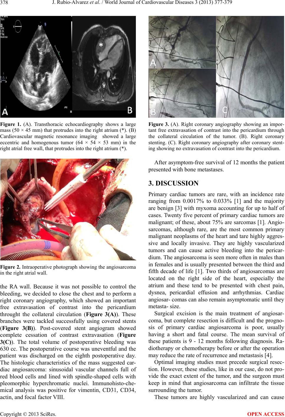
heart rate of 87 beats per minute. A transthoracic echo-
cardiogram was performed and detected a RA mass (50 ×
45 mm) that had infiltrated the free wall of and protruded
into the RA (Figure 1(A)). Left ventricular function was
normal and there were no valvular abnormalities. For
further evaluation of the RA mass magnetic resonance
imaging was performed (Figure 1(B)). This exploration
also showed a large eccentric tumor (64 × 54 × 53 mm)
in the RA free wall, protruding into the right atrium. The
tumor extended into the right atrioventricular groove, but
did not involve the right ventricle, or the annulus of the
tricuspid valve. Coronary angiography showed a right
coronary artery with collateral circulation to a large mass.
Surgery was performed under standard extracorporeal
circulation with selective cannulation of both cava veins
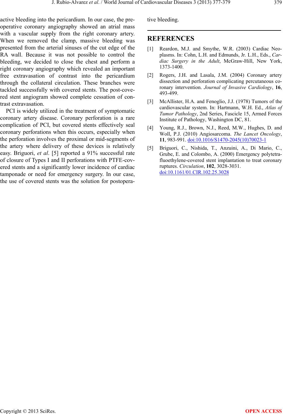
and the ascending aorta. The RA was excised (Figure 2),
but the tumor could only be partially resected because it
extended into the free wall of the right ventricle and tri-
cuspid valve annulus. The RA was reconstructed using
bovine pericardium. After declamping massive bleeding
was present from the arterial sinuses of the cut edge of
*Corresponding a uthor.
OPEN ACCESS
J. Rubio-Alvarez et al. / World Journal of Cardiovascular Diseases 3 (2013) 377-379
378
Figure 1. (A). Transthoracic echocardiography shows a large
mass (50 × 45 mm) that protrudes into the right atrium (*). (B)
Cardiovascular magnetic resonance imaging showed a large
eccentric and homogenous tumor (64 × 54 × 53 mm) in the
right atrial free wall, that protrudes into the right atrium (*).
Figure 2. Intraoperative photograph showing the angiosarcoma
in the right atrial wall.
the RA wall. Because it was not possible to control the
bleeding, we decided to close the chest and to perform a
right coronary angiography, which showed an important
free extravasation of contrast into the pericardium
throught the collateral circulation (Figure 3(A)). These
branches were tackled successfully using covered stents
(Figure 3(B)). Post-covered stent angiogram showed
complete cessation of contrast extravasation (Figure
3(C)). The total volume of postoperative bleeding was
630 cc. The postoperative course was uneventful and the
patient was discharged on the eighth postoperative day.
The histologic characteristics of the mass suggested car-
diac angiosarcoma: sinusoidal vascular channels full of
red blood cells and lined with spindle-shaped cells with
pleomorphic hyperchromatic nuclei. Immunohisto-che-
mical analysis was positive for vimentin, CD31, CD34,
actin, and focal factor VIII.
Figure 3. (A). Right coronary angiography showing an impor-
tant free extravasation of contrast into the pericardium through
the collateral circulation of the tumor. (B). Right coronary
stenting. (C). Right coronary angiography after coronary stent-
ing showing no extravasation of contrast into the pericardium.
After asymptom-free survival of 12 months the patient
presented with bone metastases.
3. DISCUSSION
Primary cardiac tumors are rare, with an incidence rate
ranging from 0.0017% to 0.033% [1] and the majority
are benign [3] with myxoma accounting for up to half of
cases. Twenty five percent of primary cardiac tumors are
malignant; of these, about 75% are sarcomas [1]. Angio-
sarcomas, although rare, are the most common primary
malignant neoplasms of the heart and tare highly aggres-
sive and locally invasive. They are highly vascularized
tumors and can cause active bleeding into the pericar-
dium. The angiosarcoma is seen more often in males than
in females and is usually presented between the third and
fifth decade of life [1]. Two thirds of angiosarcomas are
located on the right side of the heart, especially the
atrium and these tend to be presented with chest pain,
dysnea, pericardial effusion and arrhythmias. Cardiac
angiosar- comas can also remain asymptomatic until they
metasta- size.
Surgical excision is the main treatment of angiosar-
coma, but complete resection is difficult and the progno-
sis of primary cardiac angiosarcoma is poor, usually
having a short and fatal course. The mean survival of
these patients is 9 - 12 months following diagnosis. Ra-
diotherapy or chemotherapy before or after the operation
may reduce the rate of recurrence and metastasis [4].
Optimal imaging studies must precede surgical resec-
tion. However, these studies, like in our case, do not pro-
vide the exact extent of the tumor, and the surgeon must
keep in mind that angiosarcoma can infiltrate the tissue
surrounding the tumor.
These tumors are highly vascularized and can cause
Copyright © 2013 SciRes. OPEN ACCESS
J. Rubio-Alvarez et al. / World Journal of Cardiovascular Diseases 3 (2013) 377-379
Copyright © 2013 SciRes. OPEN ACCESS
379
active bleeding into the pericardium. In our case, the pre-
operative coronary angiography showed an atrial mass
with a vascular supply from the right coronary artery.
When we removed the clamp, massive bleeding was
presented from the arterial sinuses of the cut edge of the
RA wall. Because it was not possible to control the
bleeding, we decided to close the chest and perform a
right coronary angiography which revealed an important
free extravasation of contrast into the pericardium
through the collateral circulation. These branches were
tackled successfully with covered stents. The post-cove-
red stent angiogram showed complete cessation of con-
trast extravasation.
PCI is widely utilized in the treatment of symptomatic
coronary artery disease. Coronary perforation is a rare
complication of PCI, but covered stents effectively seal
coronary perforations when this occurs, especially when
the perforation involv es the proximal or mid-seg ments of
the artery where delivery of these devices is relatively
easy. Briguori, et al. [5] reported a 91% successful rate
of closure of Types I and II perforations with PTFE-cov-
ered stents and a significantly lower incidence of cardiac
tamponade or need for emergency surgery. In our case,
the use of covered stents was the solution for postopera-
tive bleeding.
REFERENCES
[1] Reardon, M.J. and Smythe, W.R. (2003) Cardiac Neo-
plasms. In: Cohn, L.H. and Edmunds, Jr. L.H., Eds., Car-
diac Surgery in the Adult, McGraw-Hill, New York,
1373-1400.
[2] Rogers, J.H. and Lasala, J.M. (2004) Coronary artery
dissection and perforation complicating percutaneous co-
ronary intervention. Journal of Invasive Cardiology, 16,
493-499.
[3] McAllister, H.A. and Fenoglio, J.J. (1978) Tumors of the
cardiovascular system. In: Hartmann, W.H. Ed., Atlas of
Tumor Pathology, 2nd Series, Fascicle 15, Armed Forces
Institute of Pathology, Washington DC, 81.
[4] Young, R.J., Brown, N.J., Reed, M.W., Hughes, D. and
Woll, P.J. (2010) Angiosarcoma. The Lancet Oncology,
11, 983-991. doi:10.1016/S1470-2045(10)70023-1
[5] Briguori, C., Nishida, T., Anzuini, A., Di Mario, C.,
Grube, E. and Colombo, A. (2000) Emergency polytetra-
fluoethylene-covered stent implantation to treat coronary
ruptures. Circulation, 102, 3028-3031.
doi:10.1161/01.CIR.102.25.3028