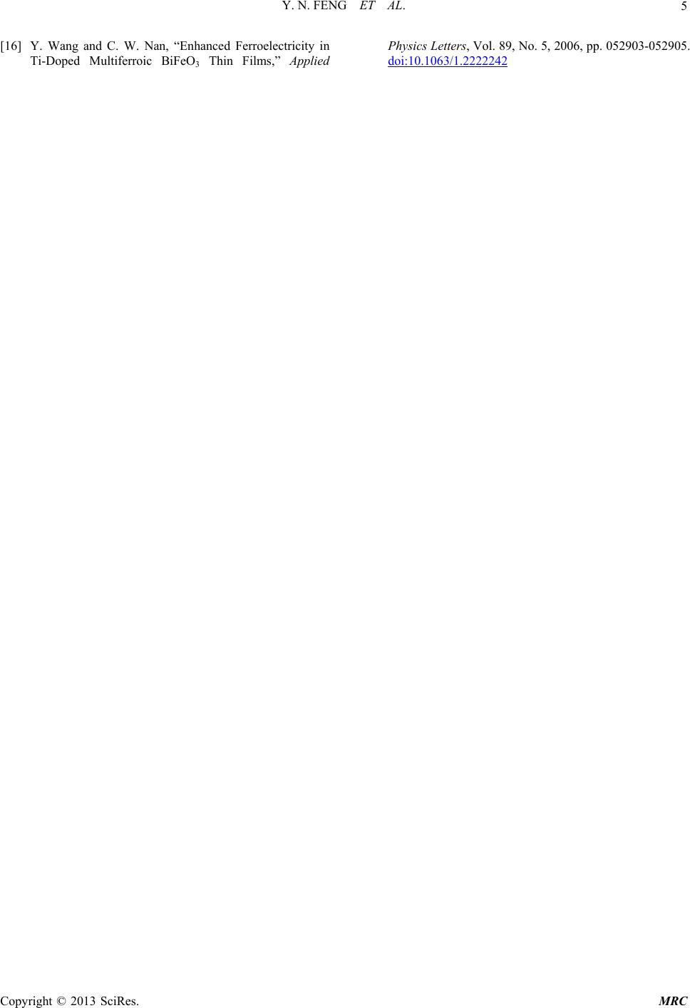
Modern Research in Catalysis, 2013, 2, 1-5
http://dx.doi.org/10.4236/mrc.2013.23A001 Published Online September 2013 (http://www.scirp.org/journal/mrc)
Magnetic and Photocatalytic Behaviors of Ca Mn
Co-Doped BiFeO3 Nanofibres
Yannan Feng, Huanchun Wang, Yang Shen, Yuanhua Lin*, Cewen Nan
State Key Laboratory of New Ceramics and Fine Processing, School of Materials Science and Engineering,
Tsinghua University, Beijing, China
Email: *linyh@tsinghua.edu.cn
Received May 29, 2013; revised June 29, 2013; accepted August 1, 2013
Copyright © 2013 Yannan Feng et al. This is an open access article distributed under the Creative Commons Attribution License,
which permits unrestricted use, distribution, and reproduction in any medium, provided the original work is properly cited.
ABSTRACT
Ca and Mn co-doped BiFeO3 ultrafine nanofibres were prepared with the purpose of improving magnetic and photo-
catalytic performances of the one-dimensional multiferroic material. Impurity phase introduced by both Bi fluctuation
and Mn substitution can be suppressed by Ca doping and a space group transition from R3c to C222 can also be trig-
gered by Bi-site doping. With co-substitution of Mn into iron site, the Ca0.15Bi0.85Mn0.05Fe0.95O3 nanofibres presented a
larger saturation magnetization than the singly Ca doping samples, possibly due to the increased double exchange in-
teration of Fe3+-O-Fe2+, strengthened by Ca and Mn. Photocatalytic degradation test witnessed a similar drop-and-rise
performance with the magnetism.
Keywords: Bismuth Ferrite; Nanofibre; Magnetic; Photocatalytic
1. Introduction
Bismuth ferrite (BiFeO3, or BFO), one of the most wide-
ly researched multiferroics, has continuously proved to
be a remarkable attraction, due to its room temperature
antiferromagnetic and ferroelectric orderings, [1] high vi-
sible light response, and consequent potentiality in mul-
tifunctional devices [2,3] and polluted water remediation
[4]. In order to promote the performance of BFO, a vari-
ety of nanostructures have been fabricated [5-7] and a
number of ions have been utilized as dopants, including
rare earth metals, [8] transition metals [9] and alkaline
earth metals [10]. However, unified theory can still be a
rather difficult issue, because the property is delicately
subject to both strain effect and doping effect. Therefore,
systematic study focused on one nanostructure and one
doping element is desired. La, Ca singly doped BFO na-
nofibres have been synthesized previously and studied
detailedly [7,11]. They did show enhanced behaviors in
magnetic and photocatalytic aspects, however none of
these individual dopants was able to modulate the prop-
erties ideally and arbitrarily. Given this fact, co-doping
has been proposed as an option. Some recent progress in
nanofibre preparation [12] has already demonstrated the
significance of co-doping and it is hence in urgent need
of extensive studies.
In this work, we fixed Mn doping content to 0.05 and
varied Ca concentration, in the hope of obtaining one-di-
mensional BFO nanofibres equipped with more excellent
capacity and exploring the interaction between dopants in
different doping sites.
2. Experimental
Ca Mn co-doped BFO (BaxBi1-xFe0.95Mn0.05O3) ultrafine
fibres with four Ca doping contents (x = 0, x = 0.05, x =
0.10, x = 0.15) were synthesized through a sol-gel based
electrospinning method. Precursor solution was prepared
with bismuth nitrate (Bi(NO3)3·5H2O) (5 mol% excess to
compensate for Bi loss during the heat treatment in air),
iron nitrate (Fe(NO3)3·9H2O), and calcium nitrate
(Ca(NO3)2·4H2O) and 50 wt.% manganous nitrate solu-
tion (Mn(NO3)2). Mixed together with the complexant,
citric acid (C6H8O7) in the solvent N, N-Dimethylfoma-
mide (DMF, C3H7NO), the homogeneous solution was
placed in the shade for 24 h aging. Afterwards, polyvi-
nylpyrrolidone (PVP, M = 1,300,000) was added to the
mixture solution, which was then continuously stirred till
PVP completely dissolved. Subsequently electrospinning
process was initiated by loading the ropy precursor solu-
tion into a syringe with its stainless-steel needle uploaded
with high voltage (13 kV). The rotating cylinder collector
10 cm away was grounded, and a static electric field of
*Corresponding author.
C
opyright © 2013 SciRes. MRC
Y. N. FENG ET AL.
2
1.3 kV/cm intensity was thus built across. The thick hy-
brid was squeezed out, drawn into ultrafine nanofibre and
started vibrating after going straight a little while, due to
the extremely high tension, which prolonged spun dura-
tion and assured DMF volatilized thoroughly. The non-
volatile, mainly the complex compound, remained on
PVP fibres which later weaved on an aluminium foil.
These as-spun fibres went through a heating process of
90 min presintering at 420˚C, and 3 h calcination at
580˚C.
X-ray diffraction (short for XRD) (Rigaku D/max 2500)
with Cu Kα (0.15418 nm) was utilized to analyze the
phase structure. Magnetic behaviors were observed in
physical property measurement system (PPMS-9T). Field
emission scan electron microscope (FESEM) (JSM-
7001F) was employed in the microstructure measurement,
and atom arrangement was approached with high resolu-
tion transmission electron microscope (HRTEM) (JOEL-
2011). Ultraviolet-visible (UV-Vis) curves were tested in
Hitachi U3310 spectrophotometer, while photocatalytic
performance was carried out under self-made reaction
and test system. The degradation of CR was determined
by the intensity of the absorption peak of CR (495 nm)
relative to its initial value (C/C0).
3. Result and Discussion
Results of phase structure analysis are shown in Figure 1.
Some typical peaks for nonstoichiometric phases can be
detected, which are possibly from Bi3.43Fe0.57O6 and
Bi25FeO40. Apart from these impurity peaks, all the other
could be ascribed to the main phase, BiFeO3, JCPDS
Card No.86 - 1518. High crystallity could be proved by
the sharp peak shape, and mergers of the doublet peaks,
for instance, (104) and (110), (006) and (202), (116) and
(122), (214) and (300), indicate a structural transition of
space group and symmetry can be obtained by Ca doping,
while Mn doping might introduce more impurities, simi-
lar phenomenon has been reported previously [13]. As
31.0 31.5 32.0 32.5 33.0
20 30 40 50 60 70
2Theta ( Deg.)
15%Ca 5%Mn
10%Ca 5%Mn
5%Ca 5%Mn
0%Ca 5%Mn
Intensity (a.u.)
2Theta (Deg.)
BiFeO3
Bi25FeO40
Figure 1. XRD patterns for pristine and doped BFO nano-
fibres. And slow scan spectra near 32˚.
Ca content grows, the peaks shifts to higher angle, indi-
cating the shrinkage of the crystal lattice.
Morphology and microstructure of these nanofibres
were observed and the results are shown in Figure 2. As
spun samples have an average diameter of 140 nm and
they are smooth in shape, owing to the polymer nature of
carrier PVP. However, after sintering process, the aver-
age diameter shrank to below 120 nm and the morphol-
ogy turned to be coarse. From the HRTEM image, we
can find that sunken area might be the grain boundary
zone and the adjacent grains stagger quite slightly in cry-
stal orientation probably because of the one-dimensional
restraint condition. In the surface area, looser atom ar-
rangement and different lattice plane can be observed,
compared with the inside bulk zone.
Mn co-doping might endowed these nanofibres enor-
mous impact on the thermal property and structure. In
fact, our previous study [11] showed that an annealing
temperature of 660˚C or a little lower was ideal for sin-
gly doped BFO nanofibres. In this co-doping case, we
had to reduce the sintering temperature to 580˚C to keep
the nanofibre shapes unbroken, though Mn concentration
was as little as 5%.
Magnetic properties of the as-prepared nanofibres are
graphed in Figure 3. When Mn is the only dopant, we
can see that the fibres show good magnetism with high
saturation magnetization, evident remnant magnetization
and coercive force. However, when Ca was doped into
the fibres, situation changed with a dramatic drop of both
saturation magnetization and coercive force. This down-
grading trend stops and turns upwards later with incre-
mental Ca content, and when Ca content reaches 10%,
saturation magnetization has exceeded that of 5% Mn
doped sample. We consider the magnetism of 5% Mn
doped fibres to be originated from the magnetic moment
of Mn itself, the charge compensation effect and the in-
cident impurity phases. Nevertheless, the pop-in of the
Ca might have indirectly conflicted with Mn somehow,
for instance, pinned the Mn valence state. Later on, when
Ca becomes majority, its impact on Fe becomes domi-
nating. At this moment, the moment of Mn can help
strengthen the magnetization instead, together with the
Fe valence state variation and Fe2+-O-Fe3+ super exchange
[12,14-16] caused by Ca doping. Our previous work has
shown saturation magnetization is positively correlated
with the Ca content, [11] while combining with this work,
we can infer that Mn should have dual effects, one will
be restrained by Ca doping, that is perhaps charge effect,
and the other, which is likely to be residual moment, will
cooperate with Ca on property improvement.
Owing to the superior specific surface area, the cata-
lytic activity is rather worthy to look forward to. Degra-
dation experiment of Congo red has demonstrated the
same trend with magnetism variation. As seen from the
Copyright © 2013 SciRes. MRC
Y. N. FENG ET AL. 3
(a)
(b)
(c)
Figure 2. Morphology and microstructure of BFO nanofi-
bres. SEM image of (a) as spun and (b) sintered sample, and
its (c) HRTEM image.
-10000 -50000500010000
-0.8
-0.4
0.0
0.4
0.8
0% Ca 5% Mn
5% Ca 5% Mn
10% Ca 5% Mn
15% Ca 5% Mn
Moment (emu /g)
Field
(
Oe
)
(a)
0.00 0.05 0.10 0.15
0
20
40
60
80
100
120
140 Coercive force
Saturation magnetization
Saturation magnet ization
Coercive forc e (O e)
0.2
0.3
0.4
0.5
0.6
0.7
0.8
Ms (emu/g)
(b)
Figure 3. (a) M-H curves and (b) magnetic properties in a
magnetic field with maximum of 10 k Oe at 300 K (room
temperature).
Figure 4, though all samples show good photocatalytic
efficiency as a result of considerably large specific sur-
face area and high visible light response, there exists a
minimum for photodecomposition. This minimum, aris-
ing when the doping concentration is equal, might simi-
larly suggest the mutual weakening effect between Ca
and Mn. When Ca doping content is more than 5%,
photocatalytic degradation shows positive correlation to
doping proportion. And with the co-existence of a small
amount of Mn, photocatalytic activity is raised markedly
and higher than Ca singly doping. This excellent per-
formance could be explained by increment of free carrier
and decreased electron-hole recombination rate, while
the excessive activity on the Mn singly doping condition
might be attributed to impurity phases and structural in-
fluence offered by Mn. In addition, when Mn was
brought in the lattice, the bandgap further narrowed
down and these nanofibres showed a bit of semi-conduc-
tivity, which was consistent with the color change from
previous Mn-free samples. We thus assumed that the
Copyright © 2013 SciRes. MRC
Y. N. FENG ET AL.
4
0.00 0.05 0.10 0.15
0.0
0.2
0.4
0.6
0.8
1.0
Residual percentage
Ca doping content
Ca doped BF O (5% Mn)
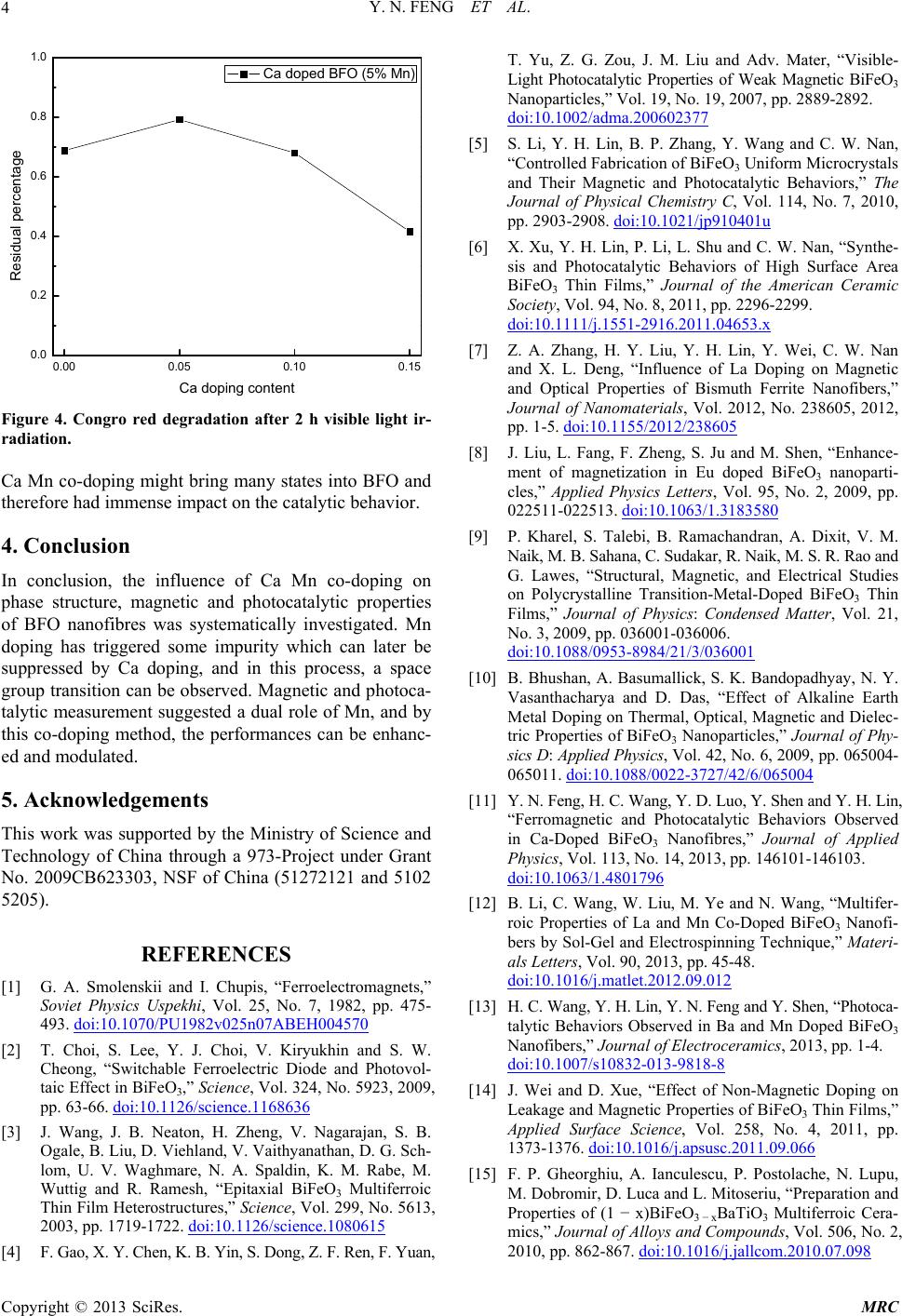
Figure 4. Congro red degradation after 2 h visible light ir-
radiation.
Ca Mn co-doping might bring many states into BFO and
therefore had immense impact on the catalytic behavior.
4. Conclusion
In conclusion, the influence of Ca Mn co-doping on
phase structure, magnetic and photocatalytic properties
of BFO nanofibres was systematically investigated. Mn
doping has triggered some impurity which can later be
suppressed by Ca doping, and in this process, a space
group transition can be observed. Magnetic and photoca-
talytic measurement suggested a dual role of Mn, and by
this co-doping method, the performances can be enhanc-
ed and modulated.
5. Acknowledgements
This work was supported by the Ministry of Science and
Technology of China through a 973-Project under Grant
No. 2009CB623303, NSF of China (51272121 and 5102
5205).
REFERENCES
[1] G. A. Smolenskii and I. Chupis, “Ferroelectromagnets,”
Soviet Physics Uspekhi, Vol. 25, No. 7, 1982, pp. 475-
493. doi:10.1070/PU1982v025n07ABEH004570
[2] T. Choi, S. Lee, Y. J. Choi, V. Kiryukhin and S. W.
Cheong, “Switchable Ferroelectric Diode and Photovol-
taic Effect in BiFeO3,” Science, Vol. 324, No. 5923, 2009,
pp. 63-66. doi:10.1126/science.1168636
[3] J. Wang, J. B. Neaton, H. Zheng, V. Nagarajan, S. B.
Ogale, B. Liu, D. Viehland, V. Vaithyanathan, D. G. Sch-
lom, U. V. Waghmare, N. A. Spaldin, K. M. Rabe, M.
Wuttig and R. Ramesh, “Epitaxial BiFeO3 Multiferroic
Thin Film Heterostructures,” Science, Vol. 299, No. 5613,
2003, pp. 1719-1722. doi:10.1126/science.1080615
[4] F. Gao, X. Y. Chen, K. B. Yin, S. Dong, Z. F. Ren, F. Yuan,
T. Yu, Z. G. Zou, J. M. Liu and Adv. Mater, “Visible-
Light Photocatalytic Properties of Weak Magnetic BiFeO3
Nanoparticles,” Vol. 19, No. 19, 2007, pp. 2889-2892.
doi:10.1002/adma.200602377
[5] S. Li, Y. H. Lin, B. P. Zhang, Y. Wang and C. W. Nan,
“Controlled Fabrication of BiFeO3 Uniform Microcrystals
and Their Magnetic and Photocatalytic Behaviors,” The
Journal of Physical Chemistry C, Vol. 114, No. 7, 2010,
pp. 2903-2908. doi:10.1021/jp910401u
[6] X. Xu, Y. H. Lin, P. Li, L. Shu and C. W. Nan, “Synthe-
sis and Photocatalytic Behaviors of High Surface Area
BiFeO3 Thin Films,” Journal of the American Ceramic
Society, Vol. 94, No. 8, 2011, pp. 2296-2299.
doi:10.1111/j.1551-2916.2011.04653.x
[7] Z. A. Zhang, H. Y. Liu, Y. H. Lin, Y. Wei, C. W. Nan
and X. L. Deng, “Influence of La Doping on Magnetic
and Optical Properties of Bismuth Ferrite Nanofibers,”
Journal of Nanomaterials, Vol. 2012, No. 238605, 2012,
pp. 1-5. doi:10.1155/2012/238605
[8] J. Liu, L. Fang, F. Zheng, S. Ju and M. Shen, “Enhance-
ment of magnetization in Eu doped BiFeO3 nanoparti-
cles,” Applied Physics Letters, Vol. 95, No. 2, 2009, pp.
022511-022513. doi:10.1063/1.3183580
[9] P. Kharel, S. Talebi, B. Ramachandran, A. Dixit, V. M.
Naik, M. B. Sahana, C. Sudakar, R. Naik, M. S. R. Rao and
G. Lawes, “Structural, Magnetic, and Electrical Studies
on Polycrystalline Transition-Metal-Doped BiFeO3 Thin
Films,” Journal of Physics: Condensed Matter, Vol. 21,
No. 3, 2009, pp. 036001-036006.
doi:10.1088/0953-8984/21/3/036001
[10] B. Bhushan, A. Basumallick, S. K. Bandopadhyay, N. Y.
Vasanthacharya and D. Das, “Effect of Alkaline Earth
Metal Doping on Thermal, Optical, Magnetic and Dielec-
tric Properties of BiFeO3 Nanoparticles,” Journal of Phy-
sics D: Applied Physics, Vol. 42, No. 6, 2009, pp. 065004-
065011. doi:10.1088/0022-3727/42/6/065004
[11] Y. N. Feng, H. C. Wang, Y. D. Luo, Y. Shen and Y. H. Lin,
“Ferromagnetic and Photocatalytic Behaviors Observed
in Ca-Doped BiFeO3 Nanofibres,” Journal of Applied
Physics, Vol. 113, No. 14, 2013, pp. 146101-146103.
doi:10.1063/1.4801796
[12] B. Li, C. Wang, W. Liu, M. Ye and N. Wang, “Multifer-
roic Properties of La and Mn Co-Doped BiFeO3 Nanofi-
bers by Sol-Gel and Electrospinning Technique,” Materi-
als Letters, Vol. 90, 2013, pp. 45-48.
doi:10.1016/j.matlet.2012.09.012
[13] H. C. Wang, Y. H. Lin, Y. N. Feng and Y. Shen, “Photoca-
talytic Behaviors Observed in Ba and Mn Doped BiFeO3
Nanofibers,” Journal of Electroceramics, 2013, pp. 1-4.
doi:10.1007/s10832-013-9818-8
[14] J. Wei and D. Xue, “Effect of Non-Magnetic Doping on
Leakage and Magnetic Properties of BiFeO3 Thin Films,”
Applied Surface Science, Vol. 258, No. 4, 2011, pp.
1373-1376. doi:10.1016/j.apsusc.2011.09.066
[15] F. P. Gheorghiu, A. Ianculescu, P. Postolache, N. Lupu,
M. Dobromir, D. Luca and L. Mitoseriu, “Preparation and
Properties of (1 x)BiFeO3 xBaTiO3 Multiferroic Cera-
mics,” Journal of Alloys and Compounds, Vol. 506, No. 2,
2010, pp. 862-867. doi:10.1016/j.jallcom.2010.07.098
Copyright © 2013 SciRes. MRC
Y. N. FENG ET AL.
Copyright © 2013 SciRes. MRC
5
[16] Y. Wang and C. W. Nan, “Enhanced Ferroelectricity in
Ti-Doped Multiferroic BiFeO3 Thin Films,” Applied
Physics Letters, Vol. 89, No. 5, 2006, pp. 052903-052905.
doi:10.1063/1.2222242